Carnegie stage 17: Difference between revisions
m (→Introduction) |
|||
| Line 2: | Line 2: | ||
== Introduction == | == Introduction == | ||
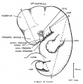
[[File:Stage17 | [[File:Stage17 bf1.jpg|left|400px]] | ||
===Facts=== | ===Facts=== | ||
Week 6, 42 - 44 days, 11 - 14 mm | Week 6, 42 - 44 days, 11 - 14 mm | ||
Revision as of 08:27, 19 January 2016
| Embryology - 14 Jun 2024 |
|---|
| Google Translate - select your language from the list shown below (this will open a new external page) |
|
العربية | català | 中文 | 中國傳統的 | français | Deutsche | עִברִית | हिंदी | bahasa Indonesia | italiano | 日本語 | 한국어 | မြန်မာ | Pilipino | Polskie | português | ਪੰਜਾਬੀ ਦੇ | Română | русский | Español | Swahili | Svensk | ไทย | Türkçe | اردو | ייִדיש | Tiếng Việt These external translations are automated and may not be accurate. (More? About Translations) |
Introduction

Facts
Week 6, 42 - 44 days, 11 - 14 mm
Gestational Age GA week 8
Events
- Ectoderm: sensory placodes, lens pit, otocyst,nasal pits moved ventrally, fourth ventricle of brain
- Mesoderm: heart prominence
- Head: 1st, 2nd and 3rd pharyngeal arch, forebrain, eye, auricular hillocks
- Body: heart, liver, umbilical cord, mesonephric ridge
- Limb: upper and lower limb buds, hand digital rays
Features
- pigmented eye, nasal pit, nasolacrimal groove, external acoustic meatus, auricular hillock, heart, digital rays, liver pronminance, thigh, ankle, foot plate, umbilical cord
- Identify: pigmented eye, nasal pit, nasolacrimal groove, external acoustic meatus, auricular hillock, heart, digital rays, liver prominence, thigh, ankle, foot plate, umbilical cord
- Links: Week 6 | System Development | Head | Lecture - Limb | Lecture - Head Development | Lecture - Sensory | Science Practical - Head | Science Practical - Sensory | Science Practical - Urogenital | Category:Carnegie Stage 17 | Stage 18
| Week: | 1 | 2 | 3 | 4 | 5 | 6 | 7 | 8 |
| Carnegie stage: | 1 2 3 4 | 5 6 | 7 8 9 | 10 11 12 13 | 14 15 | 16 17 | 18 19 | 20 21 22 23 |
- Carnegie Stages: 1 | 2 | 3 | 4 | 5 | 6 | 7 | 8 | 9 | 10 | 11 | 12 | 13 | 14 | 15 | 16 | 17 | 18 | 19 | 20 | 21 | 22 | 23 | About Stages | Timeline
Kyoto Collection
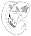
View: This is a dorsolateral view of embryo. Amniotic membrane removed.
Image source: Embryology page Created: 19.03.1999

Ventral view of head region (1 mm scale).
Image source: The Kyoto Collection images are reproduced with the permission of Prof. Kohei Shiota and Prof. Shigehito Yamada, Anatomy and Developmental Biology, Kyoto University Graduate School of Medicine, Kyoto, Japan for educational purposes only and cannot be reproduced electronically or in writing without permission.
Carnegie Collection


| iBook - Carnegie Embryos | |
|---|---|

|
|
Blechschmidt Collection
|
Model from serial section reconstruction.
|


Image source: The Blechschmidt Collection images are reproduced with the permission of Prof. Christoph Viebahn, director of the Institute of Anatomy and Embryology, , University Medical Center Göttingen. Images are for educational purposes only and cannot be reproduced electronically or in writing without permission.
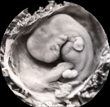
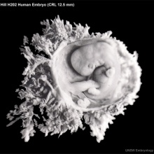
Hill Collection
| Hill H58 | |
|---|---|

|

|
| left dorsolateral | left lateral |
| Hill H202 | |

|

|
Image source: The images from the Hill Collection (part of the Embryological Collection) are reproduced with the permission of the Museum für Naturkunde, Leibniz Institute for Research on Evolution and Biodiversity. Images are for educational purposes only and must not be reproduced electronically or in writing without permission from the Museum für Naturkunde Berlin.
Embryo Virtual Slide
|
|
Scanning EM

|

|
| Ventral view of head showing upper lip, maxilla and nasal region. | Note that a ventral image of only half the head has been "mirrored" to generate this image.
Image Source: Prof Virginia Diewert |
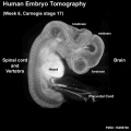
Tomography
Optical projection tomography movie of rotating stage 17 embryo. Note the detailed structural view of neural system development. |

Stage 17 Optical Projection Tomography (left) |

Stage 17 Optical Projection Tomography (right) |
Additional Images

External ear Stages 14-23 and adult
Streeter 1921 Plate 3
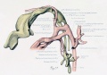
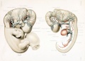
Embryo 940 cardiovascular lateral view
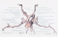
Embryo 940 cardiovascular ventrolateral view
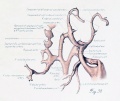
Embryo 940 cardiovascular ventral view
Sabin 1909 Fig. 4
Keith 1021 Fig. 45


- Carnegie Stages: 1 | 2 | 3 | 4 | 5 | 6 | 7 | 8 | 9 | 10 | 11 | 12 | 13 | 14 | 15 | 16 | 17 | 18 | 19 | 20 | 21 | 22 | 23 | About Stages | Timeline
Cite this page: Hill, M.A. (2024, June 14) Embryology Carnegie stage 17. Retrieved from https://embryology.med.unsw.edu.au/embryology/index.php/Carnegie_stage_17
- © Dr Mark Hill 2024, UNSW Embryology ISBN: 978 0 7334 2609 4 - UNSW CRICOS Provider Code No. 00098G