Implantation: Difference between revisions
No edit summary |
|||
| Line 10: | Line 10: | ||
[[Image:Week1_summary.jpg|400|Week 1 Human Development Overview]] | [[Image:Week1_summary.jpg|400|Week 1 Human Development Overview]] | ||
[[Development_Animation_-_Implantation| | |||
==Implantation Cartoon== | |||
{| | |||
| <qt>file=Week2_001.mov|width=250px|height=240px|controller=true|autoplay=false</qt> | |||
| This animation shows the process of implantation, occurring during week 2 of development in humans. | |||
The beginning of the animation shows: the uterus lining (endometrium epithelium), the hatched [[B#blastocyst|blastocyst]] with a flat outer layer of trophoblast cells (green), the inner cell mass which has formed into the bilaminar embryo ([[E#epiblast|epiblast]] and [[H#hypoblast|hypoblast]]) and the large fluid-filled space ([[B#blastocoel|blastocoel]]). | |||
'''green cells''' - trophoblast layer of the conceptus | |||
'''blue cells''' - [[E#epiblast|epiblast layer]] of the bilaminar embryo | |||
'''yellow cells''' - [[H#hypoblast|hypoblast layer]] of the bilaminar embryo | |||
'''white cells''' - uterine endometrium epithelium | |||
'''red''' - maternal blood vessel | |||
Links: [[Quicktime_Development_Animation_-_Implantation|Quicktime version]] | [[Development_Animation_-_Implantation|Flash Version]] | [[Media:Week2_001.mov|Quicktime movie]] | |||
|} | |||
The second week of human development is concerned with the process of implantation and the differentiation of the blastocyst into early embryonic and placental forming structures. | The second week of human development is concerned with the process of implantation and the differentiation of the blastocyst into early embryonic and placental forming structures. | ||
Revision as of 06:41, 12 October 2010
Introduction
The term used to describe process of attachment and invasion of the uterus endometrium by the blastocyst (conceptus) in placental animals. Abnormal implantation is where this process does not occur in the body of the uterus (ectopic) or where the placenta forms incorrectly.
--Mark Hill 19:10, 5 August 2009 (EST) Page under development - notice removed when completed.
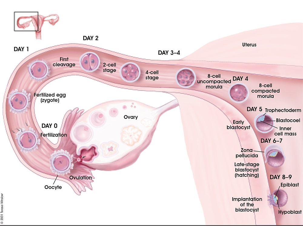
Week 1 and 2 Human Development Overview
Implantation Cartoon
| width=250px|height=240px|controller=true|autoplay=false</qt> | This animation shows the process of implantation, occurring during week 2 of development in humans.
The beginning of the animation shows: the uterus lining (endometrium epithelium), the hatched blastocyst with a flat outer layer of trophoblast cells (green), the inner cell mass which has formed into the bilaminar embryo (epiblast and hypoblast) and the large fluid-filled space (blastocoel). green cells - trophoblast layer of the conceptus blue cells - epiblast layer of the bilaminar embryo yellow cells - hypoblast layer of the bilaminar embryo white cells - uterine endometrium epithelium red - maternal blood vessel Links: Quicktime version | Flash Version | Quicktime movie |
The second week of human development is concerned with the process of implantation and the differentiation of the blastocyst into early embryonic and placental forming structures.
- implantation commences about day 6 to 7
- Adplantation - begins with initial adhesion to the uterine epithelium
- blastocyst then slows in motility, "rolls" on surface, aligns with the inner cell mass closest to the epithelium and stops
- Implantation - migration of the blastocyst into the uterine epithelium, process complete by about day 9
- coagulation plug - left where the blastocyst has entered the uterine wall day 12
Normal Implantation Sites - in uterine wall superior, posterior, lateral
Abnormal Implantation
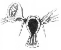
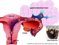
Abnormal implantation sites or Ectopic Pregnancy occurs if implantation is in uterine tube or outside the uterus.
- sites - external surface of uterus, ovary, bowel, gastrointestinal tract, mesentry, peritoneal wall
- If not spontaneous then, embryo has to be removed surgically
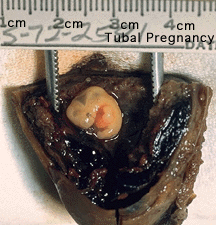
Tubal pregnancy - 94% of ectopic pregnancies
- if uterine epithelium is damaged (scarring, pelvic inflammatory disease)
- if zona pellucida is lost too early, allows premature tubal implantation
- embryo may develop through early stages, can erode through the uterine horn and reattach within the peritoneal cavity

|

|
Embryo Week: Week 1 | Week 2 | Week 3 | Week 4 | Week 5 | Week 6 | Week 7 | Week 8 | Week 9
- Carnegie Stages: 1 | 2 | 3 | 4 | 5 | 6 | 7 | 8 | 9 | 10 | 11 | 12 | 13 | 14 | 15 | 16 | 17 | 18 | 19 | 20 | 21 | 22 | 23 | About Stages | Timeline
Glossary Links
- Glossary: A | B | C | D | E | F | G | H | I | J | K | L | M | N | O | P | Q | R | S | T | U | V | W | X | Y | Z | Numbers | Symbols | Term Link
Cite this page: Hill, M.A. (2024, June 2) Embryology Implantation. Retrieved from https://embryology.med.unsw.edu.au/embryology/index.php/Implantation
- © Dr Mark Hill 2024, UNSW Embryology ISBN: 978 0 7334 2609 4 - UNSW CRICOS Provider Code No. 00098G

