Somitogenesis: Difference between revisions
No edit summary |
|||
| Line 1: | Line 1: | ||
<div style="background:#F5FFFA; border: 1px solid #CEF2E0; padding: 1em; margin: auto; width: 95%; float:left;"><div style="margin:0;background-color:#cef2e0;font-family:sans-serif;font-size:120%;font-weight:bold;border:1px solid #a3bfb1;text-align:left;color:#000;padding-left:0.4em;padding-top:0.2em;padding-bottom:0.2em;">Notice - Mark Hill</div>Currently this page is only a template and will be updated (this notice removed when completed).</div> | |||
==Introduction== | ==Introduction== | ||
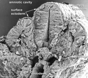
[[File:Stage11 sem100c.jpg|thumb|Somites (week 4)]] | [[File:Stage11 sem100c.jpg|thumb|Somites (week 4)]] | ||
| Line 5: | Line 7: | ||
A [[S#somite|somite]] is added either side of the [[N#notochord|notochord]] ([[A#axial mesoderm|axial mesoderm]]) to form a somite pair. The segmentation does not occur in the head region, and begins cranially (head end) and extends caudally (tailward) adding a somite pair at regular time intervals. The process is sequential and therefore used to stage the age of many different species embryos based upon the number visible somite pairs. | A [[S#somite|somite]] is added either side of the [[N#notochord|notochord]] ([[A#axial mesoderm|axial mesoderm]]) to form a somite pair. The segmentation does not occur in the head region, and begins cranially (head end) and extends caudally (tailward) adding a somite pair at regular time intervals. The process is sequential and therefore used to stage the age of many different species embryos based upon the number visible somite pairs. | ||
: | :'''Links:''' [[Musculoskeletal System Development]] | ||
Revision as of 10:02, 27 August 2010
Introduction
The term used to describe the process of segmentation of the paraxial mesoderm within the trilaminar embryo body to form pairs of somites, or balls of mesoderm. In humans, the first somite pair appears at day 20 and adds caudally at 1 somite pair/90 minutes until on average 44 pairs eventually form.
A somite is added either side of the notochord (axial mesoderm) to form a somite pair. The segmentation does not occur in the head region, and begins cranially (head end) and extends caudally (tailward) adding a somite pair at regular time intervals. The process is sequential and therefore used to stage the age of many different species embryos based upon the number visible somite pairs.
Embryo Week: Week 1 | Week 2 | Week 3 | Week 4 | Week 5 | Week 6 | Week 7 | Week 8 | Week 9
- Carnegie Stages: 1 | 2 | 3 | 4 | 5 | 6 | 7 | 8 | 9 | 10 | 11 | 12 | 13 | 14 | 15 | 16 | 17 | 18 | 19 | 20 | 21 | 22 | 23 | About Stages | Timeline
Glossary Links
- Glossary: A | B | C | D | E | F | G | H | I | J | K | L | M | N | O | P | Q | R | S | T | U | V | W | X | Y | Z | Numbers | Symbols | Term Link
Cite this page: Hill, M.A. (2024, June 9) Embryology Somitogenesis. Retrieved from https://embryology.med.unsw.edu.au/embryology/index.php/Somitogenesis
- © Dr Mark Hill 2024, UNSW Embryology ISBN: 978 0 7334 2609 4 - UNSW CRICOS Provider Code No. 00098G
