Embryology History - Julius Kollmann: Difference between revisions

From Embryology
No edit summary
mNo edit summary
 
(21 intermediate revisions by 2 users not shown)
Line 1: Line 1:
{{Header}}
==Introduction==
==Introduction==
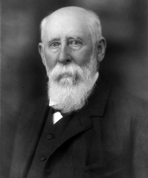
[[File:Julius_kollmann.jpg|thumb| Julius Konstantin Ernst Kollmann (1834-1918)]]
[[File:Julius_kollmann.jpg|thumb| Julius Konstantin Ernst Kollmann (1834-1918)]]
Line 5: Line 6:
Professor extraordinarius, Munich University, 1870–8; professor of anatomy, Basel University, 1878 – 1913.


Images from Kollmann's atlas of embryology were extensively reused in embryology textbooks and are the basis of many modern drawings.
Images from Kollmann's 2 volume Atlas of the Development of Man (Handatlas der entwicklungsgeschichte des menschen) were extensively reused in other embryology textbooks and are also the basis of many modern drawings.


[[:Category:Kollmann|Category:Kollmann]]
:'''Links:'''  [[Atlas_of_the_Development_of_Man_1|Atlas (Volume 1)]] | [[Atlas_of_the_Development_of_Man_2|Atlas (Volume 2)]] | [[:Category:Kollmann|Category:Kollmann]] | [[Embryology History - Julius Kollmann|Julius Kollmann]]


:{{Template:History People}}
{{History People}}


==Atlas of the Development of Man (Volume 2)==
Contents
===Contents===
 
[[Atlas of the Development of Man 2]]
* [[Atlas of the Development of Man 2 - Gastrointestinal]]
* [[Atlas of the Development of Man 2 - Gastrointestinal|Gastrointestinal]]
* [[Atlas of the Development of Man 2 - Respiratory]]
* [[Atlas of the Development of Man 2 - Respiratory|Respiratory]]
* [[Atlas of the Development of Man 2 - Urogenital]]
* [[Atlas of the Development of Man 2 - Urogenital|Urogenital]]
* [[Atlas of the Development of Man 2 - Cardiovascular]]
* [[Atlas of the Development of Man 2 - Cardiovascular|Cardiovascular]]
* [[Atlas of the Development of Man 2 - Neural]]
* [[Atlas of the Development of Man 2 - Neural|Neural]]
* [[Atlas of the Development of Man 2 - Integumentary]]
* [[Atlas of the Development of Man 2 - Integumentary|Integumentary]]
* [[Atlas of the Development of Man 2 - Smell]]
* [[Atlas of the Development of Man 2 - Smell|Smell]]
* [[Atlas of the Development of Man 2 - Vision]]
* [[Atlas of the Development of Man 2 - Vision|Vision]]
* [[Atlas of the Development of Man 2 - Hearing]]
* [[Atlas of the Development of Man 2 - Hearing|Hearing]]
 
==References==
<references/>
 
Handatlas der entwicklungsgeschichte des menschen: Volume 1 (Atlas of the Development of Man Volume 1)
 
Handatlas der entwicklungsgeschichte des menschen: Volume 2 (Atlas of the Development of Man Volume 2)
 
==Images==
===Head===
<gallery>
File:Kollmann341.jpg|Fig. 341.  Branchial gill arch and columns, fissures branchiales in an embryo Shark (''Callorhynchus antarcticus'')
File:Kollmann342.jpg|Fig. 342. The anterior end of an embryo of Shark (Callorhynchus antarcticus
File:Kollmann343.jpg|Fig. 343. Head of a Lizard Embryo (''Sphenodon punctatum Hatteria'')
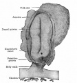
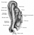
File:Kollmann344.jpg|Fig. 344. Mouth opening and its boundary in a human embryo of 19-20 days
File:Kollmann345.jpg
File:Kollmann346.jpg
File:Kollmann347.jpg
File:Kollmann348.jpg
File:Kollmann349.jpg
File:Kollmann350.jpg
File:Kollmann351.jpg
File:Kollmann352.jpg
File:Kollmann353.jpg
File:Kollmann354.jpg
File:Kollmann355.jpg
File:Kollmann356.jpg
File:Kollmann357.jpg
File:Kollmann358.jpg
File:Kollmann359.jpg
File:Kollmann360.jpg
File:Kollmann361.jpg
File:Kollmann362.jpg
File:Kollmann363.jpg
File:Kollmann364.jpg
File:Kollmann365.jpg
File:Kollmann366.jpg
File:Kollmann367.jpg
File:Kollmann368.jpg
File:Kollmann369.jpg
File:Kollmann370.jpg
File:Kollmann371.jpg
File:Kollmann372.jpg
File:Kollmann373.jpg
File:Kollmann374.jpg
File:Kollmann375.jpg
File:Kollmann376.jpg
File:Kollmann377.jpg
File:Kollmann378.jpg
</gallery>
 
:Links: [[Head Development]]
 
===Gastrointestinal===
<gallery>
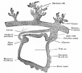
File:Kollmann379.jpg|Fig. 379. The yolk sac, saccule vitellinus, the first organ of nutrition of the embryo.
File:Kollmann380.jpg|Fig. 380. The germ of the embryo as in Fig. 379.
File:Kollmann381.jpg|Fig. 381. Intestines a human embryo 2.4 mm in length
File:Kollmann382.jpg
File:Kollmann383.jpg
File:Kollmann384.jpg
File:Kollmann385.jpg
File:Kollmann386.jpg
File:Kollmann387.jpg
File:Kollmann388.jpg
File:Kollmann389.jpg
File:Kollmann390.jpg
File:Kollmann391.jpg
File:Kollmann392.jpg
File:Kollmann393.jpg
File:Kollmann394.jpg
File:Kollmann395.jpg
File:Kollmann396.jpg
File:Kollmann397.jpg
File:Kollmann398-403.jpg|Fig. 398-403.
File:Kollmann404.jpg
</gallery>
===Respiratory===
<gallery>
File:Kollmann405-406.jpg|Fig. 405-406.
File:Kollmann407-409.jpg|Fig. 407-409.
File:Kollmann410.jpg
File:Kollmann411.jpg
File:Kollmann412.jpg
File:Kollmann413.jpg
File:Kollmann414.jpg
File:Kollmann415.jpg
File:Kollmann416.jpg
File:Kollmann417.jpg
File:Kollmann418.jpg
File:Kollmann419.jpg
File:Kollmann420.jpg
File:Kollmann421.jpg
File:Kollmann422.jpg
File:Kollmann423.jpg
File:Kollmann424.jpg
</gallery>
 
===Urogenital===


<gallery>
===Sample Images from the Atlas===
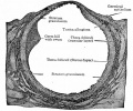
File:Kollmann425.jpg|Fig. 425. Middle plate (intermediate mesoderm) human embryo of 13 somites, and 2.4 mm CRL.
{|
File:Kollmann426.jpg|Fig. 426. Mesonephroi, a human embryo mesonephros of 9 mm CRL (week 5).
| width="300px"|[[File:Kollmann344.jpg|300px]]
File:Kollmann427.jpg
| width="300px"|[[File:Kollmann407-409.jpg|200px]]
File:Kollmann428.jpg
| width="300px"|[[File:Kollmann425.jpg|300px]]
File:Kollmann429.jpg
|-
File:Kollmann430.jpg
| [[Atlas of the Development of Man 2 - Gastrointestinal|Gastrointestinal]] Fig. 344. Mouth opening and its boundary in a human embryo of 19-20 days.
File:Kollmann431.jpg
| [[Atlas of the Development of Man 2 - Respiratory|Respiratory]] Fig. 407-409. Pulmonary system of a human embryo.
File:Kollmann432.jpg
| [[Atlas of the Development of Man 2 - Urogenital|Urogenital]] Fig. 425. Middle plate (intermediate mesoderm) human embryo of 13 somites, and 2.4 mm CRL.
File:Kollmann433.jpg
|-
File:Kollmann434.jpg
| [[File:Kollmann511.jpg|300px]]
File:Kollmann435.jpg
| [[File:Kollmann607.jpg|300px]]
File:Kollmann436.jpg
| [[File:Kollmann666.jpg|200px]]
File:Kollmann437.jpg
|-
File:Kollmann438.jpg|Fig. 438. The urogenital inside the peritoneal cavity in a human embryo of 11 weeks.
| [[Atlas of the Development of Man 2 - Cardiovascular|Cardiovascular]] Fig. 511. Vascular system of a human embryo of 1.3 mm.
File:Kollmann439.jpg
| [[Atlas of the Development of Man 2 - Neural|Neural]] Fig. 607. Neural tube of a human embryo of 22 mm CRL about 7 weeks old.
File:Kollmann440.jpg
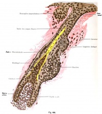
| [[Atlas of the Development of Man 2 - Integumentary|Integumentary]] Fig. 666. Development of the wool hair (lanugo) in a human fetus of 5.5 months.
File:Kollmann441.jpg
|-
File:Kollmann442.jpg
| [[File:Kollmann683.jpg|300px]]
File:Kollmann443.jpg
| [[File:Kollmann709.jpg|300px]]
File:Kollmann444.jpg|Fig. 444. Descent of testicles
| [[File:Kollmann738.jpg|300px]]
File:Kollmann445.jpg
|-
File:Kollmann446.jpg
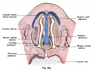
| [[Atlas of the Development of Man 2 - Smell|Smell]] Fig. 683.
File:Kollmann447.jpg
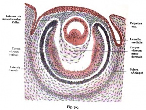
| [[Atlas of the Development of Man 2 - Vision|Vision]]  Fig. 709.
File:Kollmann448.jpg
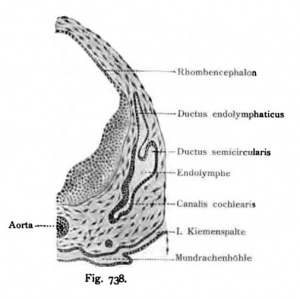
| [[Atlas of the Development of Man 2 - Hearing|Hearing]] Fig. 738. Typical auditory vesicle in a human embryo of 10.2 mm CRL.
File:Kollmann449.jpg
|}
File:Kollmann450.jpg
File:Kollmann451.jpg
File:Kollmann452.jpg
File:Kollmann453.jpg
File:Kollmann454.jpg
File:Kollmann455.jpg
File:Kollmann456.jpg
File:Kollmann457.jpg
File:Kollmann458.jpg
File:Kollmann459.jpg
File:Kollmann460.jpg
File:Kollmann461.jpg
File:Kollmann462.jpg
File:Kollmann463.jpg
File:Kollmann464.jpg
File:Kollmann465.jpg
File:Kollmann466.jpg
File:Kollmann467.jpg
File:Kollmann468.jpg
File:Kollmann469.jpg
File:Kollmann470.jpg
File:Kollmann471.jpg
File:Kollmann472.jpg
File:Kollmann473.jpg
File:Kollmann474.jpg
File:Kollmann475.jpg
File:Kollmann476-477.jpg|Fig. 476-477.
File:Kollmann478.jpg
File:Kollmann479.jpg
File:Kollmann480.jpg
File:Kollmann481.jpg
File:Kollmann482.jpg
File:Kollmann483.jpg
File:Kollmann484.jpg
File:Kollmann485-490.jpg|Fig. 485-490
File:Kollmann491-496.jpg|Fig. 491-496
File:Kollmann497-500.jpg|Fig. 497-500
File:Kollmann501.jpg
File:Kollmann502.jpg
File:Kollmann503.jpg
File:Kollmann504.jpg
File:Kollmann505.jpg
File:Kollmann506.jpg
File:Kollmann507.jpg
File:Kollmann508.jpg
File:Kollmann509.jpg
File:Kollmann510.jpg
</gallery>


===Cardiovascular===
{{KollmannAtlas2}}
Embryology the heart and vessels (Embryologia cordis et vasorum)


For the embryology of the heart, wherever possible, human hearts were used. Where this was not possible, the models of the mammalian heart by Born been used since the development of the heart of man
==Images used in Bailey Textbook==
coincides with that of the rabbit.


The coloring of the vessels is the same as in representations of the circulation is otherwise common, that is, those vessels that lead to the birth of venous blood are blue, those who after the birth of arterial
Bailey, F.R. and Miller, A.M. (1921). Text-Book of Embryology. New York: William Wood and Co.
blood are painted red.
 
The embryonic vessels had received clear walls to represent graphically. In reality they have only limited endothelial lining, as indicated in many sections.
 
<gallery>
File:Kollmann511.jpg|Fig. 511. Vascular system of a human embryo of 1.3 mm.
File:Kollmann512.jpg|Fig. 512. Vascular system of a human embryo of 1.3 mm.
File:Kollmann513.jpg|Fig. 513. Human embryo heart of 2.11 mm in length.
File:Kollmann514.jpg
File:Kollmann515.jpg
File:Kollmann516.jpg
File:Kollmann517.jpg
File:Kollmann518.jpg
File:Kollmann519.jpg
File:Kollmann520.jpg
File:Kollmann521.jpg
File:Kollmann522.jpg
File:Kollmann523.jpg
File:Kollmann524.jpg
File:Kollmann525.jpg
File:Kollmann526.jpg
File:Kollmann527.jpg
File:Kollmann528.jpg|Fig. 528. Heart of a Rabbit Embryo seen from behind at 3.4 mm head length
File:Kollmann529.jpg|Fig. 529. The heart of a 24 mm Embryo
File:Kollmann530.jpg|Fig. 530. Fetal heart (6 months) in normal situation
File:Kollmann531.jpg|Fig. 531. Heart included in the pericardium of a human embryo of 7.5 mm body length
File:Kollmann532.jpg|Fig. 532. Development of the heart chambers and septa
File:Kollmann533.jpg|Fig. 533. Heart of a newborn viewed from the front and placed in the vertical direction
File:Kollmann534.jpg|Fig. 534. Fetal heart, dorsal half with the afferent paths, open and colored according to the physiological condition of the blood
File:Kollmann535.jpg|Fig. 535. The aortic arch in the shark embryo (Pristiurus)
File:Kollmann536.jpg|Fig. 536. The arteries of the gill arch region of a shark embryo (Pristiurus)
File:Kollmann537.jpg|Fig. 537. Aortic arch of mammals and man
File:Kollmann538.jpg|Fig. 538. Arteries in mammals and humans from the Aortic Arch
File:Kollmann539.jpg
File:Kollmann540.jpg
File:Kollmann541.jpg
File:Kollmann542.jpg
File:Kollmann543.jpg
File:Kollmann544.jpg
File:Kollmann545.jpg
File:Kollmann546.jpg
File:Kollmann547.jpg
File:Kollmann548.jpg
File:Kollmann549.jpg
File:Kollmann550.jpg
File:Kollmann551.jpg
File:Kollmann552.jpg
File:Kollmann553.jpg
File:Kollmann554.jpg
File:Kollmann555.jpg
File:Kollmann556.jpg
File:Kollmann557.jpg
File:Kollmann558.jpg
File:Kollmann559.jpg
File:Kollmann560.jpg
File:Kollmann561.jpg
File:Kollmann562.jpg
File:Kollmann563.jpg
File:Kollmann564.jpg
Kollmann565-569.jpg|Fig. 565-569.
File:Kollmann570.jpg
File:Kollmann571.jpg
File:Kollmann572.jpg
File:Kollmann573.jpg
File:Kollmann574.jpg
File:Kollmann575.jpg
File:Kollmann576.jpg
File:Kollmann577.jpg
File:Kollmann578.jpg
File:Kollmann579.jpg
File:Kollmann580.jpg
File:Kollmann581.jpg
File:Kollmann582.jpg
</gallery>
 
===Neural===
<gallery>
File:Kollmann583.jpg|Fig. 583. Human embryo of 1.3 mm length
File:Kollmann584.jpg|Fig. 584. The first unit of the nervous system in a human embryo
File:Kollmann585.jpg|Fig. 585. The formation of the nervous system in a human embryo of 2.69 mm length
File:Kollmann586.jpg|Fig. 586. The primordial central nervous system in a human embryo of 2.11 mm length
File:Kollmann587.jpg
File:Kollmann588.jpg
File:Kollmann589.jpg
File:Kollmann590.jpg
File:Kollmann591.jpg
File:Kollmann592.jpg
File:Kollmann593.jpg
File:Kollmann594.jpg
File:Kollmann595.jpg
File:Kollmann596.jpg
File:Kollmann597.jpg
File:Kollmann598.jpg
File:Kollmann599.jpg
File:Kollmann600.jpg
File:Kollmann601.jpg
File:Kollmann602.jpg
File:Kollmann603.jpg
File:Kollmann604.jpg
File:Kollmann605.jpg
File:Kollmann606.jpg
File:Kollmann607.jpg
File:Kollmann608.jpg
File:Kollmann609.jpg
File:Kollmann610.jpg
File:Kollmann611.jpg
File:Kollmann612.jpg
File:Kollmann613.jpg
File:Kollmann614.jpg
File:Kollmann615.jpg
File:Kollmann616.jpg
File:Kollmann617.jpg
File:Kollmann618.jpg
File:Kollmann619.jpg
File:Kollmann620.jpg
File:Kollmann621.jpg
File:Kollmann622.jpg
File:Kollmann623.jpg
File:Kollmann624.jpg
File:Kollmann625.jpg
File:Kollmann626.jpg
File:Kollmann627.jpg
File:Kollmann628.jpg
File:Kollmann629.jpg
File:Kollmann630.jpg
File:Kollmann631.jpg
File:Kollmann632.jpg
File:Kollmann633.jpg
File:Kollmann634.jpg
File:Kollmann635.jpg
File:Kollmann636.jpg
File:Kollmann637.jpg
File:Kollmann638.jpg
File:Kollmann639.jpg
File:Kollmann640-641.jpg
File:Kollmann642.jpg
File:Kollmann643.jpg
File:Kollmann644.jpg
File:Kollmann645.jpg
File:Kollmann646.jpg
File:Kollmann647.jpg
File:Kollmann648.jpg
File:Kollmann649.jpg
File:Kollmann650.jpg
File:Kollmann651.jpg
</gallery>
===Integumentary===
<gallery>
File:Kollmann652.jpg
File:Kollmann653.jpg
File:Kollmann654.jpg
File:Kollmann655-656.jpg
File:Kollmann657.jpg
File:Kollmann658.jpg
File:Kollmann659.jpg
File:Kollmann660.jpg
File:Kollmann661.jpg
File:Kollmann662.jpg
File:Kollmann663.jpg
File:Kollmann664.jpg
File:Kollmann665.jpg
File:Kollmann666.jpg
File:Kollmann667.jpg
File:Kollmann668-670.jpg
</gallery>
 
===Smell===
<gallery>
File:Kollmann671.jpg|
File:Kollmann672.jpg|
File:Kollmann673.jpg|
File:Kollmann674.jpg|
File:Kollmann675.jpg|
File:Kollmann676.jpg|
File:Kollmann677.jpg|
File:Kollmann678.jpg|
File:Kollmann679.jpg|
File:Kollmann680.jpg|
File:Kollmann681.jpg|
File:Kollmann682.jpg|
File:Kollmann683.jpg|
File:Kollmann684.jpg|
File:Kollmann685.jpg|
File:Kollmann686.jpg|
File:Kollmann687.jpg|
File:Kollmann688.jpg|
File:Kollmann689.jpg|
File:Kollmann690.jpg|
</gallery>
 
===Vision===
<gallery>
File:Kollmann691.jpg|
File:Kollmann692.jpg|
File:Kollmann693.jpg|
File:Kollmann694.jpg|
File:Kollmann695.jpg|
File:Kollmann696.jpg|
File:Kollmann697.jpg|
File:Kollmann698.jpg|
File:Kollmann699.jpg|
File:Kollmann700.jpg|
File:Kollmann701.jpg|
File:Kollmann702.jpg|
File:Kollmann703.jpg|
File:Kollmann704.jpg|
File:Kollmann705.jpg|
File:Kollmann706.jpg|
File:Kollmann707.jpg|
File:Kollmann708.jpg|
File:Kollmann709.jpg|
File:Kollmann710.jpg|
File:Kollmann711.jpg|
File:Kollmann712.jpg|
File:Kollmann713.jpg|
File:Kollmann714.jpg|
File:Kollmann715.jpg|
File:Kollmann716.jpg|
File:Kollmann717.jpg|
File:Kollmann718.jpg|
File:Kollmann719.jpg|
File:Kollmann720.jpg|
File:Kollmann721.jpg|
File:Kollmann722.jpg|
File:Kollmann723.jpg|
File:Kollmann724.jpg|
File:Kollmann725.jpg|
File:Kollmann726.jpg|
File:Kollmann727.jpg|
File:Kollmann728.jpg|
File:Kollmann729.jpg|
File:Kollmann730.jpg|
</gallery>
 
===Hearing===
<gallery>
File:Kollmann731.jpg|
File:Kollmann732.jpg|
File:Kollmann733.jpg|
File:Kollmann734.jpg|
File:Kollmann735.jpg|
File:Kollmann736-737.jpg|
File:Kollmann738.jpg|
File:Kollmann739.jpg|
File:Kollmann740.jpg|
File:Kollmann741.jpg|
File:Kollmann742.jpg|
File:Kollmann743.jpg|
File:Kollmann744.jpg|
File:Kollmann745.jpg|
File:Kollmann746.jpg|
File:Kollmann747.jpg|
File:Kollmann748.jpg|
File:Kollmann749.jpg|
File:Kollmann750.jpg|
File:Kollmann751.jpg|
File:Kollmann752.jpg|
File:Kollmann753.jpg|
File:Kollmann754.jpg|
File:Kollmann755.jpg|
File:Kollmann756.jpg|
File:Kollmann757.jpg|
File:Kollmann758.jpg|
File:Kollmann759.jpg|
File:Kollmann760.jpg|
File:Kollmann761.jpg|
File:Kollmann762-765.jpg|
File:Kollmann766-769.jpg|
 
</gallery>
 
===Bailey===


<gallery>
<gallery>
Line 493: Line 72:
File:Bailey308.jpg
File:Bailey308.jpg
</gallery>
</gallery>
Bailey, F.R. and Miller, A.M. (1921). '''Text-Book of Embryology.''' New York: William Wood and Co.
:[[Book_-_Text-Book_of_Embryology_(1921)|'''Text-Book of Embryology''']]: [[Book_-_Text-Book_of_Embryology_1|Germ cells]] | [[Book_-_Text-Book_of_Embryology_2|Maturation]] | [[Book_-_Text-Book_of_Embryology_3|Fertilization]] | [[Book_-_Text-Book_of_Embryology_4|Amphioxus]] | [[Book_-_Text-Book_of_Embryology_5|Frog]] | [[Book_-_Text-Book_of_Embryology_6|Chick]] | [[Book_-_Text-Book_of_Embryology_7|Mammalian]] | [[Book_-_Text-Book_of_Embryology_8|External body form]] | [[Book_-_Text-Book_of_Embryology_9|Connective tissues and skeletal]] | [[Book_-_Text-Book_of_Embryology_10|Vascular]] | [[Book_-_Text-Book_of_Embryology_11|Muscular]] | [[Book_-_Text-Book_of_Embryology_12|Alimentary tube and organs]] | [[Book_-_Text-Book_of_Embryology_13|Respiratory]] | [[Book_-_Text-Book_of_Embryology_14|Coelom, Diaphragm and Mesenteries]] | [[Book_-_Text-Book_of_Embryology_15|Urogenital]] | [[Book_-_Text-Book_of_Embryology_16|Integumentary]] | [[Book_-_Text-Book_of_Embryology_17|Nervous System]] | [[Book_-_Text-Book_of_Embryology_18|Special Sense]] | [[Book_-_Text-Book_of_Embryology_19|Foetal Membranes]] | [[Book_-_Text-Book_of_Embryology_20|Teratogenesis]]


==External Links==
==External Links==

Latest revision as of 09:07, 19 February 2018

Embryology - 16 Jun 2024    Facebook link Pinterest link Twitter link  Expand to Translate  
Google Translate - select your language from the list shown below (this will open a new external page)

العربية | català | 中文 | 中國傳統的 | français | Deutsche | עִברִית | हिंदी | bahasa Indonesia | italiano | 日本語 | 한국어 | မြန်မာ | Pilipino | Polskie | português | ਪੰਜਾਬੀ ਦੇ | Română | русский | Español | Swahili | Svensk | ไทย | Türkçe | اردو | ייִדיש | Tiếng Việt    These external translations are automated and may not be accurate. (More? About Translations)

Introduction

Julius Konstantin Ernst Kollmann (1834-1918)

Kollmann, Julius Konstantin Ernst, 1834 - 1918 Professor extraordinarius, Munich University, 1870–8; professor of anatomy, Basel University, 1878 – 1913.

Images from Kollmann's 2 volume Atlas of the Development of Man (Handatlas der entwicklungsgeschichte des menschen) were extensively reused in other embryology textbooks and are also the basis of many modern drawings.


Links: Atlas (Volume 1) | Atlas (Volume 2) | Category:Kollmann | Julius Kollmann


Embryologists: William Hunter | Wilhelm Roux | Caspar Wolff | Wilhelm His | Oscar Hertwig | Julius Kollmann | Hans Spemann | Francis Balfour | Charles Minot | Ambrosius Hubrecht | Charles Bardeen | Franz Keibel | Franklin Mall | Florence Sabin | George Streeter | George Corner | James Hill | Jan Florian | Thomas Bryce | Thomas Morgan | Ernest Frazer | Francisco Orts-Llorca | José Doménech Mateu | Frederic Lewis | Arthur Meyer | Robert Meyer | Erich Blechschmidt | Klaus Hinrichsen | Hideo Nishimura | Arthur Hertig | John Rock | Viktor Hamburger | Mary Lyon | Nicole Le Douarin | Robert Winston | Fabiola Müller | Ronan O'Rahilly | Robert Edwards | John Gurdon | Shinya Yamanaka | Embryology History | Category:People
Related Histology Researchers  
Santiago Ramón y Cajal | Camillo Golgi

Atlas of the Development of Man (Volume 2)

Contents

Atlas of the Development of Man 2

Sample Images from the Atlas

Kollmann344.jpg Kollmann407-409.jpg Kollmann425.jpg
Gastrointestinal Fig. 344. Mouth opening and its boundary in a human embryo of 19-20 days. Respiratory Fig. 407-409. Pulmonary system of a human embryo. Urogenital Fig. 425. Middle plate (intermediate mesoderm) human embryo of 13 somites, and 2.4 mm CRL.
Kollmann511.jpg Kollmann607.jpg Kollmann666.jpg
Cardiovascular Fig. 511. Vascular system of a human embryo of 1.3 mm. Neural Fig. 607. Neural tube of a human embryo of 22 mm CRL about 7 weeks old. Integumentary Fig. 666. Development of the wool hair (lanugo) in a human fetus of 5.5 months.
Kollmann683.jpg Kollmann709.jpg Kollmann738.jpg
Smell Fig. 683. Vision Fig. 709. Hearing Fig. 738. Typical auditory vesicle in a human embryo of 10.2 mm CRL.
Kollmann Atlas 2: Gastrointestinal | Respiratory | Urogenital | Cardiovascular | Neural | Integumentary | Smell | Vision | Hearing | Kollmann Atlas 1 | Kollmann Atlas 2 | Julius Kollmann

Images used in Bailey Textbook

Bailey, F.R. and Miller, A.M. (1921). Text-Book of Embryology. New York: William Wood and Co.

Bailey, F.R. and Miller, A.M. (1921). Text-Book of Embryology. New York: William Wood and Co.

Text-Book of Embryology: Germ cells | Maturation | Fertilization | Amphioxus | Frog | Chick | Mammalian | External body form | Connective tissues and skeletal | Vascular | Muscular | Alimentary tube and organs | Respiratory | Coelom, Diaphragm and Mesenteries | Urogenital | Integumentary | Nervous System | Special Sense | Foetal Membranes | Teratogenesis

External Links

External Links Notice - The dynamic nature of the internet may mean that some of these listed links may no longer function. If the link no longer works search the web with the link text or name. Links to any external commercial sites are provided for information purposes only and should never be considered an endorsement. UNSW Embryology is provided as an educational resource with no clinical information or commercial affiliation.

http://catalog.hathitrust.org/Record/002076161

http://www.archive.org/details/handatlasderent00unkngoog

http://www.archive.org/details/lehrbuchderentwi00kolluoft

Google Search

Glossary Links

Glossary: A | B | C | D | E | F | G | H | I | J | K | L | M | N | O | P | Q | R | S | T | U | V | W | X | Y | Z | Numbers | Symbols | Term Link

Cite this page: Hill, M.A. (2024, June 16) Embryology Embryology History - Julius Kollmann. Retrieved from https://embryology.med.unsw.edu.au/embryology/index.php/Embryology_History_-_Julius_Kollmann

What Links Here?
© Dr Mark Hill 2024, UNSW Embryology ISBN: 978 0 7334 2609 4 - UNSW CRICOS Provider Code No. 00098G