File:Pohlman1911 plate3E.jpg: Difference between revisions
mNo edit summary |
mNo edit summary |
||
| Line 1: | Line 1: | ||
==Plate 3E== | ==Plate 3E== | ||
[[:File:Pohlman1911 plate3E.jpg|'''Model E''']]. {No. 19; Mall no. | [[:File:Pohlman1911 plate3E.jpg|'''Model E''']]. {No. 19; Mall no. {{CE221}}; 12.0 mm.) This stage shows the cloaca about half divided along the furrow line indicated in Model A. The cloacal membrane has thickened but mostly through addition of ectodermal cells, and has been displaced through development of the ' precloacal mesoderm' from its primitive position parallel to the dorsal line of the cloaca to one more nearly at right angles to it. In so far as it was possible to ascertain, this downward displacement of the cloacal membrane in no way affects its caudal limit. The dorsal segment has retained the original proportions while the ventral segment has widened - most marked again at the level of the Wolffian orifices. The further development of the kidney and ureter will be noted and the gradual approach of the ureteral orifice to the cloaca proper is evident. The ureter shows signs of shifting from its primitive dorsal position on the Wolffian duct to a more lateral one. The peritoneum has descended to the level of the ducts while the cloacal division is relatively far advanced. The model agrees with Keibel's model of an 11.5 mm. embryo. | ||
No. 19 (Mall 221; 12.0 mm.; = ; 20; Good). [[:File:Pohlman1911 plate3E.jpg|Model E]] x 100. All traces of tail gut lost. Cloacal membrane somewhat depressed from surface. Cloacal segment of Wolffian duct shortened and opening of ureter and duct common into ventral cloacal segment. Beginning formation of genital eminence and lengthening of the ventral cloacal wall. Cloacal membrane intact. [[Carnegie stage 16]] | No. 19 (Mall 221; 12.0 mm.; = ; 20; Good). [[:File:Pohlman1911 plate3E.jpg|Model E]] x 100. All traces of tail gut lost. Cloacal membrane somewhat depressed from surface. Cloacal segment of Wolffian duct shortened and opening of ureter and duct common into ventral cloacal segment. Beginning formation of genital eminence and lengthening of the ventral cloacal wall. Cloacal membrane intact. [[Carnegie stage 16]] | ||
<br> | |||
{{Carnegie stage 16 links}} | |||
<br> | |||
{{Carnegie_stage_table_1}} | |||
<br> | |||
{{Pohlman1911 figures}} | {{Pohlman1911 figures}} | ||
[[Category:Carnegie Stage 16]] | [[Category:Carnegie Stage 16]] | ||
[[Category:Carnegie Embryo 221]] | [[Category:Carnegie Embryo 221]] | ||
Latest revision as of 15:46, 24 May 2017
Plate 3E
Model E. {No. 19; Mall no. 221; 12.0 mm.) This stage shows the cloaca about half divided along the furrow line indicated in Model A. The cloacal membrane has thickened but mostly through addition of ectodermal cells, and has been displaced through development of the ' precloacal mesoderm' from its primitive position parallel to the dorsal line of the cloaca to one more nearly at right angles to it. In so far as it was possible to ascertain, this downward displacement of the cloacal membrane in no way affects its caudal limit. The dorsal segment has retained the original proportions while the ventral segment has widened - most marked again at the level of the Wolffian orifices. The further development of the kidney and ureter will be noted and the gradual approach of the ureteral orifice to the cloaca proper is evident. The ureter shows signs of shifting from its primitive dorsal position on the Wolffian duct to a more lateral one. The peritoneum has descended to the level of the ducts while the cloacal division is relatively far advanced. The model agrees with Keibel's model of an 11.5 mm. embryo.
No. 19 (Mall 221; 12.0 mm.; = ; 20; Good). Model E x 100. All traces of tail gut lost. Cloacal membrane somewhat depressed from surface. Cloacal segment of Wolffian duct shortened and opening of ureter and duct common into ventral cloacal segment. Beginning formation of genital eminence and lengthening of the ventral cloacal wall. Cloacal membrane intact. Carnegie stage 16
| Week: | 1 | 2 | 3 | 4 | 5 | 6 | 7 | 8 |
| Carnegie stage: | 1 2 3 4 | 5 6 | 7 8 9 | 10 11 12 13 | 14 15 | 16 17 | 18 19 | 20 21 22 23 |
| Key to Lettering | ||
|---|---|---|
|
A., Allantois K., Kidney A.M., Position of anal membrane P.T., Precloacal mesodermic tissue C., Cloaca |
R.B., Renal bud CM., Cloacal membrane T.G., Tail cut C.S., Cloacal segment of Wolffian duct U., Ureter |
G.E., Genital eminence U.G., Urogenital sinus H.G., Hind gut U.M., Position of urogenital membrane * Probable position of point where allantois joins cloaca. |
All drawings represent 100 diameters enlargement except of Model F which is 50 diameters.
- Links: plate 1 | 1 A | 1 B | plate 2 | plate 2C | plate 2D | plate 3 | plate 3E | plate 3F | Pohlman 1911
Reference
Pohlman AG. The development of the cloaca in human embryos. (1911) Amer. J Anat. 12: 1-26.
Cite this page: Hill, M.A. (2024, June 14) Embryology Pohlman1911 plate3E.jpg. Retrieved from https://embryology.med.unsw.edu.au/embryology/index.php/File:Pohlman1911_plate3E.jpg
- © Dr Mark Hill 2024, UNSW Embryology ISBN: 978 0 7334 2609 4 - UNSW CRICOS Provider Code No. 00098G
File history
Click on a date/time to view the file as it appeared at that time.
| Date/Time | Thumbnail | Dimensions | User | Comment | |
|---|---|---|---|---|---|
| current | 14:55, 15 June 2016 | 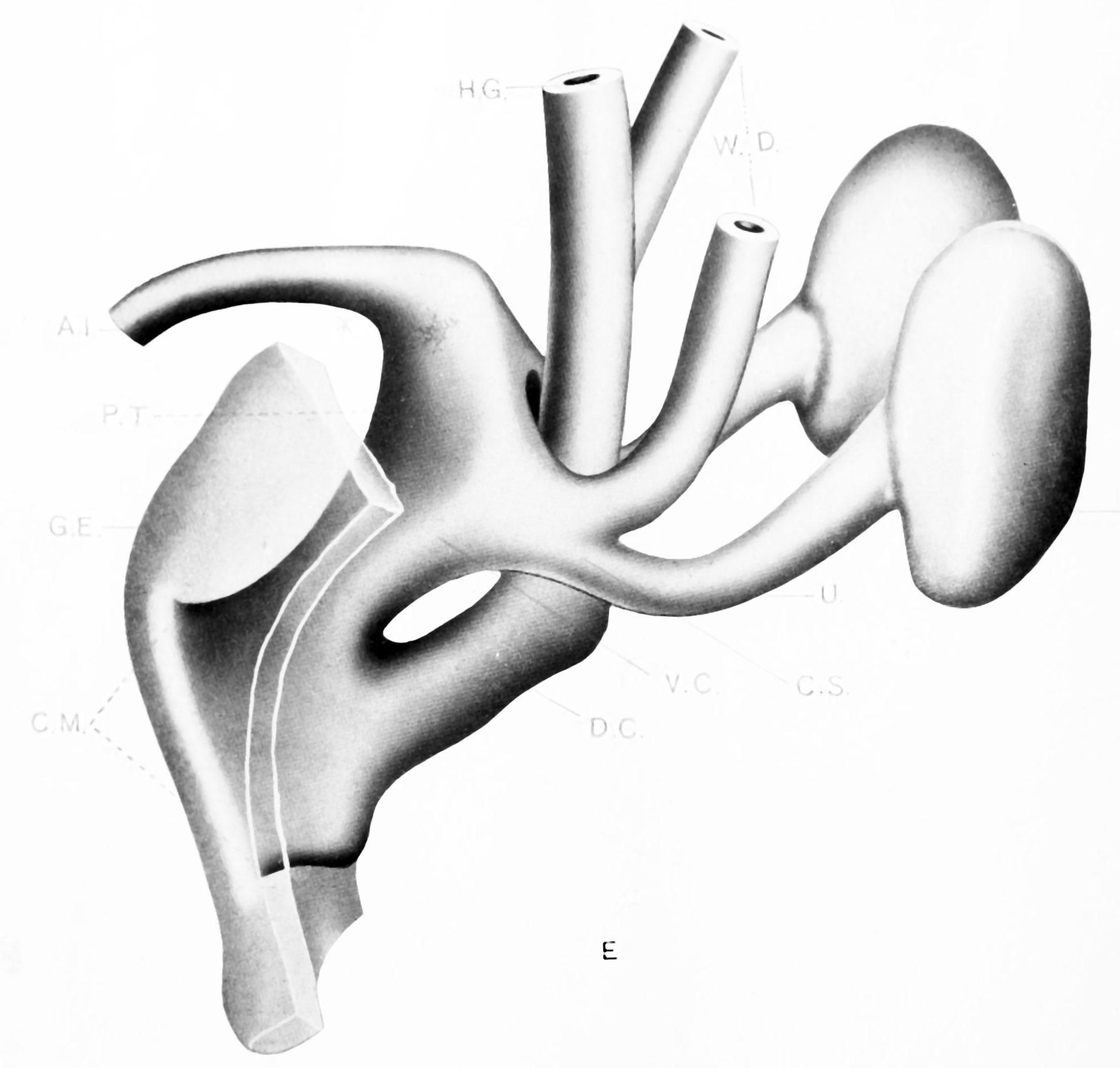 | 1,988 × 1,895 (198 KB) | Z8600021 (talk | contribs) | ==Plate 3E== {{Pohlman1911 figures}} |
You cannot overwrite this file.
File usage
The following page uses this file: