File:Stage11 sem100.jpg: Difference between revisions
mNo edit summary |
|||
| (4 intermediate revisions by the same user not shown) | |||
| Line 1: | Line 1: | ||
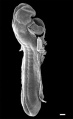
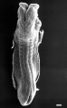
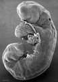
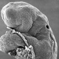
==Human Embryo Carnegie stage 11== | ==Human Embryo Carnegie stage 11== | ||
[[Week 4]] [[Carnegie stage 11]] 25 days, 19 somite pairs. This is a scanning EM of the embryo fractured to show the neural tube, notochord and somites. | |||
Features shown include the surface ectoderm, neural tube, notochord, somites, somitocoels, dorsal aortas, and gastrointestinal tract. | |||
:'''Links:''' [[:File:Stage11 sem10.jpg|unlabeled version]] | [[:File:Stage11 sem100.jpg|labeled version]] | [[Somitogenesis]] | |||
{{Stage11SEM}} | |||
''' | :[[Somitogenesis|'''Somite Cartoons''']]: [[:File:Somite cartoon1.png|'''1''' paraxial]] | [[:File:Somite cartoon2.png|'''2''' early somite]] | [[:File:Somite cartoon3.png|'''3''' sclerotome and dermomyotome]] | [[:File:Somite cartoon4.png|'''4''' dermatome and myotome]] | [[:File:Somite cartoon5.png|'''5''' somite spreading]] | ||
| Line 20: | Line 19: | ||
{{SEM}} | {{SEM}} | ||
'''Image version links:''' [[:File:Stage11 sem100.jpg|Large 1000px]] | [[:File:Stage11 sem100a.jpg| 800px]] | [[:File:Stage11 sem100b.jpg|Medium 600px]] | [[:File:Stage11 sem100c.jpg|Small 400px]] | |||
{{Carnegie_stages}} | {{Carnegie_stages}} | ||
| Line 27: | Line 27: | ||
[[Category:Carnegie Stage 11]] | [[Category:Carnegie Stage 11]] | ||
[[Category:Week 4]] | [[Category:Week 4]] | ||
[[Category:Somite]][[Category:Notochord]][[Category:Neural]] | |||
Latest revision as of 12:53, 15 May 2017
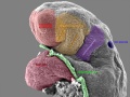
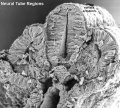
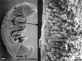
Human Embryo Carnegie stage 11
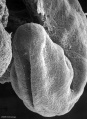
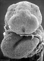
Week 4 Carnegie stage 11 25 days, 19 somite pairs. This is a scanning EM of the embryo fractured to show the neural tube, notochord and somites.
Features shown include the surface ectoderm, neural tube, notochord, somites, somitocoels, dorsal aortas, and gastrointestinal tract.
- Links: unlabeled version | labeled version | Somitogenesis
- Stage 11 SEM Images: dorsolateral whole embryo | dorsal embryo | lateral embryo | lateral head | lateral head with overlay | embryo cross-section | ventrolateral head | ventrolateral head with overlay | ventral head | buccopharyngeal membrane | neural crest | posterior neuropore | anterior neuropore | Carnegie stage 11
- Human Embryo (stage 11)
dorsolateral whole embryo
dorsal embryo
lateral embryo
lateral head
lateral head with overlay

embryo cross-section

embryo cross-section label
neural cross-section label

ventrolateral head
ventrolateral head with overlay

ventral head

buccopharyngeal membrane
neural crest
posterior neuropore
anterior neuropore


- Somite Cartoons: 1 paraxial | 2 early somite | 3 sclerotome and dermomyotome | 4 dermatome and myotome | 5 somite spreading
- Stage 11 SEM Images: dorsolateral whole embryo | dorsal embryo | lateral embryo | lateral head | lateral head with overlay | embryo cross-section | ventrolateral head | ventrolateral head with overlay | ventral head | buccopharyngeal membrane | neural crest | posterior neuropore | anterior neuropore | Carnegie stage 11
- Human Embryo (stage 11)
dorsolateral whole embryo
dorsal embryo
lateral embryo
lateral head
lateral head with overlay
embryo cross-section
embryo cross-section label
neural cross-section label
ventrolateral head
ventrolateral head with overlay
ventral head
buccopharyngeal membrane
neural crest
posterior neuropore
anterior neuropore
Image Source: Scanning electron micrographs of the Carnegie stages of the early human embryos are reproduced with the permission of Prof Kathy Sulik, from embryos collected by Dr. Vekemans and Tania Attié-Bitach. Images are for educational purposes only and cannot be reproduced electronically or in writing without permission.
Image version links: Large 1000px | 800px | Medium 600px | Small 400px
- Carnegie Stages: 1 | 2 | 3 | 4 | 5 | 6 | 7 | 8 | 9 | 10 | 11 | 12 | 13 | 14 | 15 | 16 | 17 | 18 | 19 | 20 | 21 | 22 | 23 | About Stages | Timeline
Cite this page: Hill, M.A. (2024, June 25) Embryology Stage11 sem100.jpg. Retrieved from https://embryology.med.unsw.edu.au/embryology/index.php/File:Stage11_sem100.jpg
- © Dr Mark Hill 2024, UNSW Embryology ISBN: 978 0 7334 2609 4 - UNSW CRICOS Provider Code No. 00098G
File history
Yi efo/eka'e gwa ebo wo le nyangagi wuncin ye kamina wunga tinya nan
| Gwalagizhi | Nyangagi | Dimensions | User | Comment | |
|---|---|---|---|---|---|
| current | 10:57, 27 August 2010 | 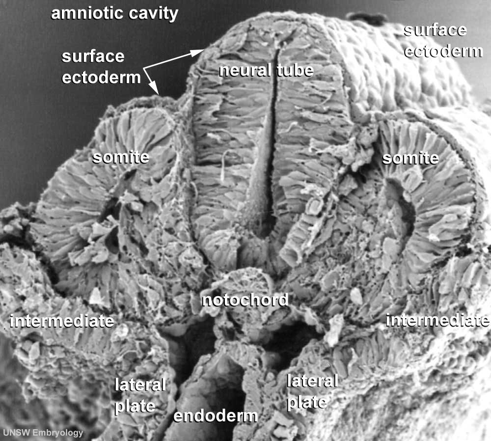 | 1,000 × 898 (109 KB) | S8600021 (talk | contribs) | |
| 11:36, 6 May 2010 |  | 1,000 × 898 (107 KB) | S8600021 (talk | contribs) | '''Human Embryo''' Carnegie stage 11 25 days, 19 somite pairs Facts: Week 4, 23 - 26 days, 2.5 - 4.5 mm, Somite Number 13 - 20 View: This is a labeled scanning EM of the embryo fractured to show the neural tube, notochord and somites. Features: surfac |
You cannot overwrite this file.
File usage
The following 64 pages use this file:
- 2011 Lab 3 - Week 4
- ANAT2341 Lab 3 - Week 4
- BGDA Lecture - Development of the Embryo/Fetus 2
- BGDA Lecture - Development of the Nervous System
- BGDA Practical 7 - Week 3
- BGD Lecture - Gastrointestinal System Development
- Brain Awareness Week 2012
- Carnegie stage 11
- Developmental Mechanism - Mesenchymal Epithelial Transition
- Human Embryo SEM
- K12 Brain Awareness Week
- Lecture - Gastrointestinal Development
- Lecture - Gastrointestinal Development 2013
- Lecture - Mesoderm Development
- Maternal-Fetal Medicine Trainees - Renal
- Musculoskeletal System - Muscle Development
- Musculoskeletal System Development
- Notochord
- S
- Somitogenesis
- File:Stage11 sem10.jpg
- File:Stage11 sem100.jpg
- File:Stage11 sem100a.jpg
- File:Stage11 sem100b.jpg
- File:Stage11 sem100c.jpg
- File:Stage11 sem101.jpg
- File:Stage11 sem10a.jpg
- File:Stage11 sem10b.jpg
- File:Stage11 sem10c.jpg
- File:Stage11 sem13.jpg
- File:Stage11 sem13a.jpg
- File:Stage11 sem13b.jpg
- File:Stage11 sem13c.jpg
- File:Stage11 sem2.jpg
- File:Stage11 sem21.jpg
- File:Stage11 sem2a.jpg
- File:Stage11 sem2b.jpg
- File:Stage11 sem2c.jpg
- File:Stage11 sem3.jpg
- File:Stage11 sem3a.jpg
- File:Stage11 sem3b.gif
- File:Stage11 sem3b.jpg
- File:Stage11 sem3c.jpg
- File:Stage11 sem4.jpg
- File:Stage11 sem4a.jpg
- File:Stage11 sem4b.jpg
- File:Stage11 sem4c.jpg
- File:Stage11 sem5.jpg
- File:Stage11 sem5a.jpg
- File:Stage11 sem5b.jpg
- File:Stage11 sem5c.jpg
- File:Stage11 sem6.jpg
- File:Stage11 sem7.jpg
- File:Stage11 sem7a.jpg
- File:Stage11 sem7b.jpg
- File:Stage11 sem8.jpg
- File:Stage11 sem81.jpg
- File:Stage11 sem82.jpg
- File:Stage11 sem8a.jpg
- File:Stage11 sem8b.jpg
- File:Stage11 sem9.jpg
- File:Stage11 sem9a.jpg
- File:Stage11 sem9b.jpg
- Template:Stage11SEM