Category:Carnegie Stage 7: Difference between revisions
From Embryology
mNo edit summary |
|||
| Line 14: | Line 14: | ||
* '''HEB-37.''' Summarized by Mazanec (1959)<ref name=Mazanec1959>Mazanec, K. 1959. Blastogenese des Metjschen. Fischer, Jena.</ref>. Chorionic cavity, 2.25 x 1.29 x 0.4 mm. Embryonic disc, 0.4 mm. Primitive streak, 0.104 mm, and node, 0.04 mm. Notochordal process, 0.032 mm. Stalk of umbilical vesicle (ibid., fig. 77). Median projection published (ibid., fig. 45). | * '''HEB-37.''' Summarized by Mazanec (1959)<ref name=Mazanec1959>Mazanec, K. 1959. Blastogenese des Metjschen. Fischer, Jena.</ref>. Chorionic cavity, 2.25 x 1.29 x 0.4 mm. Embryonic disc, 0.4 mm. Primitive streak, 0.104 mm, and node, 0.04 mm. Notochordal process, 0.032 mm. Stalk of umbilical vesicle (ibid., fig. 77). Median projection published (ibid., fig. 45). | ||
* '''H. R. 1.''' Described by Johnston (1940)<ref>Johnston, T. B. 1940. [[Paper - An Early Human Embryo, with 0.55 mm long Embryonic Shield|An early human embryo, with 0.55 mm. long embryonic shield]]. J. Anat, 75, 1-49. | * [[Paper_-_An_Early_Human_Embryo,_with_0.55_mm_long_Embryonic_Shield|'''H. R. 1.''']] Described by Johnston (1940)<ref>Johnston, T. B. 1940. [[Paper - An Early Human Embryo, with 0.55 mm long Embryonic Shield|An early human embryo, with 0.55 mm. long embryonic shield]]. J. Anat, 75, 1-49.</ref>, who believed that a notochordal process (0.04 mm) and a prechordal plate (0.075 mm) were present. Florian in an appendix to the article disagreed, and his interpretation is followed here (see stage 6). | ||
</ref>, who believed that a notochordal process (0.04 mm) and a prechordal plate (0.075 mm) were present. Florian in an appendix to the article disagreed, and his interpretation is followed here (see stage 6). | |||
* [[Paper - Two Early Human Embryos|'''Biggart''']]. Described by Morton (1949)<ref>Morton, W. R. M. 1949. [[Paper - Two Early Human Embryos|Two early human embryos]]. J. Anat., 83, 308-314.</ref>. Curettage. Embryonic disc (narrow type), 0.27 x 0.16 mm. Primitive streak and node, 0.059 mm. A notochordal process is not referred to in the text but is mapped on a dorsal projection of the embryo (ibid., fig. 2) and is approximately 0.04 mm in length. The specimen is said to resemble the Yale embryo. | * [[Paper - Two Early Human Embryos|'''Biggart''']]. Described by Morton (1949)<ref>Morton, W. R. M. 1949. [[Paper - Two Early Human Embryos|Two early human embryos]]. J. Anat., 83, 308-314.</ref>. Curettage. Embryonic disc (narrow type), 0.27 x 0.16 mm. Primitive streak and node, 0.059 mm. A notochordal process is not referred to in the text but is mapped on a dorsal projection of the embryo (ibid., fig. 2) and is approximately 0.04 mm in length. The specimen is said to resemble the Yale embryo. | ||
* '''Guá''' (Guálberto). Described by Lordy (1931)<ref>Lordy, C. 1931. A human ovum in its early phases of development. Ann. Fac. Med. Sao Paulo, 6, 29-35.</ref>. Hysterectomy. Chorionic cavity, 8 x 7.5 mm. Embryonic disc, 0.776 x 0.0465 mm. Primitive streak, 0.09 mm. Notochordal process, 0.045 mm. Possible notochordal canal. Said to resemble Hugo embryo. Probably belongs either to stage 7 or to stage 8. | * '''Guá''' (Guálberto). Described by Lordy (1931)<ref>Lordy, C. 1931. A human ovum in its early phases of development. Ann. Fac. Med. Sao Paulo, 6, 29-35.</ref>. Hysterectomy. Chorionic cavity, 8 x 7.5 mm. Embryonic disc, 0.776 x 0.0465 mm. Primitive streak, 0.09 mm. Notochordal process, 0.045 mm. Possible notochordal canal. Said to resemble Hugo embryo. Probably belongs either to stage 7 or to stage 8. | ||
Revision as of 09:57, 10 August 2015
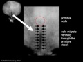
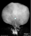
This Embryology category shows pages and media related to Carnegie stage 7 of embryonic development. In human development this stage occurs during week 3 (post-fertilisation) or gestational age GA week 5 (LMP).
- Links: Carnegie stage 7 | Week 3
| Week: | 1 | 2 | 3 | 4 | 5 | 6 | 7 | 8 |
| Carnegie stage: | 1 2 3 4 | 5 6 | 7 8 9 | 10 11 12 13 | 14 15 | 16 17 | 18 19 | 20 21 22 23 |
| Carnegie Collection - Stage 7 | ||||||||||
|---|---|---|---|---|---|---|---|---|---|---|
| Serial No. | Grade | Fixative | Embedding Medium | Thinness (µm) | Stain | Year | Notes | |||
| 7802 | Exc. | Alc. & Bouin | C-P | 6 | (Stain - Haematoxylin Eosin) | 1940 | Heuser et al. (1945) | |||
| 8206 | Good | p | C-P | 6 | (Stain - Haematoxylin Eosin) | 1943 | ||||
| 8361 | Good | Bouin | C-P | 10 | p | 1946 | Abnormal | |||
| 8602 | Exc. | Alc. | C-P | 8 | (Stain - Haematoxylin Eosin) | 1948 | ||||
| 8752 | Exc. | ? | C-P | 10 | (Stain - Haematoxylin Eosin) | 1950 | ||||
| 8755 | Exc. | Formol | C-P | 10 | (Stain - Haematoxylin Eosin) | 1950 | ||||
| 9217 | Exc. | p | P | 10 | (Stain - Haematoxylin Eosin) | 1954 | ||||
Abbreviations
| ||||||||||
Embryo Examples
(listed in order of length of notochordal process)
- HEB-37. Summarized by Mazanec (1959)[1]. Chorionic cavity, 2.25 x 1.29 x 0.4 mm. Embryonic disc, 0.4 mm. Primitive streak, 0.104 mm, and node, 0.04 mm. Notochordal process, 0.032 mm. Stalk of umbilical vesicle (ibid., fig. 77). Median projection published (ibid., fig. 45).
- H. R. 1. Described by Johnston (1940)[2], who believed that a notochordal process (0.04 mm) and a prechordal plate (0.075 mm) were present. Florian in an appendix to the article disagreed, and his interpretation is followed here (see stage 6).
- Biggart. Described by Morton (1949)[3]. Curettage. Embryonic disc (narrow type), 0.27 x 0.16 mm. Primitive streak and node, 0.059 mm. A notochordal process is not referred to in the text but is mapped on a dorsal projection of the embryo (ibid., fig. 2) and is approximately 0.04 mm in length. The specimen is said to resemble the Yale embryo.
- Guá (Guálberto). Described by Lordy (1931)[4]. Hysterectomy. Chorionic cavity, 8 x 7.5 mm. Embryonic disc, 0.776 x 0.0465 mm. Primitive streak, 0.09 mm. Notochordal process, 0.045 mm. Possible notochordal canal. Said to resemble Hugo embryo. Probably belongs either to stage 7 or to stage 8.
- Carnegie No. 7802 An important specimen described and illustrated by Heuser, Rock, and Hertig (1945)[5]. Hysterectomy. Chorion, 3.75 x 2.35 x 2.2 mm. Chorionic cavity, 2.3 x 1.4 x 1.1 mm. Embryonic disc (broad type), 0.42 x 0.35 x 0.05 mm. Primitive streak, 0.11 mm, and node, 0.03 mm. Notochordal process, 0.048 mm. Presumed age, 16 days. Median projection published (ibid., plate 6; Mazanec, 1959, fig. 46) and dorsal projection has been prepared by the present writers.
- P.M. Described by Meyer (1924)[6]. Curettage. Measurements have been criticized by Stieve (1926) but defended by Mazanec (1959)[1]. Chorion, 3.9 x 3.77 x 2.5 mm. Chorionic cavity, 2.7 x 2.6 x 2.1 mm. Embryonic disc (circular), 0.41 x 0.41 mm. Primitive streak, 0.12 mm, and node, 0.02 mm. Notochordal process, 0.06 mm, acknowledged by Mazanec (1959)[1]. although denied by Fahrenholz (1927)[7]. No notochordal canal. Median projection published (Mazanec, 1959, fig. 47).
- Hugo. Described by Stieve (1926)[8], who reproduced a photomicrograph of every second section. Hysterectomy. Surrounded by 942 chorionic villi ranging in length from 0.3 to 1 mm. Chorion, 6.4 x 5.9 x 5.6 mm. Chorionic cavity, 4.7 x 4.4 x 3.8 mm. Embryonic disc (broad type), 0.635 (Florian, 1931) x 0.63 mm. Primitive streak, 0.245 mm, and node, 0.05 mm. Notochordal process, 0.07 (0.11?) mm. (Florian, 1934c). No notochordal canal. Prechordal plate probably not yet developed (Hill and Florian, 1931a). Dorsal and median projections published (Florian, 1934c[9], fig. 1; Hill and Florian, 1931b[10], figs. 44 and 11; Mazanec, 1959[1], fig. 49).
- Robertson, O’Neill, and Chappell (1948)[11] described a hysterectomy specimen that possessed a chorion of 3.816 x 3.639 x 2.687 mm. Chorionic cavity, 2.718 x 2.239 x 1.679 mm. Embryonic disc (broad type), 0.462 x 0.485 mm. Primitive streak, 0.138 mm, and node (situated halfway), 0.03 mm. Notochordal process, 0.072 mm. Suggestion of notochordal canal in one or two sections. Assigned to horizon VIII by authors but probably belongs to stage 7. Median projection published (ibid., fig. 12).
- D’Arrigo (1961)[12] described an embryonic disc of 0.47 mm, which showed a notochordal process of 0.075 mm. Canalization of the process is “doubtful,” but the presence of a prechordal plate is “probable.” The specimen “could be recorded in Streeter’s horizon VII.”
- Goodwin. Described by Kindred (1933)[13]. Tubal. Chorion, 5.8 x 2.72 x 2.25 mm. Chorionic cavity, 2.44 x 2.25 x 0.75 mm. Embryonic disc, 0.588 mm in width. Primitive streak, 0.215 mm, and node, 0.078 mm. Notochordal process, 0.078 mm. No notochordal canal and no prechordal plate.
- Pha I. Described by Mazanec (1949)[1]. Chorionic cavity, 7.872 x 5.475 x 2.032 mm. Embryonic disc, 0.66 x 0.52 mm. Primitive streak, 0.145 mm, and node, 0.06 mm. Notochordal process, 0.09 mm. No prechordal plate. Median projection published (ibid., fig. 51).
- H. Schm. 10 (H. Schmid). Described briefly by Grosser (1931)[14]. Embryonic disc (almost circular), 0.51 x 0.58 mm. Primitive streak, 0.14 mm, and node, 0.1 mm. Notochordal process, 0.1 mm. Probably belongs to stage 7, although a cavity in one section was thought to represent “Lieberkühn’s canal.”
- Bi 24 (Bittmann). Described by Hill and Florian (1931b)[10]. Chorionic cavity, 3.05 x 3.036 x 3.029 mm. Embryonic disc (narrow type), 0.62 x 0.39 mm. Primitive streak, 0.28 mm, and node (Mazanec, 1959, fig. 105), 0.05 mm. Notochordal process, 0.105 mm, consists of median chord and lateral mesoblastic wings. Prechordal plate, 0.03 mm. Possible primordial germ cells in endoderm of region of cloacal membrane and in endoderm of umbilical vesicle caudally (Florian, 1931). Politzer (1933)[15] counted 41 germ cells in the region of the allanto-enteric diverticulum in this embryo, and 19 such cells in another presomite specimen (Bi 25). Dorsal and median projections published (Hill and Florian, 1931b, figs. 4 and 12; Florian, 1945, plate 5, fig. 43; Mazanec, 1959, fig. 50).
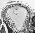
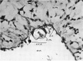
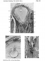
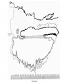
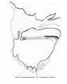
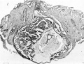
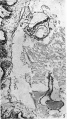
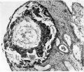
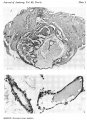
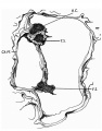
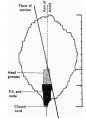
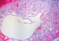
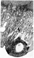
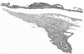
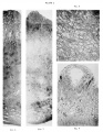
- Manchester No. 1285 (fig. 7-1). Described by Florian and Hill (1935)[16],. Hysterectomy. Chorionic cavity, 4.28 x 3.28 mm. Embryonic disc (narrow type), 0.87 x 0.625 mm. Primitive streak, 0.39 mm, and node, 0.05 mm. Notochordal process, 0.125 mm. Prechordal plate, 0.03 mm. Connecting stalk attached to chorion at decidua capsularis (suggesting polar variety of velamentous insertion of umbilical cord). Dorsal and median projections published (Hill and Florian, 1931b, figs. 5 and 13; Florian and Hill, 1935, figs. 1-3; Mazanec, 1959[1], fig. 52). Specimen is housed in Department of Anatomy, University of Manchester.
- Pha II. Summarized by Mazanec (1959)[1]. Chorionic cavity, 4.985 x 3.882 x 3.52 mm. Embryonic disc, 0.895 x 0.62 mm. Primitive streak, 0.37 mm, and node, 0.06 mm. Notochordal process, 0.13 mm. No prechordal plate. Median projection published (ibid., fig. 53).
- Thompson and Brash (1923)[17] described a specimen that showed a notochordal process of 0.3 mm.
Following embryo examples do not have measurements of the notochordal process, they are listed by the year of publication.
- Debeyre (1912)[18] described in detail a specimen that possessed a chorionic cavity of 5.6 x 2.1 mm. Embryonic disc, 0.9 x 0.6 x 0.95 mm. Primitive streak stated to be 0.54 mm in length. Chorionic villi (0.4-1.6 mm) showed some branching. Unsuitable plane of section makes it impossible to assess the specimen precisely.
- Carnegie No. 1399, Mateer. Described by Streeter (1920). Hysterectomy. Angiogenesis in villi described by Hertig (1935). Chorion, 9 x 8 x 3.5 mm. Chorionic cavity, 6.1 x 5.6 x 2.5 mm. Embryonic disc, 1 x 0.75 mm. Primitive streak and groove present. Although the notochordal process was originally thought probably to be absent, Hill and Florian (1931b) have no doubt that it is present. More advanced than Hugo (Florian and Völker, 1929). A very small twin embryo was originally described but that “interpretation has become open to doubt” (Corner, 1955). Drawing of every section reproduced by Turner (1920). A median drawing (Davis, 1927, fig. 5A) and a projection have been published (Mazanec, 1959, fig. 56).
- Ho (Hodiesne). Described by Fahrenholz (1927). Abortion. Chorionic cavity, 6.5 x 6 x 3 mm. Embryonic disc (deformed), 0.6 mm (0.725 mm by flexible scale). Primitive streak, 0.22 mm (0.345 mm by flexible scale). Notochordal process just beginning (“undoubtedly present,” Hill and Florian, 1931b). Possible prechordal plate claimed (disputed by Waldeyer, 1929a, but supported by Hill and Florian, 1963). “Lieberkühn’s canal” (0.065 mm) is an artificial folding of the embryonic disc (Hill and Florian, 1931b). Dorsal and median projections published (Fahrenholz, 1927, figs. 32, 6 and 7; Mazanec, 1959, fig. 54).
- Debeyre (1933)[19] described a specimen (0.9 mm) that possessed a primitive streak and probably belonged to stage 6 or stage 7. Large cells near the opening of the allantois were identified as primordial germ cells.
- Falkiner. Described by Martin and Falkiner (1938)[20]. Curettage. Embryo damaged and not in good condition. Measurements seem too small (see Mazanec, 1959). Chorionic cavity, 1.5 x 1.4 mm. Embryonic disc, 0.15 x 0.29 mm. Primitive streak, 0.07 mm. Notochordal process contains “no definite lumen.” Cells rostral to notochordal process are “probably” the prechordal plate. Development “agrees most closely” with that of Bi I. Median projection published (Martin and Falkiner, 1938, fig. 8; Mazanec, 1959, fig. 39).
- Gar (Green-Armytage). Described by West (1952)[21]. Hysterectomy. Chorionic cavity, 3 x 2.6 x 2 mm. Embryonic disc (broad type), 0.56 x 0.69 mm. Primitive streak, node, and groove present. Short notochordal process. No notochordal plate. Said to resemble Hugo embryo. Trophoblast described by Hamilton and Boyd (1960).
- Mal (Maliphant). Described by West (1952[21]). Hysterectomy. Chorionic cavity, 3 x 1.8 mm. Embryonic disc (broad type), 0.45 x 0.6 mm. Primitive streak, node, and groove present. Notochordal process present (Mazanec, 1959); small cavity in node (West, 1952)[21] or in notochordal process (Mazanec, 1959) “hardly sufficient to warrant the name chorda canal.” No prechordal plate. Belongs either to stage 7 or to stage 8 (said to resemble Jones-Brewer I, which is in stage 8).
- Carnegie No. 8602. Photomicrograph reproduced by Hertig, Rock, and Adams (1956, plate 10, fig. 53)[22]. Chorion, 2.73 x 2.43 mm. Chorionic cavity, 1.83 x 1.33 mm. Embryonic disc, 0.3 x 0.06 mm. Presumed age, 16-17 days.
- Missen. Trophoblast described by Hamilton and Boyd (1960)[23]. Curettage. Chorion, 1.66 x 1.43 mm. Embryonic disc, 0.28 x 0.214 mm. Primitive streak and node. Notochordal process. Said to resemble No. 7801 and Edwards Jones-Brewer (stage 6). Presumed age, about 14 days.
References
- ↑ 1.0 1.1 1.2 1.3 1.4 1.5 1.6 Mazanec, K. 1959. Blastogenese des Metjschen. Fischer, Jena. Cite error: Invalid
<ref>tag; name 'Mazanec1959' defined multiple times with different content Cite error: Invalid<ref>tag; name 'Mazanec1959' defined multiple times with different content Cite error: Invalid<ref>tag; name 'Mazanec1959' defined multiple times with different content - ↑ Johnston, T. B. 1940. An early human embryo, with 0.55 mm. long embryonic shield. J. Anat, 75, 1-49.
- ↑ Morton, W. R. M. 1949. Two early human embryos. J. Anat., 83, 308-314.
- ↑ Lordy, C. 1931. A human ovum in its early phases of development. Ann. Fac. Med. Sao Paulo, 6, 29-35.
- ↑ Heuser, C. H., Rock, J., and Hertig, A. T. 1945. Two human embryos showing early stages of the definitive yolk sac. Carnegie Instn. Wash. Publ. 557, Contrib. Embryol., 31, 85-99.
- ↑ Meyer, P. 1924. Ein junges menschliches Ei mit 0,4 mm Iangem Embryonalschild. Arch. Gyndk, 122, 38-87.
- ↑ Fahrenholz, C. 1927. Ein junges menschliches Abortivei. Z mikr.-anat. Forsch., 8, 250-324.
- ↑ Stieve, H. 1926a. Ein 13V2-Tage altes, in der Gebarmutter erhaltenes und durch Eingriff gewonnenes menschliches Ei. Z mikr.-anat. Forsch., 7, 295-402.
- ↑ Florian, J. 1934c. Uber einige bisher unkorrigiert gebliebene fehlerhafte Angaben der junge menschliche Embryonen beschreibenden Arbeiten. Anat. Anz., 78, 445-450.
- ↑ 10.0 10.1 Hill, J. P., and Florian, J. 1931b. A young human embryo (embryo Dobbin) with head-process and prochordal plate. Phil. Tram. Roy. Soc. London B, 219, 443-486.
- ↑ Robertson, G. G., O'Neill, S. L, and Chappell, R. H. 1948. On a normal human embryo of 17 days development. Anat. Rec, 100, 9-28. PMID 18917638
- ↑ D'Arrigo, S. 1961. Illustrazione di un embrione umano al principio della terza settimana di sviluppo. Riv. Patol. Clin. Sper., 2, 1-15.
- ↑ Kindred, J. E. 1933. A human embryo of the presomite period from the uterine tube. Amer.J. Anat., 53, 221-241.
- ↑ Grosser, O. 1931. Weiteres uber den Primitivstreifen des Menschen. Verb. Anat. Ges., Erg. Heft Anat. Anz., 72, 42-44.
- ↑ Politzer, G. 1933. Die Keimbahn des Menschen. Z. Anat. Entw., 100, 331-361.
- ↑ Florian, J., and Hill, J. P. 1935. An early human embryo (No. 1285, Manchester Collection), with capsular attachment of the connecting stalk. J. Anat,, 69, 399-411.
- ↑ Thompson, P., and Brash, J. C. 1923. A human embryo with head-process and commencing arch enteric canal. J. Anat., 58, 1-20. PMID 17103992
- ↑ Debeyre, A. 1912. Description d'un embryon humain de 0-mm.9. J. Anat. Physiol, Paris, 48, 448-515.
- ↑ Debeyre, A. 1933. Sur la presence de gonocytes chez un embryon humain au stade de la ligne primitive. C. R. Ass. Anat., 28, 240-250.
- ↑ Martin, C. P., and Falkiner, N. Mel. 1938. The Falkiner ovum. Amer.f. Anat, 63, 251-271.
- ↑ 21.0 21.1 21.2 West, C. M. 1952. Two presomite human embryos. J. Of eat Gynaecol. Brit. Emp., 59, 336-351.
- ↑ Hertig, A. T., Rock, J., and Adams, E. C. 1956. A description of 34 human ova within the first 17 days of development. Amer. J. Anat., 98, 435-493.
- ↑ Hamilton, W. J., and Boyd, J. D. 1950. Phases of human development. Ch. 8 in K. Bowes (ed), Modem Trends in Obstetrics and Gynaecology Butterworth, London, pp. 114-137.
Subcategories
This category has the following 9 subcategories, out of 9 total.
Pages in category 'Carnegie Stage 7'
The following 55 pages are in this category, out of 55 total.
C
P
- Paper - A human embryo of the pre-somite period from the uterine tube
- Paper - A human embryo with head-process and commencing arch enteric canal
- Paper - A Note on the Development of the Septum Transversum and the Liver
- Paper - A presomite human embryo showing a yolk-sac duct
- Paper - A presomite human embryo showing an early stage of the primitive streak
- Paper - An early human embryo (no. 1285, Manchester Collection) with capsular attachment of the connecting stalk (1935)
- Paper - An Early Human Embryo (No. 1285, Manchester Collection), with Capsular Attachment of the Connecting Stalk
- Paper - An Early Human Embryo, with 0.55 mm long Embryonic Shield
- Paper - Description of a Human Embryo of Twenty-three Paired Somites
- Paper - Normal development of early human embryos: Observation of 90 specimens at Carnegie stages 7 to 13
- Paper - Six normal and complete presomite human ova
- Paper - The Falkiner ovum
- Paper - Two Early Human Embryos
- Paper - Two presomite human embryos
R
- Template:Ref-Brewer1937
- Template:Ref-BrewerFitzgerald1937
- Template:Ref-FlorianHill1935
- Template:Ref-George1942
- Template:Ref-Kindred1933
- Template:Ref-Mazanec1949
- Template:Ref-Mazanec1959
- Template:Ref-PMID13044724
- Template:Ref-Stieve1926a
- Template:Ref-Stieve1926b
- Template:Ref-Thompson1923
- Template:Ref-West1952
S
Media in category 'Carnegie Stage 7'
The following 84 files are in this category, out of 84 total.
 Florian1935 chorionic vesicle fig11.jpg 1,009 × 952; 248 KB
 Florian1935 fig01.jpg 774 × 405; 115 KB
 Florian1935 fig02.jpg 790 × 405; 117 KB
 Florian1935 fig03.jpg 800 × 455; 130 KB
 Florian1935 fig04.jpg 798 × 443; 120 KB
 Florian1935 fig05.jpg 803 × 227; 58 KB
 Florian1935 fig06.jpg 770 × 573; 145 KB
 Florian1935 fig07.jpg 597 × 812; 206 KB
 Florian1935 fig08.jpg 691 × 480; 114 KB
 Florian1935 fig09.jpg 510 × 765; 168 KB
 Florian1935 fig10.jpg 464 × 721; 157 KB
 Florian1935 fig11.jpg 1,280 × 1,427; 443 KB
 Florian1935 plate01.jpg 1,481 × 1,825; 529 KB
 Florian1935 plate02.jpg 1,481 × 1,806; 578 KB
 Florian1935 plate03.jpg 1,481 × 2,000; 735 KB
 Florian1935 textfig01.jpg 1,120 × 1,202; 136 KB
 Florian1935 textfig02.jpg 1,579 × 2,000; 342 KB
 Florian1935 textfig03.jpg 1,579 × 2,000; 212 KB
 MartinFalkiner1938 fig01.jpg 800 × 931; 133 KB
 MartinFalkiner1938 fig02.jpg 600 × 886; 76 KB
 MartinFalkiner1938 fig03.jpg 700 × 1,000; 89 KB
 MartinFalkiner1938 fig04.jpg 700 × 974; 82 KB
 MartinFalkiner1938 fig05.jpg 700 × 1,022; 88 KB
 MartinFalkiner1938 fig06.jpg 700 × 1,009; 87 KB
 MartinFalkiner1938 fig07.jpg 700 × 951; 63 KB
 MartinFalkiner1938 fig08.jpg 670 × 840; 46 KB
 MartinFalkiner1938 plate01.jpg 1,280 × 1,803; 344 KB
 MartinFalkiner1938 plate02.jpg 1,280 × 1,840; 247 KB
 Morton1949 fig01.jpg 1,673 × 1,282; 615 KB
 Morton1949 fig02.jpg 652 × 786; 79 KB
 Morton1949 fig03.jpg 1,016 × 786; 108 KB
 Morton1949 fig04.jpg 629 × 1,316; 332 KB
 Morton1949 fig05.jpg 738 × 1,316; 383 KB
 Morton1949 fig06.jpg 1,012 × 868; 253 KB
 Morton1949 plate01.jpg 1,784 × 2,457; 975 KB
 Morton1949 text-fig01.jpg 847 × 1,084; 111 KB
 Morton1949 text-fig02.jpg 778 × 1,078; 70 KB
 Stage7 800x700px.jpg 690 × 800; 69 KB
 Stage7 axes.jpg 500 × 375; 9 KB
 Stage7 axial process.jpg 500 × 375; 11 KB
 Stage7 bf5.jpg 1,712 × 1,206; 875 KB
 Stage7 bf51.jpg 600 × 450; 96 KB
 Stage7 bf52.jpg 600 × 450; 114 KB
 Stage7 bf53.jpg 500 × 375; 74 KB
 Stage7 bf5a.jpg 1,024 × 721; 690 KB
 Stage7 bf5b.jpg 500 × 352; 216 KB
 Stage7 bf6.jpg 347 × 599; 69 KB
 Stage7 bf7.jpg 400 × 341; 22 KB
 Stage7 bf8.jpg 400 × 341; 18 KB
 Stage7 bf9.jpg 1,200 × 1,039; 501 KB
 Stage7 cloacal-oral-membranes.jpg 690 × 800; 70 KB
 Stage7 features.jpg 500 × 375; 9 KB
 Stage7 folding.jpg 500 × 375; 10 KB
 Stage7 intermediate-mesoderm.jpg 690 × 800; 67 KB
 Stage7 lateral-plate.jpg 690 × 800; 65 KB
 Stage7 mesoderm.jpg 690 × 800; 67 KB
 Stage7 notochord.jpg 690 × 800; 70 KB
 Stage7 paraxial-mesoderm.jpg 690 × 800; 69 KB
 Stage7 primitive streak labelled.jpg 500 × 375; 13 KB
 Stage7 primitive-streak-node.jpg 690 × 800; 69 KB
 Stage7-bf1.jpg 600 × 676; 34 KB
 Stage7-bf2.jpg 800 × 579; 53 KB
 Stage7-bf3.jpg 523 × 600; 45 KB
 Stage7-bf4.jpg 800 × 695; 53 KB
 Stage7-sem1.jpg 814 × 600; 100 KB
 Stage7-sem2.jpg 590 × 800; 98 KB
 Stage7-sem4.jpg 937 × 595; 79 KB
 Stage7-sem5.jpg 1,000 × 735; 202 KB
 Stage7.jpg 312 × 427; 7 KB
 ThompsonBrash1923 fig01.jpg 961 × 917; 212 KB
 ThompsonBrash1923 fig02.jpg 910 × 1,345; 146 KB
 ThompsonBrash1923 fig03.jpg 1,327 × 1,131; 135 KB
 ThompsonBrash1923 fig04.jpg 1,453 × 806; 166 KB
 ThompsonBrash1923 fig05.jpg 1,420 × 655; 226 KB
 ThompsonBrash1923 fig06.jpg 1,348 × 865; 166 KB
 ThompsonBrash1923 fig07.jpg 1,440 × 872; 324 KB
 ThompsonBrash1923 fig08.jpg 1,395 × 1,222; 299 KB
 ThompsonBrash1923 fig09.jpg 1,145 × 1,151; 518 KB
 ThompsonBrash1923 fig10.jpg 1,342 × 875; 249 KB
 ThompsonBrash1923 fig11.jpg 1,336 × 1,344; 385 KB
 ThompsonBrash1923 fig12.jpg 1,018 × 680; 160 KB
 West1952 plate01.jpg 1,280 × 1,797; 251 KB
 West1952 plate02.jpg 1,280 × 1,651; 284 KB
 Yolk sac and amniotic cavity volume graph.jpg 719 × 1,000; 50 KB


