Paper - Description of a Human Embryo of Twenty-three Paired Somites
| Embryology - 28 Apr 2024 |
|---|
| Google Translate - select your language from the list shown below (this will open a new external page) |
|
العربية | català | 中文 | 中國傳統的 | français | Deutsche | עִברִית | हिंदी | bahasa Indonesia | italiano | 日本語 | 한국어 | မြန်မာ | Pilipino | Polskie | português | ਪੰਜਾਬੀ ਦੇ | Română | русский | Español | Swahili | Svensk | ไทย | Türkçe | اردو | ייִדיש | Tiếng Việt These external translations are automated and may not be accurate. (More? About Translations) |
Thompson P. Description of a human embryo of twenty-three paired somites. (1907) J Anat Physiol, 41(3):159-71. PMID 17232726
| Online Editor |
|---|
| Peter Thompson worked in embryology at King’s College in the United Kingdom in the early 1900's with J. Ernest Frazer.
Other papers by Peter Thompson:
|
| Historic Disclaimer - information about historic embryology pages |
|---|
| Pages where the terms "Historic" (textbooks, papers, people, recommendations) appear on this site, and sections within pages where this disclaimer appears, indicate that the content and scientific understanding are specific to the time of publication. This means that while some scientific descriptions are still accurate, the terminology and interpretation of the developmental mechanisms reflect the understanding at the time of original publication and those of the preceding periods, these terms, interpretations and recommendations may not reflect our current scientific understanding. (More? Embryology History | Historic Embryology Papers) |
Description of a Human Embryo of Twenty-three Paired Somites
Peter Thompson
Carnegie Staging Comparison: A 23 somite stage embryo would be similar to a Carnegie stage 12 (26 - 30 days), caudal neuropore closes, Somite Number 21-29.
- Historic Paper Links: 13-14 Somites | 22 Somites | 23 Somites | 25 Somites | 27 Somites | Mall Human Embryo Collection | Embryology History | Carnegie stage 11 | Carnegie stage 12 | Journal of Anatomy | Embryonic Development | Category:Historic Embryology
Introduction
The embryo which forms the basis of this work came from Dr Robert Meyer's collection in Berlin. Sent to Professor Keibel, who was accumulating material for his forthcoming Normentafeln of human embryos, the specimen was most kindly lent to me with the object of making a model whilst I was at the Anatomical Institute in Freiburg during the summer of 1906. Working with this specimen, I had an excellent opportunity of becoming acquainted with the reconstruction of embryos by the wax-plate method, as carried out so successfully in that University.
The embryo, obtained at an operation, was recorded as 2.5 mm. long, and was cut transversely into 488 sections, each 5 , in thickness, and stained with borax-carmine. In making the model, every other section was drawn, enlarged 100 diameters, and the wax plates were made 1 millimetre in thickness. When the plates had been cut and laid in position, it was found, owing probably to the hot weather and the weight of wax, that the total height of the model was only 220 millimetres, and, in order to correct the error, 24 additional wax plates were made, duplicates of every tenth section, and introduced into the series. In this way the total length of the model was brought up to 244 millimetres, corresponding to the 244 sections used, which indicates a shortage of less than 3 per cent, when compared with the 250 millimetres, the absolutely accurate measurement which the enlargement should have been, taking the length of embryo as 25 min. The difference is probably due to a slight shrinkage, which would most probably occur in preparing the specimen for cutting. It may be stated here that the embryo is histologically in excellent condition, mitosis being readily observed in the multiplying cells, and there seems no valid reason for doubting that the specimen is a normal one. Inaddition to the model of the whole embryo and its yolk sac, other models were made of special organs, namely, the heart and its endothelial tube, the brain, a part of the alimentary canal, and the septum transversum.
It may be noted that the embryo described in this paper resembles in many ways His's embryo Lg., which was 2-15 mm. in length and estimated to be about fifteen days old.
Descriptions of Models
The Embryo and its Yolk Sac
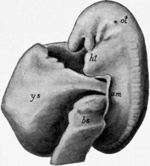
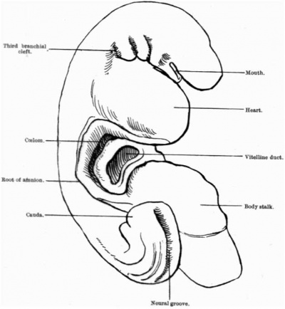
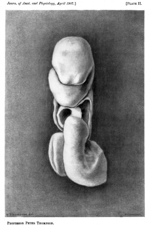
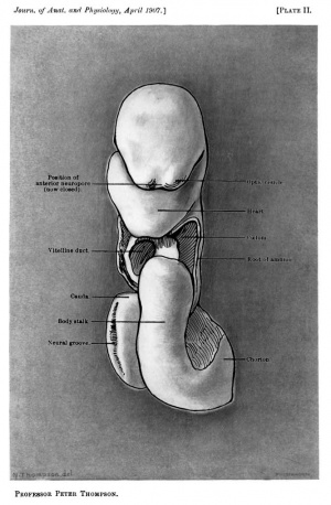
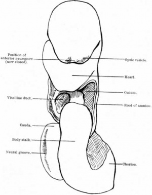
The head is small, somewhat flattened from above downwards, and pointed. At the side, the opening of the otocyst externally is seen over the upper end of the second post-oral bronchial cleft. Anteriorly, two slight but well-marked bulgings indicate the position of the optic vesicles, between which is a depressed area corresponding to the anterior neuropore, com- pletely closed, but stil recognizable in the sections by the continuity of the general ectoderm and the neural ectoderm. The mouth is a narrow transverse cleft, at the bottom of which the bucco-pharyngeal membrane is seen, perforated in two or three places. Behind the mouth three bronchial clefts are seen externally, and the large prominence below it indicates the position of the heart. There is no trace of limbs.
The alimentary canal is in wide communication with the yolk sac through the vitelline duct, which, with the body stalk, occupies the umbilical orifice. The yolk sac, about as large as the embryo, is globular in form and presents two marked depressions, an upper one for the accommodation of the head, and the prominence of the heart, and a lower one for the tail and the body stalk. The body stalk,about the same thickness as the vitelline duct, below and to the left side of which it is placed as it emerges from the umbilical orifice, is directed forwards, downwards, and outwards to join the chorion. In the caudal region the back of the embryo curves to end in the prominent tail which is sharply flexed and turned to the right, so that the embryo appears to be somewhat spirally twisted. Here the neural groove is not yet closed, giving rise to a posterior neuro- pore, a gutter, shallow at first but gradually deepening up to the point where it becomes continuous with the central canal of the spinal cord just in front of the bend of the rump. The cloacal membrane is clearly distinguishable on the side opposite to the neural groove, and the free extremity of the caudal is received into a depression on the right side of the body stalk, intoalitlecul-de-sac formed by the amnion. At the curve of the rump the mesoblastic somites are recognizable as distinct protuberances, whereas in the region of the back and the neck they are much less prominent.
Root of Amnion. - Caudalwards, the root of the amnion is prolonged on to the dorsal aspect of the body stalk, where it has a V-shaped attachment. The two limbs are continued upwards on either side of the umbilical orifice, that on the right passing between the tail and the stalk and forming, as mentioned above, the recess into which the tip of the tail isreceived. At the upper (anterior) boundary of the umbilical orifice the amnion is reflected along a line which runs transversely across the embryo at the lower end of the heart prominence, and in this way the attachment of the root of the amnion is completed. It will thus be seen that, in this embryo, a very small part only of the body stalk, on its dorsal surface, is covered with ectoderm.
Nervous System
The nervous system is closed except in the region of the tail, where, as already noted, there is a posterior neuropore. Whilst, therefore, the caudal portion of the neural tube is the last part to close in this embryo,this is not invariably the rule. Indeed there seems to be considerable variations both with regard to the last place of closure of the tube, and the time at which the closure is complete.
According to Hertwig's Hanzdbuch, the medullary groove in the human embryo at the end of the second week is not yet closed, and attention is drawn to the fact that the series, from the second week to the time when the closure is complete, have not yet been described. The specimen under consideration forms another link in that series, of which a few may be briefly noted.
In Eternod's (1) embryo of eight paired somites and a length of 2-1 mm., the medullary folds are united in the cervical and thoracic regions, but the groove is open in front and behind. In Kollmann's embryo (vonBulle)(2) of thirteen paired somites and a length of 2 3 mm., the groove is closed posteriorly, but open for a considerable distance in front. In Janosik's (3) embryo of twenty-four paired somites and a length of 3 mm., the whole length of the medullary canal is closed except in front, in the neighbour- hood of the cerebral vesicles, where an incomplete fusion of the edges of the medullary tube is visible. In the embryo described by His (4), 2.4mm. long, the medullary groove, with the exception of a short portion, is closed. Lastly, J. L. Bremer (5), in an embryo of 4mm., which he has recently described, found the neural groove open both in front and behind. With differences such as these, it seems impossible to state any definite time, length of embryo, or the number of somites with which complete closure might be associated.
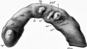
The brain consists of three cerebral vesicles. The fore-brain, with its optic outgrowths, but with no trace of cerebral hemispheres or hypophysis; the mid-brain, the smallest of the three, and distinctly marked of, both in front and behind, by two constrictions; and the hind-brain, larger than either of the preceding vesicles, and more over distinguished by exhibiting a series of neuromeres. These are seven in number, of which the second, third, fourth, and fifth are the most prominent.
Certain important structures are in relationship with these neuromeres. The trigeminal ganglion,some what pear-shaped, is placed opposite the second; the ganglion agoustico-facialis is placed opposite the fourth; and the otocyst opposite the fifth. The otocyst is a hollow rounded vesicle, with a small aperture opening on to the surface at the side of the head: it is in contact medially with the hind-brain. The ganglion of the ninth and tenth cranial nerves isnot yet visible,and this may explain the imperfect differentiation of the sixth and seventh neuromeres, opposite which the ganglia of these nerves normally develop.
The number of cases in which seven neuromeres have been observed in association with the hind-brain are now so numerous that the arrangement may be considered normal. C. Bradley (6) in 1904 described the series in the pig, and Broman (7) in 1895 fully described the neuromeres of the hind-brain in a human embryo of three millimetres.
The flexures of the brain in this specimen are so different from those usually described that a brief reference must now be made to them.
At the isthmus rhombencephali, the district between the mid- and hind-brains, there is a prominent flexure, which also involves the anterior extremity of the notochord. The mid-brain and the fore-brain are bent downwards, but there is no flexure of the fore-brain round the anterior end of the notochord. Its place is taken by the flexure at the isthmus, and the head-bend of the embryo corresponds with it.
A pontine flexure occurs between the anterior part of the hind-brain, which is placed horizontally, and the posterior portion which slopes down- wards and backwards. The angle of the bend is open downwards, and is placed opposite the fourth neuromere.
This bend does not correspond with the pons flexure formed later as a result of a ventral bending of the floor of the hind-brain.
The neck flexure is a very gradual one, and there is no indication of any constriction separating the brain from the cord.
Two of the flexures just described are so different from those universally regarded as primary cerebral flexures, that one hesitates to go further than simply place them on record. One point, however, may perhaps be added. The model shows the brain in a very early stage of development, before the hemispheres or the hypophysis have appeared. Is it not possible that there may be certain flexures, of a temporary character, which precede the primary flexures usually described? It will be interesting to see if future models of embryos at this age exhibit any flexures at all resembling them.
Notochord and Somites
The notochord lies close to the ventral surface of the spinal cord and brain, the general curve of which it follows. At its cephalic end it terminates opposite the bucco-pharyngeal membrane under the mid-brain, at its junction with the fore-brain. This part is distinctly flexed in association with the bend of the neural tube at the isthmus rhombencephali, and a little further back, i.e. under the foremost neuromeres of the hind-brain, it is thicker than elsewhere, and here it retains a direct connection with theentoderm. At its caudal end the notochord can be traced round the region of the rump into the tail, where it ends indistinctly by joining the undifferentiated mesoderm of that structure.
There are twenty-three pairs of somites, of which the first three, or three and a half,may be regarded as occipital, the remainder constituting the trunk somites, of which there are twenty. The twenty-first trunk somite is just beginning to form, but there is a region, including the tail, in which the mesoderm is not yet segmented.
Alimentary Canal
The pharynx is separated from the mouth by the bucco-pharyngeal membrane, which is already perforated, but there is no trace of the diverticulum of Rathke. Behind the membrane the pharynx is relatively capacious in a trans- verse direction, and exhibits on each side four pouches, of which the first is the largest, the succeeding ones diminishing in size from before backwards. They are compressed antero-posteriorly, and the first three terminate in blunt extremities, which are more or less vertically disposed. The fourth pouch is very small and pointed. As stated above, there are only three depressions externally behind the mouth, forming the bronchial clefts.
In the roof of the pharynx there is a prominent ridge continuous with the notochord, and connected with the floor is a small, hollow, rounded diverticulum, the median rudiment of the thyroid body. Itopensintothe pharynx by an exceedingly small aperture some distance behind the remains of the bucco-pharyngeal membrane, and opposite to the first pharyngeal pouch. There is no indication of the tuberculum impar or the furcula.
Behind the fourth pair of pharyngeal pouches the alimentary tube suddenly narrows, and becoming compressed laterally, forms a cleft-like lumen directed antero-posteriorly. The part of the tube,however,where the gradual change from a transversely disposed lumen to an antero- posterior one is manifest, is particularly interesting and important, as being the region at which the anlage of the lungs maybe recognized. Thetwo lung buds are already indicated as outgrowths from the entodermal tube, the epithelium of which is markedly thickened in this situation, and they are situated a short distance behind the fourth pair of pouches. The left lung bud, more prominent, is placed in advance of the light, and both are growing towards the pleural passages, the communications between the pericardial and peritoneal portions of the coelom. The lung rudiments are placed opposite either the hindmost occipital or first trunk somite in front (cephalad) of the dorsal inesocardium of the heart.
It is significant that the lung buds in such an early embryo should already be bilateral, particularly in connection with the debatable question of the nature of the lung anlage and the changes leading to the formation of the lung sacs. Whilst, as J.W. Flint (8) has recently pointed out, there is almost unanimity of opinion on the question of an unpaired origin, amongst those who have contributed to our knowledge of the development of the lungs, there are, however, a few who believe in a paired anlage, and regard the mammalian respiratory apparatus as arising from primitively paired structures. In so much that the paired buds are already present in this embryo, it lends some support to the latter view.
It is a very interesting question, seeing that a paired anlage for the lungs would bring them into line with the bronchial pouches. "The lungs," writes J. M. Flint (8) when dealing with this view, " while not representing actually existing bronchial pouches (would) indicate the reappearance of endoderimic evaginations of the head gut which have carried gils among the ancestors of vertebrates."
Below the origin of the lung buds is the anlage of the Esophagus and stomach, and in this part of the tube, beginning with the region where the lung rudiments are found, and extending backwards as far as the liver bud, the epithelial lining is markedly thickened. Opposite the third and fourth trunk somites the liver is seen growing into the septum transversum. The hepatic bud is a median structure with thick walls enclosing a cavity in communication with the alimentary canal. There is no trace of the pancreas.
The succeeding part of the alimentary canal is in wide communication with the yolk sac, and then follows the hind gut, a very narrow part with a rounded lumen. This terminates in the cloaca, relatively capacious cavity, pear-shaped in transverse section, opposite the point of junction with the allantois, triangular in section,beyond this,towards the tailgut. The Wolffian ducts have not yet reached the cloaca, but terminate in connection with the ectoderin some distance fromt it.
Along its greater curvature the cloaca is in relation with the notochord, and along its lesser curvature it comes into contact with the ectoderm at the root of the tail to form the cloacal membrane. The terminal partof the gut extends beyond the ineimbrane into the tail,and forms the post- analogue.
Connected with the cloaca is the allantois, an extremely small tubular structure which extends outwards for some distance through the umbilical orifice embedded in the mesoderm of the body stalk. At its cloacal end it is somewhat funnel-shaped, and at the opposite extremity, where it ends blindly, is a small swelling bent upon itself.
The allantois at its origin is situated, like the -cloaca, between the two primarycaudalarches. From this origin it is directed dorsalwards, and forwards, towards the umbilical orifice, being placed between the umbilical arteries. This part of the duct is very narrow and of fairly uniform calibre until it changes its direction, and runs ventralwards through the umbilical orifice. At the bend is a well-marked swelling, and another one is present just outside the orifice, a short distance from the blind extremity. This part of the tube is also accompanied by the umbilical arteries, which fuse to form a single vessel soon after they pass out of the embryo into the body stalk.
Excretory System
The excretory system, which is not shown in the model, consists of three parts (1) the Wolftian duct, (2) the rudimentary pronephric anlage, and (3) the mesonephric anlage.
The Wolfian Duct is situated close to the ectoderm, as is shown in Keibel's model of an embryo 15-18 days old. It begins as a duct with a distinct lumen opposite the eighth trunk somite and extends uninterruptedly as far as the twentieth trunk somite, where it ends by joining the ectoderm. This point is some distance from the cloaca, and coincides with the end of the differentiated muscle somites and the mesonephric anlage. Neither the muscle segments nor the mesonephric anlage are differentiated beyond the twentieth trunk somite, but as the formation of these structures is continued, the Wolffian duct, it may be assumed, likewise becomes differentiated, and, extending caudally, opens into the cloaca. In the embryo described by Bremer (5), which measured 4 mm., the duct of the right side opened into the cloaca; on the left side, however, it did not reach so far,but ended blindly.
The Rudimentary Pronephros. - Situated in the neck region, opposite the sixth, seventh, and eighth trunk somites,thereis,on each side,two or three rudimentary tubules, not connected by any duct, but communicating with the ccelom, each by a funnel-shaped passage or "trichter." This group of rudimentary tubules probably represents the pronephros, and itis separated by a distinct interval from the anlage of the mesonephros, which lies posterior to it.
Pronephric rudiments have been described by Keibel (9) in the embryos of apes, tarsius, and Man; by Tandler (10) in eight out of twelve human embryos of 5-20 mm. always at the level of the sixth and seventh segments; by S. P. Gage (11), in a human embryo of 3 mm.; and also by M'Callum (12), Bremer (5) and others.
The Anlage of the Mesomephros begins opposite the eighth trunk somite, i.e.the same segment opposite which the Wolffian duct begins to have a lumen. At this stage of development the mesonephros consists of a series of segmental vesicles with rudimentary nephrostomes. They are close together and connected with the Wolffian duct, but there is no definite numerical agreement with the mesoblastic somites. The mesonephric tissue is connected with the somites, and can be traced caudally as far as the latter are differentiated. There are no glomeruli.
The Vascular System
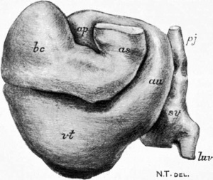
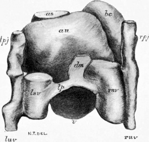
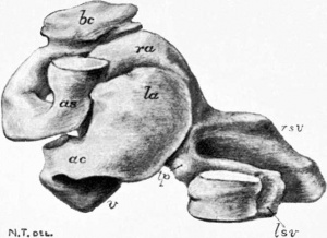
The Heart. - The specially interesting feature of this model is the clear indication of a fourth chamber- the bulbus cordis*- to which A. Keith (13) has recently drawn attention. Besides the sinus venous, auricle, and ventricle, the three parts which are supposed to give rise to the whole heart in the adult, there is undoubtedly a chamber on the right side, extending between the ventricle and the aortic stem. Its summit forms the highest part of the heart, and rises to the right side of the neck region. A. Greil (14) of Innsbruck has already published an account of its embryology in birds and reptiles, and traced its subsequent history; and A. Keith (13) has shown that many malformations of the human heart, hitherto believed to be the result of fetal endocarditis, are really due to mal-development, or arrest of development of this fourth chamber. In its general configuration the heart has an S-shaped form, the various parts of which are already foreshadowed, and disposed to some extent in the same relative position as in the adult. The four parts of the tube which can be recognized are the sinus venosus, the auricle, the ventricle, and the bulbus cordis, aud it will perhaps be most advantageous if each of the divisions be separately considered.
*bulbus cords - The name proposed by A. Langer for the two homologous structures, the conus arterioles of an anamniota and the bulbus arterioles of the amniote.
Sinus venous. - The sinus venosus consists of two horns, right and left,united by a transverse connecting piece. It constitutes the hindmost part of the heart, and is in intimate relation with the septum transversum, the latter forming a distinct shelf between the two horns dorsally, and the ventricle ventrally. The right horn is larger than the left, and on each side the summit or blind end extends into the pleural passage. Each horn receives, on its lateral aspect, a very short duct of Cuvier, and the umbilical vein. The vitelline vein,on the other hand, opens into the posterior (caudal) end of the sinus venosus, which appears to be a direct continuation of it. The transverse connecting piece runs from the left horn to the right horn; it is placed immediately ventral to the gut, and between the dorsal mesocardium in front and the septum transversum behind. The right horn communicates freely with the right side of the common auricle, but there is no indication of the formation of the right and left venous valves.
The Auricle. - The auricular chamber is placed dorsal to the aorticstein, which is received into a concavity on its ventral surface. The auricular appendices are already indicated, particularly the right,which projects ventrally between the bulbus cordis and the aortic stem.
On the right side of the auricle is the bulbus cords, a narrow cleft separating the two structures,and on its dorsal surface it gives attachment over a somewhat oblong-shaped area to the dorsal mesocardium. Asthe auricle is traced leftwards and downwards, the tube somewhat narrows, and a slight constriction indicates the site of the auricular canal.
The Ventricle. - The ventricle begins on the left side and forms the lowest part of the heart or apex. From this it passes to the right in contact with the septum transversujn, which is sloped for its reception, and finally it bends forward to become continuous with the bulbus cords.
The Bulbus cordis. - This is the part of the tube situated between the ventricle and the aortic stem. At its commencement it is a fairly capacious chamber, but traced onwards it narrows to become continuous with the aortic stem, the two forming a V-shaped figure on the anterior (ventral) aspect of the heart. According to Greil (14) and Keith(13) this fourth chamber of the heart becomes completely incorporated in the right ventricle to form the infundibulum.
Endothelial Tube. - A special model of this was made (fig. 5) to show its relationships to the muscular tube of the heart. The two are separated by an interval, particularly in the ventricle, the bulbus cordis, and the aortic stem, so that at this stage of development there are really two tubes, endothelial and muscular, one inside the other. The endothelial tube shows constrictions at the auricular canal,at the ventricular end of the bulbus cordis,and at the aorticend ofthe bulbus cordis. There is also a most obvious constriction in the bulbus cordis,not however at right angles to the tube, but in line with it, and disposed in such a way as to constrict the entrance of a pouch which occupies the highest part of the bulbus, and opens into the rest of the tube by a narrow orifice. It is difficult to say what the significance of this constriction maybe, but it is possibly associated with the series of events leading to the incorporation of the bulb with the right ventricle. Finally, it may be noted that the endothelial tube rises higher on the right side of the auricle than on the left.
Arteries. - From the aortic stem, the arterial arches, two in number on each side, have a radial disposition as they pass into the first and second visceral arches. The dorsal aorta pass backwards on either side of the notochord: at first separated, but gradually approaching one another, they eventually fuse to form a single vessel, ventral to the notochord. The fusion takes place opposite the seventh trunk somite, and the- two vessels continue as a single stem up to the thirteenth trunk somite, when they again separate to form the two primary caudal arches. The umbilical arteries accompany the allantois, the right distinctly smaller than the left, and finally the two vessels join together to form a single trunk within the bodystalk.
Veins. - The vitelline veins, returning the blood from the plexus on the yolk sac, form two well-developed vessels placed one on either side of the alimentary canal, and in close relation with the dorsal part of the septum transversum. They open into the hind end of the two horns of the sinus venous, medial to the termination of the umbilical veins.
The umbilical veins entering the embryo by the body stalk run in the body wall on either side of the umbilical orifice. Reaching the venous end of the heart, each opens into the lateral aspect of the sinus venosus in common with the duct of Cuvier. The two vessels are nearly equal in size, and it is worthy of mention that the vein on each side receives a small tributary, soon after it enters the embryo, from that part of the body wall corresponding to the site at which the bud of the hind limb subsequently grows out.
The primitive jugular vein begins in the neighbourhood of the otocyst, and, becoming more superficial as it is traced backwards, is finally joined by the posterior cardinal vein to form the duct of Cuvier. The latter is a short trunk, which opens into the sinus venous.
A further note on the septum transversum and the liver will be published subsequently.
In conclusion, I should like to express my warmest thanks to Professor R. Wiedersheim for placing at my disposal a place in his laboratory, and especially to Professor Keibel for lending me this rare and valuable embryo, and for his kindly advice and criticism whilst the models were being made. My thanks are also due to Dr Robert Meyer, to whom the specimen belongs.
Explanation of Plates
Plate I
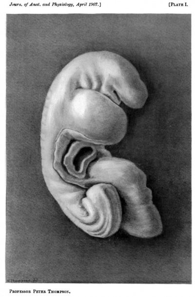
Drawing of a plaster cast of the reconstructed embryo, right side (nearly half size).
|
|
Plate II
Drawing of a plaster cast of the reconstructed embryo, frontal view (nearly half size).
|
|
|
Plate III
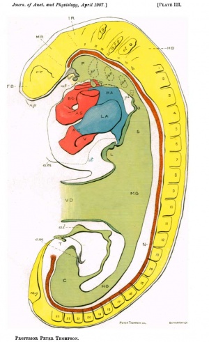
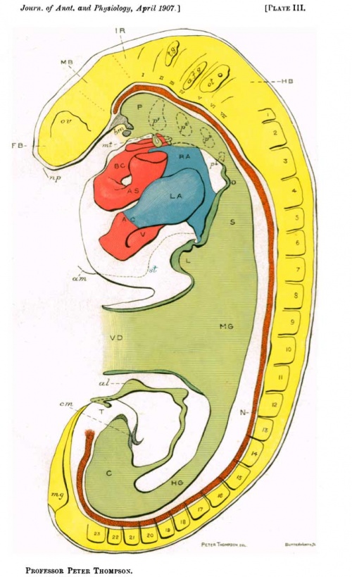
Graphic reconstruction of embryo from serial sections; the endothelial tube only of the heart is shown.
Bibliography
(1) ETERNOD, A. C. F., " Communication sur un oauf humain avec embryon excessivementjeune," Auch. Ital.deBiologie, xi., 1895.
(2) KOLLMANN, J., " Die Korperform menschlicher normaler und pathologischer Embryonen, ArchivftirAnat. und Physiol. Anat., Abth. Supplem., 1889.
(4) His, W., Anatomie menschlicher Embryonen, 1880-1885.
(5) BREMER, J. L., Description of a 4 mm. Human Embryo, vol. v., No. 4.
(6) BRADLEY, C., " Neuromeres of the Rhombencephalon of the Pig," Review of Neurol. and Psych., Sept. 1904.
(7) BROMAN, J., "Beobachtung eines menschlichen Embryos von beinahe 3 mm. Lange., etc," Morph. Arbeiten, v. 1895.
(8) FLINT, J. W., "The Development of the Lungs," American Journal of Anatomy, vol.vi.,No. 1.
(9) KEIBEL, F., " Zur embryologie des Menschen, der Affen und der Halbaffen," Anat.Anz.,vol.xxvii.,1905. GenevaProceedings,p.39.
(10) TANDLER, J., "Ueber Vornierenrudimente beim menschlichen Embryo," CentralblattfiirPhysiologie, vol.xvi.,1904, pp. 582-583.
(11) GAGE, S. P., "A Three-weeks Human Embryo, with especial reference to the Brain and Nephric System," American Journal of Anatomy, Vol. iv., No. 4, 1905.
(12) MACCALLUM, J. B., "Notes on the Wolffian Bodies of Higher Mammals," American Journal of Anatomy, Vol. i., No. 3, 1902.
(13) KEITH, A., "Malformations of the Bulbus cordis: an unrecognised division of the Human Heart," Studies in Pathology, Aberdeen Quatercentenary, 1906.
(14) GREIL, A. SeeHochstetter'sReviewinJertwig'sHandbuch;alsoMorph. Jahr., xxxi., 1903.
(15) JANOSIK, "Zwei junge menschliche Embryonen," Arch. fur Mic. Anal., XXX.,1887.
| Historic Disclaimer - information about historic embryology pages |
|---|
| Pages where the terms "Historic" (textbooks, papers, people, recommendations) appear on this site, and sections within pages where this disclaimer appears, indicate that the content and scientific understanding are specific to the time of publication. This means that while some scientific descriptions are still accurate, the terminology and interpretation of the developmental mechanisms reflect the understanding at the time of original publication and those of the preceding periods, these terms, interpretations and recommendations may not reflect our current scientific understanding. (More? Embryology History | Historic Embryology Papers) |
The information below was not part of the above original historic article.
- Carnegie Stages: 1 | 2 | 3 | 4 | 5 | 6 | 7 | 8 | 9 | 10 | 11 | 12 | 13 | 14 | 15 | 16 | 17 | 18 | 19 | 20 | 21 | 22 | 23 | About Stages | Timeline
Glossary Links
- Glossary: A | B | C | D | E | F | G | H | I | J | K | L | M | N | O | P | Q | R | S | T | U | V | W | X | Y | Z | Numbers | Symbols | Term Link
Cite this page: Hill, M.A. (2024, April 28) Embryology Paper - Description of a Human Embryo of Twenty-three Paired Somites. Retrieved from https://embryology.med.unsw.edu.au/embryology/index.php/Paper_-_Description_of_a_Human_Embryo_of_Twenty-three_Paired_Somites
- © Dr Mark Hill 2024, UNSW Embryology ISBN: 978 0 7334 2609 4 - UNSW CRICOS Provider Code No. 00098G


