Horse Development
| Embryology - 27 Jul 2026 |
|---|
| Google Translate - select your language from the list shown below (this will open a new external page) |
|
العربية | català | 中文 | 中國傳統的 | français | Deutsche | עִברִית | हिंदी | bahasa Indonesia | italiano | 日本語 | 한국어 | မြန်မာ | Pilipino | Polskie | português | ਪੰਜਾਬੀ ਦੇ | Română | русский | Español | Swahili | Svensk | ไทย | Türkçe | اردو | ייִדיש | Tiếng Việt These external translations are automated and may not be accurate. (More? About Translations) |
Introduction
Equine development (Latin, equus = "horse")
| Horse Links: horse | Category:Horse |
| Historic Papers: 1897 Critical Period in Horse Development | 1925 Organ of Jacobson | 1945 Cleavage Stages of the Horse Ova |
| Animal Development: axolotl | bat | cat | chicken | cow | dog | dolphin | echidna | fly | frog | goat | grasshopper | guinea pig | hamster | horse | kangaroo | koala | lizard | medaka | mouse | opossum | pig | platypus | rabbit | rat | salamander | sea squirt | sea urchin | sheep | worm | zebrafish | life cycles | development timetable | development models | K12 |
Some Recent Findings
|
| More recent papers |
|---|
 This table allows an automated computer search of the external PubMed database using the listed "Search term" text link.
More? References | Discussion Page | Journal Searches | 2019 References | 2020 References Search term: Equine Development | Equine Embryology | Horse Development |
| Older papers |
|---|
| These papers originally appeared in the Some Recent Findings table, but as that list grew in length have now been shuffled down to this collapsible table.
See also the Discussion Page for other references listed by year and References on this current page.
|
Taxon
Equine Development
338 days
| Animal Development Time | ||||||||||||||||||||||||||||||||||||||||||||||||||||||||||||||||||||||||||||||||||||||||||||||||||||||||||||||||||||||||||||||||||||||||||||||
|---|---|---|---|---|---|---|---|---|---|---|---|---|---|---|---|---|---|---|---|---|---|---|---|---|---|---|---|---|---|---|---|---|---|---|---|---|---|---|---|---|---|---|---|---|---|---|---|---|---|---|---|---|---|---|---|---|---|---|---|---|---|---|---|---|---|---|---|---|---|---|---|---|---|---|---|---|---|---|---|---|---|---|---|---|---|---|---|---|---|---|---|---|---|---|---|---|---|---|---|---|---|---|---|---|---|---|---|---|---|---|---|---|---|---|---|---|---|---|---|---|---|---|---|---|---|---|---|---|---|---|---|---|---|---|---|---|---|---|---|---|---|---|
| ||||||||||||||||||||||||||||||||||||||||||||||||||||||||||||||||||||||||||||||||||||||||||||||||||||||||||||||||||||||||||||||||||||||||||||||
Animal Notes and Table Data Sources
|
Genetics
Chromosomes: 1 | 2 | 3 | 4 | 5 | 6 | 7 | 8 | 9 | 10 | 11 | 12 |
31 | X | 11 12 13 14 15 16 17 18 19 20 21 22 23 24 25 26 27 28 29 30 31 X
Genome: Equus caballus
Morula and Blastocyst
The following images are from a historic (1945) article on early horse development.[6]
- Links: Fig. 1 | Fig. 2 | Fig. 3 | Fig. 4 | Fig. 5 | Plate 1 | Fig 6 | Fig 7 | Fig 8 | Fig 9 | Fig 10 | Fig 11 | Plate 2 | Fig 12 | Fig 13 | Fig 14 | Fig 15 | Plate 3
| Historic Disclaimer - information about historic embryology pages |
|---|
|
Plate 1

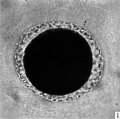
Fig. 1. Photograph of a living unsegmented ovum of the pony (P 1) in Locke’s fluid. Granular material is seen in the perivitelline space. X 480.
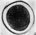
Fig. 2: Photograph of an unsegmented ovum of the pony (P 1) in agar. The nucleus is just visible. x 480.
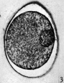
Fig. 3. Section of an unsegmented ovum of the pony (P1) with a large vesicular nucleus. x 520.
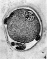
Fig. 4. Section of an unsegmented ovum of the pony (P 8) showing deutoplasmolysis. x 520.
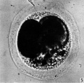
Fig. 5. Photograph of a living two-cell stage ovum of the pony (P 5). Granular material is seen in the lower part of the perivitelline space. x 480.


Plate 2

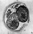
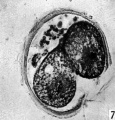
Figs. 6, 7. Two consecutive sections through the two-cell ovum shown in fig. 5. x 520.
Figs. 6, 7. Two consecutive sections through the two-cell ovum shown in fig. 5. x 520.
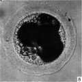
Fig. 8. Photograph of a living four-cell stage ovum of the pony (P 7). x 480.
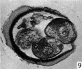
Figs. 9, 10. Two consecutive sections through the four-cell ovum shown in fig. 8. x 520.
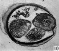
Figs. 9, 10. Two consecutive sections through the four-cell ovum shown in fig. 8. x 520.
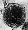
Fig. 11. Photograph of a five-cell stage ovum of the pony (P 4) in agar. x 480.


Plate 3

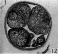
Fig. 12. Section through the five-cell ovum shown in fig. 11. x 520.
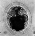
Fig. 13. Photograph of the living morula. of the pony (P 6). it shows extensive deutoplasmolysis. x 480.
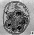
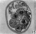
Figs. 14, 15. Two consecutive sections through the morula. shown in fig. 13. x 520.
Figs. 14, 15. Two consecutive sections through the morula. shown in fig. 13. x 520.


Genital Development
Gastrointestinal Tract
Prenatal Development of the Digestive System in the Horse.[7]
- 21 days - oral cavity was an empty space, and the liver contained proliferating endodermal cells.
- 25 days - fusiform stomach and the pancreatic bud were present.
- 28 days - small tongue and the esophagus occurred.
- 30 days - primary and secondary palates were developed, the liver contained cords of hepatocytes, and the pancreas was triangular.
- 40 days - crypts had formed in the intestinal loops, cell differentiation was observed in the hepatic parenchyma, and the pancreas was elongated.
- 50 days - Pancreatic acini and islets and intestines were highly convoluted.
- 75 days - Three segments of the pharynx
- 105 days - intestinal villi were wide with round tips; especially, the liver, stomach, and oral cavity showed key steps of anatomical and cellular differentiation.
Neural
Neonatal encephalopathy is the most common neurologic condition affecting newborn foals.[8] This neural abnormality has been compared to perinatal asphyxia syndrome of human infants.
References
- ↑ Agnew ME, Slack J, Stefanovski D, Linton JK & Sertich PL. (2019). Sonographic appearance of the late gestation equine fetal intestine. Theriogenology , 138, 121-126. PMID: 31326658 DOI.
- ↑ Fages A, Hanghøj K, Khan N, Gaunitz C, Seguin-Orlando A, Leonardi M, McCrory Constantz C, Gamba C, Al-Rasheid KAS, Albizuri S, Alfarhan AH, Allentoft M, Alquraishi S, Anthony D, Baimukhanov N, Barrett JH, Bayarsaikhan J, Benecke N, Bernáldez-Sánchez E, Berrocal-Rangel L, Biglari F, Boessenkool S, Boldgiv B, Brem G, Brown D, Burger J, Crubézy E, Daugnora L, Davoudi H, de Barros Damgaard P, de Los Ángeles de Chorro Y de Villa-Ceballos M, Deschler-Erb S, Detry C, Dill N, do Mar Oom M, Dohr A, Ellingvåg S, Erdenebaatar D, Fathi H, Felkel S, Fernández-Rodríguez C, García-Viñas E, Germonpré M, Granado JD, Hallsson JH, Hemmer H, Hofreiter M, Kasparov A, Khasanov M, Khazaeli R, Kosintsev P, Kristiansen K, Kubatbek T, Kuderna L, Kuznetsov P, Laleh H, Leonard JA, Lhuillier J, Liesau von Lettow-Vorbeck C, Logvin A, Lõugas L, Ludwig A, Luis C, Arruda AM, Marques-Bonet T, Matoso Silva R, Merz V, Mijiddorj E, Miller BK, Monchalov O, Mohaseb FA, Morales A, Nieto-Espinet A, Nistelberger H, Onar V, Pálsdóttir AH, Pitulko V, Pitskhelauri K, Pruvost M, Rajic Sikanjic P, Rapan Papeša A, Roslyakova N, Sardari A, Sauer E, Schafberg R, Scheu A, Schibler J, Schlumbaum A, Serrand N, Serres-Armero A, Shapiro B, Sheikhi Seno S, Shevnina I, Shidrang S, Southon J, Star B, Sykes N, Taheri K, Taylor W, Teegen WR, Trbojević Vukičević T, Trixl S, Tumen D, Undrakhbold S, Usmanova E, Vahdati A, Valenzuela-Lamas S, Viegas C, Wallner B, Weinstock J, Zaibert V, Clavel B, Lepetz S, Mashkour M, Helgason A, Stefánsson K, Barrey E, Willerslev E, Outram AK, Librado P & Orlando L. (2019). Tracking Five Millennia of Horse Management with Extensive Ancient Genome Time Series. Cell , 177, 1419-1435.e31. PMID: 31056281 DOI.
- ↑ Smits K, Willems S, Van Steendam K, Van De Velde M, De Lange V, Ververs C, Roels K, Govaere J, Van Nieuwerburgh F, Peelman L, Deforce D & Van Soom A. (2018). Proteins involved in embryo-maternal interaction around the signalling of maternal recognition of pregnancy in the horse. Sci Rep , 8, 5249. PMID: 29588480 DOI.
- ↑ Blanke A, Aupperle H, Seeger J, Kubick C & Schusser GF. (2015). Histological study of the external, middle and inner ear of horses. Anat Histol Embryol , 44, 401-9. PMID: 25283481 DOI.
- ↑ Wang X, Miller DC, Clark AG & Antczak DF. (2012). Random X inactivation in the mule and horse placenta. Genome Res. , 22, 1855-63. PMID: 22645258 DOI.
- ↑ Hamilton WJ. Cleavage Stages of the Ova of the Horse, with Notes on Ovulation. J Anat. 1945 Jul; 79(Pt 3): 127–130.3.
- ↑ <pubmed>24778084</pubmed>
- ↑ Tennent-Brown BS, Morrice AV & Reed S. (2015). The Equine Neonatal Central Nervous System: Development and Diseases. Vet. Clin. North Am. Equine Pract. , 31, 587-600. PMID: 26612749 DOI.
Reviews
Miraglia N, Salimei E & Fantuz F. (2020). Equine Milk Production and Valorization of Marginal Areas-A Review. Animals (Basel) , 10, . PMID: 32098374 DOI.
Fowden AL, Giussani DA & Forhead AJ. (2020). Physiological development of the equine fetus during late gestation. Equine Vet. J. , 52, 165-173. PMID: 31721295 DOI.
Roser JF & Meyers-Brown G. (2019). Enhancing Fertility in Mares: Recombinant Equine Gonadotropins. J. Equine Vet. Sci. , 76, 6-13. PMID: 31084750 DOI.
Tennent-Brown BS, Morrice AV & Reed S. (2015). The Equine Neonatal Central Nervous System: Development and Diseases. Vet. Clin. North Am. Equine Pract. , 31, 587-600. PMID: 26612749 DOI.
Perkins GA & Wagner B. (2015). The development of equine immunity: Current knowledge on immunology in the young horse. Equine Vet. J. , 47, 267-74. PMID: 25405920 DOI.
Articles
Rigoglio NN, Smith OE, Matias GSS, Miglino MA & Smith LC. (2019). Development of the central nervous system in equine twin fetuses derived by somatic cell nuclear transfer. Reprod. Fertil. Dev. , 31, 941-952. PMID: 30689958 DOI.
Rodrigues MN, Carvalho RC, Franciolli AL, Rodrigues RF, Rigoglio NN, Jacob JC, Gastal EL & Miglino MA. (2014). Prenatal development of the digestive system in the horse. Anat Rec (Hoboken) , 297, 1218-27. PMID: 24778084 DOI.
Wang X, Miller DC, Clark AG & Antczak DF. (2012). Random X inactivation in the mule and horse placenta. Genome Res. , 22, 1855-63. PMID: 22645258 DOI.
Fahiminiya S, Labas V, Roche S, Dacheux JL & Gérard N. (2011). Proteomic analysis of mare follicular fluid during late follicle development. Proteome Sci , 9, 54. PMID: 21923925 DOI.
Klein C & Troedsson MH. (2011). Transcriptional profiling of equine conceptuses reveals new aspects of embryo-maternal communication in the horse. Biol. Reprod. , 84, 872-85. PMID: 21209420 DOI.
Search Pubmed
Search Pubmed: equine development
External Links
External Links Notice - The dynamic nature of the internet may mean that some of these listed links may no longer function. If the link no longer works search the web with the link text or name. Links to any external commercial sites are provided for information purposes only and should never be considered an endorsement. UNSW Embryology is provided as an educational resource with no clinical information or commercial affiliation.
- Oklahoma State University Learning Reproduction in Farm Animals
| Animal Development: axolotl | bat | cat | chicken | cow | dog | dolphin | echidna | fly | frog | goat | grasshopper | guinea pig | hamster | horse | kangaroo | koala | lizard | medaka | mouse | opossum | pig | platypus | rabbit | rat | salamander | sea squirt | sea urchin | sheep | worm | zebrafish | life cycles | development timetable | development models | K12 |
Glossary Links
- Glossary: A | B | C | D | E | F | G | H | I | J | K | L | M | N | O | P | Q | R | S | T | U | V | W | X | Y | Z | Numbers | Symbols | Term Link
Cite this page: Hill, M.A. (2026, July 27) Embryology Horse Development. Retrieved from https://embryology.med.unsw.edu.au/embryology/index.php/Horse_Development
- © Dr Mark Hill 2026, UNSW Embryology ISBN: 978 0 7334 2609 4 - UNSW CRICOS Provider Code No. 00098G