Book - Manual of Human Embryology 16-1
| Embryology - 30 Apr 2024 |
|---|
| Google Translate - select your language from the list shown below (this will open a new external page) |
|
العربية | català | 中文 | 中國傳統的 | français | Deutsche | עִברִית | हिंदी | bahasa Indonesia | italiano | 日本語 | 한국어 | မြန်မာ | Pilipino | Polskie | português | ਪੰਜਾਬੀ ਦੇ | Română | русский | Español | Swahili | Svensk | ไทย | Türkçe | اردو | ייִדיש | Tiếng Việt These external translations are automated and may not be accurate. (More? About Translations) |
Keibel F. The Development of the Sense Organs. (1912) chapter 16, vol. 2, in Keibel F. and Mall FP. Manual of Human Embryology II. (1912) J. B. Lippincott Company, Philadelphia.
The Gustatory Organ
The tongue is frequently spoken of simply as the organ of taste, and one may say of a person that he has a good tongue just as one might say he "has a good eye ; but on the one hand the tongue is not merely a gustatory organ, and on the other hand, the organs that are receptive for taste, the taste-buds, are not limited in their distribution to the territory of the tongue. Accordingly the development of the tongue will be treated along with that of the digestive tract, and we have to discuss here in the first place the development of the taste-buds. Brief consideration must also be given to those papillae of the tongue which are to be regarded as accessory organs of the gustatory sense. The territory within which taste-buds are found in man is quite extensive. Their principal situation in the adult individual is on the papillae vallatae; but they have also been described as occurring on the papillae foliatae and the papillae fungiformes, on the under surface of the tongue on the plica fimbriata (Ponzo, 1905 1 , 1905 2 , and 1907), on both surfaces of the epiglottis and also in the mucous membrane of the larynx itself, and in the region of the arytenoid cartilages (Davis, 1877). They are also said to occur in the anterior surface of the soft palate, especially in the neighborhood of the uvula (A. Hoffmann, 1875, W. Krause, 1876). Von Ebner (1899) has not been able to find them in this situation, and J. Schaffer (1898) believes that the thickened ends of papillae have been confused with taste-buds; Ponzo (1907), however, has recently stated that he has found them in the human fetus on the palatine tonsil, on the palatine arches, and on both surfaces of the soft palate. Such contradictions are probably to be explained by the fact that, as will be seen, the taste-buds are at first more widely distributed than they are later on, and undergo a partial retrogression which apparently is not of quite the same extent in all individuals. Very generally the gustatory organs have been regarded as being of ectodermal origin (see Schwalbe, Lehrbuch der Anatomie der Sinnesorgane, p. 36) ; this view is not, however, free from objection. It is not possible, it is true, to delimit exactly in the mouth cavity the ectoblastic and entoblastic territories; but the majority of the tastebuds lie undoubtedlv within the entoblastic territorv, and even although epithelial encroachments are possible, yet it seems difficult to suppose that the ectoblast has penetrated into the region of the larynx. The first thorough investigations of the development of the taste-buds in man were undertaken by Tuckermann (1889, 1890, 1890 2 ), who also reviewed the older literature, for Hoffmann (1875) and Lustig (1884) had already published statements concerning the fetal conditions. Tuckermann failed to find taste-buds in a fetus of ten weeks, but did find them in one of fourteen weeks ; he concluded, therefore, that they were formed during the twelfth week of intra-uterine life. The first statements, accompanied by figures, concerning the actual histological differentiation of the buds were made by Qraberg (1898), and his account will lie followed here.
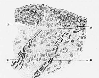
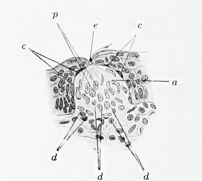
In a fetus of about three months (11 cm vertex-sole length) this author could not recognize any anlagen of the papillae vallatae, but he found two ridges of the mucous membrane at the back part of the tongue which were placed obliquely and met in the median line to form an angle open anteriorly. These ridges are the foundations for the papillae vallatae. The epithelium covering the ridges is growing even at this stage into the stratum proprium in the form of simple invaginations and is dividing the ridges into papillae. The first anlagen of the taste-buds are accordingly to be found in this fetus, but they are not as yet clearly denned (Fig. 121). The basal cells of the epithelium have lost their usual low cylindrical form and have increased in size noticeably. It is noteworthy that already in this earliest stage of development a nerve is in connection with that portion of the epithelium from which a taste-bud is differentiating, and Graberg believes that it may have a direct influence on the differentiation. Fig. 122 shows a further developed, well-defined taste-bud with a gustatory pore, from a fetus of 21.3 cm. vertex-rump measurement; a differentiation of the cells of the taste-bud into extrabulbar, basal, and pillar cells has also taken place to some extent, and, in addition, there are also present cells of an indifferent nature. So soon as they have become well differentiated the cells of the taste-bud reach the surface of the epithelium; then the gustatory pore is formed by the epithelium at the sides of the taste-bud increasing in thickness while the cells of the bud itself have almost completed their growth in length. The appearance of the taste-buds and their degree of development are in general subject to great variation. In the new-born child the different kinds of cells which constitute a bud are readily recognizable in transverse sections, neuro-epithelial cells, pillar cells, basal cells, and the extrabulbar cells; Graberg failed to find only the rod-shaped cells of Hermann, but, on the other hand, he believed that he could distinguish the striated margin of the inner gustatory pore, or ciliary corona of Schwalbe, as well as the hairs projecting into the pore. Many taste-buds undergo degeneration during the latter portions of intra-uterine life and after birth, and the degeneration process affects the firstformed buds situated on the upper free surfaces of the papillae vallatae, furthermore those on the papilla? fungiformes and those on the anterior surface of the epiglottis, on the tonsils, the soft palate, and on the plica fimbriata. Thus, Kiesow (1902) in almost mature fetuses in the majority of cases found taste-buds on the lingual surface of the epiglottis; after birth they vanish. According to Stahr (1901) the abundance of buds on the different kinds of papillae is associated with the degree of elaboration of the form of the papillae, their size and number. The vallate papillae are late to become fully formed, and when they do the papillae fungiformes assume a less definite form; they become relatively smaller and less numerous, and their epithelium partly loses its buds and becomes cornified. In the new-born child taste-buds occur on all the fungiform papillae. The significance of the different kinds of papillae for the function of taste alters during the life of the individual. The fungiform papillae have their greatest abundance of taste-buds, and with that their chief period for functioning as taste-organs, in the new-born child ; for the vallatae and the f oliatae these conditions occur in the adult. As regards the degeneration of the taste-buds on the upper surfaces of the papillae vallatae, Graberg finds that in fetuses of 24.5 and 39.5 cm. their outlines become indistinct and the nuclei of their cells shrivel. Occasionally leucocytes seem to invade them. The degenerated buds are carried to the surface and thrown off by the growth of the epithelium.
Fig. 121. Frontal section through a papilla vallata of an 11 cm human fetus. (After Graberg. Schwalbe's Morphol. Arbeiten, vol. 8, 1898, PI. 11, Fig 1.) a, places at which the formation of tastebuds is beginning; n, nerves.
Fig. 122. Frontal section through a papilla vallata of a 21.3 cm human fetus. (After Graberg, Schwalbe's Morphol. Arbeiten, vol. 8, 1898, PI. 11, Fig. 4.) o. indifferent cells; c, extrabulbar cells; d, basal cells; e, gustatory pore; p, pillar cells.
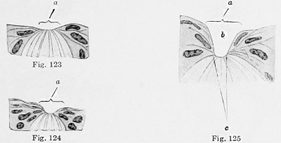
Figs. 123-125. The summits of three taste-buds from a human fetus of 21.3 cm a, in Fig. 123, indication of the gustatory pore, in Fig. 124, its anlage, and in Fig. 125, outer gustatory pore; b, gustatory canal; c, inner gustatory pore. (After Graberg, Schwalbe's Morphol. Arbeiten, vol. 8, 1898, PL 11, Figs. 8-10.)
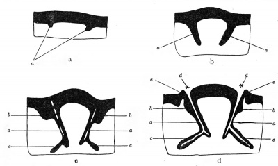
The papillae vallatae, f oliatae, and fungiformes may properly be regarded as organs accessory to the taste-buds. The first appearance of the fungiform papillae is shown in the Normentaf el of Keibel and Elze in Plates 62, 64, and 66, in embryos less than 20 mm. in length. Graberg has described and illustrated by some diagrams (Fig. 126, a-d) the development of the papillae vallatse. The ridges that precede the anlagen of these papillae have alreadybeen described, and also the process by which they are broken up into the individual papillae. The annular walls are formed from definite epithelial growths, derived from the epithelial ridges that bound the primitive papillae, and, lateral to these, extending down into the stratum proprium. The stratum proprium on its part then projects up into the epithelial thickening, raising the epithelium over it and thus producing on the free surface of the mucous membrane surrounding the papillae a slight elevation, which is the anlage of the annular wall. The fossae are formed by the fusion of small clefts that develop in the epithelial downgrowths that separate the papillae. The glands of Ebner appear as solid outgrowths which extend laterally from the lower edges of the epithelial downgrowths. Later they acquire a lumen by the degeneration of their central cells, but even in the new-born child they are not everywhere fully developed. Until after birth the growth of the fungiform papillae is mainly in length (Stahr, 1901), the fungus shape being acquired with the development of secondary crops of papillae, by which a second stage in the growth of the papillae is characterized; now for the first time can the papillae be said to have a foot and a head. The secondary papillae have already appeared in children of a few months, and in these all fungiform papillae still bear taste-buds, whereas in the tongue of the adult fungiform papillae without buds occur. In the adult the epithelium often cornifies to form long tips, yet papillae also occur whose epithelium is in part cornified and in part bears buds. Transitions between vallate and fungiform and between fungiform and filiform papillae do not occur.
Fig. 126, a-d. Diagrams illustrating the development of the vallate papillas and their adnexae. (After Graberg. Schwalbe's Morphol. Arbeiten. vol. 8, 1898, p. 121-122.) a, "primary," b, "secondary" epithelial downgrowths: c, anlagen of glands of von Ebner; d, fossae: e, annular wall.
| Embryology - 30 Apr 2024 |
|---|
| Google Translate - select your language from the list shown below (this will open a new external page) |
|
العربية | català | 中文 | 中國傳統的 | français | Deutsche | עִברִית | हिंदी | bahasa Indonesia | italiano | 日本語 | 한국어 | မြန်မာ | Pilipino | Polskie | português | ਪੰਜਾਬੀ ਦੇ | Română | русский | Español | Swahili | Svensk | ไทย | Türkçe | اردو | ייִדיש | Tiếng Việt These external translations are automated and may not be accurate. (More? About Translations) |
Keibel F. The Development of the Sense Organs. (1912) chapter 16, vol. 2, in Keibel F. and Mall FP. Manual of Human Embryology II. (1912) J. B. Lippincott Company, Philadelphia.
| XVI. The Development of the Sense Organs: General Considerations | Touch Cells | Epibranchial Sense Organs | Gustatory Organ | Olfactory Organ | Eye | Ear | Manual of Human Embryology II | ||||||||||||||||||||||
|
| |||||||||||||||||||||
Bibliography
(For the lamellate corpuscles, touch-corpuscles, and the gustatory organ)
Davis, C. : Die becherfbrmigen Organe des Kehlkopfes, Arch, fur mikr. Anat., vol. 14, 1877.
Davydow : Materialien zur Kenutnis der Entwicklung des peripheren Nerven systems, Dissert., Moskow, 1903.
von Ebner, V.: in Kolliker's Handbuch der Gewebelehre, vol. 3, p. 21, 1899.
Graberg, J.: Beitrage zur Genese des Geschmacksorganes des Mensehen, Schwalbe's Morphol. Arbeiten, vol. 8, p. 117 (12 plates and 4 text figures), 1898.
Henle, J., and Kolliker, A. : Ueber die Pacini' schen Korperchen an den Nerven des Mensehen und der Saugetiere, with 3 plates, gr. 4to. Meyer and Zeller. Zurich, 1844.
Hoffmann, A. : Ueber die Verbreitung der Geschmaeksorgane beim Mensehen, Virchow's Arch. f. path. Anat., vol. 62, 1875.
Keibel, F., and Elze, C. : Norrnentafel zur Entwicklungsgesckichte des Mensehen, Jena, 1908.
Kiesow, F. : Sur la presence des boutons gustatifs a la surface linguale de 1'epiglotte humaine avec quelques reflexions sur les rneines organes, qui se trouvent dans la muqueuse du larynx, Arch. Ital. Biol., vol. 37, p. 234. 1902.
Krause, W. : Die tenninalen Korperchen der einfach sensiblen Nerven, Hannover, 1860. (Compare Hertwig's Handbuch, vol. 2, p. 330.) Die motorischen Endplatten der quergestreiften Muskelfasern, Hannovei*, 1869. Anatomie, vol. 1, 1876.
Langerhans, P.: Ueber Tastkorperchen und rete Malpighii, Arch, fiir mikr. Anat., vol. 9, 1873.
Lustig, A. : Beitrage zur Kenntnis der Entwicklung der Geschmacksknospen, Sitzungsber. Akad. Wiss. Wien, vol. 89, part 3, p. 308, 1884.
Ponzo, M. : Sulla presenza di calici gustativi in alcune parti della retrobocca e nella parte •nasale della faringe del feto umano, Giornale d. R. Accad. di Med. Torino, vol. 68, also in Monit. Zool. Ital., vol. 16, 1905a
- Sur la presence de bourgeons gustatifs dans quelques parties de l'arriere bouche et dans la partie nasale du pharynx du foetus humain, Arch. Ital. Biol., vol. 43, 1905b
- In torn o alia presenza di organi gustativi sulla faccia inferiore della lingua del feto umano, Anat. Anz., vol. 30, 1907.
Ramstrom, M. : Anatomische und experimentelle Untersuchungen iiber die lamellosen Endkorperchen im peritoneum parietale des Mensehen. Anat. Hefte. vol. 36, Part 109, 1908.
Ranvier, L. : Nouvelles recherches sur les organes du tact, Comptes Rend. Acad Sci. Paris, vol. 91, p. 1087, 1880. Traite technique d'histologie, p. 706-707, 1880.
Schumacher, S. von : Ueber das glomus coccygeum des Mensehen und die glomeruli caudales der Saugetiere, Verhandl. Anat. Ges. "Wiirzburg, Anat. Anz., vol. 30, Suppl., 1907.
Schaffer. J. : Beitrage zur Histologie menschlichen Organe. V. Mundhohle. Schlundkopf, Sitzungsber. Akad. Wiss. Wien.. vol. 106 (Oct.. 1897), 1898.
Stahr, H. : Ueber die papillae fungiformes der Kinderzunge und ihre Bedeutung als Geschniacksorgan. With 4 plates. Zeit. fur Morphol. und Anthrop., vol. 41, p. 199, 1901.
Tuckermann F. Further Observations on the Development of the Taste-organs of Man (1890) Journ. of Anat. and Physiol. 24: 130.
Tuckermann F. On the Gustatory Organs of the Mammalia (1890) Proc. Boston Soc. Natural Hist. 24: 470.
| Embryology - 30 Apr 2024 |
|---|
| Google Translate - select your language from the list shown below (this will open a new external page) |
|
العربية | català | 中文 | 中國傳統的 | français | Deutsche | עִברִית | हिंदी | bahasa Indonesia | italiano | 日本語 | 한국어 | မြန်မာ | Pilipino | Polskie | português | ਪੰਜਾਬੀ ਦੇ | Română | русский | Español | Swahili | Svensk | ไทย | Türkçe | اردو | ייִדיש | Tiếng Việt These external translations are automated and may not be accurate. (More? About Translations) |
Keibel F. The Development of the Sense Organs. (1912) chapter 16, vol. 2, in Keibel F. and Mall FP. Manual of Human Embryology II. (1912) J. B. Lippincott Company, Philadelphia.
| XVI. The Development of the Sense Organs: General Considerations | Touch Cells | Epibranchial Sense Organs | Gustatory Organ | Olfactory Organ | Eye | Ear | Manual of Human Embryology II | ||||||||||||||||||||||
|
| |||||||||||||||||||||
| Manual of Human Embryology II 1912: Nervous System | Chromaffin Organs and Suprarenal Bodies | Sense-Organs | Digestive Tract and Respiration | Vascular System | Urinogenital Organs | Figures 2 | Manual of Human Embryology 1 | Figures 1 | Manual of Human Embryology 2 | Figures 2 | Franz Keibel | Franklin Mall | Embryology History |
Cite this page: Hill, M.A. (2024, April 30) Embryology Book - Manual of Human Embryology 16-1. Retrieved from https://embryology.med.unsw.edu.au/embryology/index.php/Book_-_Manual_of_Human_Embryology_16-1
- © Dr Mark Hill 2024, UNSW Embryology ISBN: 978 0 7334 2609 4 - UNSW CRICOS Provider Code No. 00098G


