BGDA Practical 7 - Week 4
Introduction
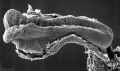
Stage 10
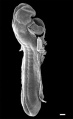
Stage 11

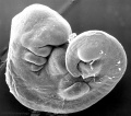
Stage 12
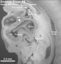
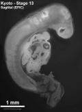
Stage 13


| Week 4 Embryo - Stage 13 Movies | ||||||||||||||||||
|---|---|---|---|---|---|---|---|---|---|---|---|---|---|---|---|---|---|---|
|
<html5media height="600" width="500">File:Stage 13 MRI 3D04.mp4</html5media> |
Week 4 Embryo surface view
Week 4 Stage 13 embryo shown as rotating with surface details. Note:
| |||||||||||||||||
| <html5media height="700" width="500">File:Stage 13 MRI 3D03.mp4</html5media> | Central Nervous System
Week 4 Stage 13 embryo shown as rotating with details of the CNS brain and spinal cord. Note:
| |||||||||||||||||
| <html5media height="600" width="500">File:Stage 13 MRI_S02.mp4</html5media> | Heart
This movie shows sagittal sections through the Stage 13 week 4 human embryo region of the pharyngeal arches (contributing to head and neck development) and the heart. | |||||||||||||||||
|
| |||||||||||||||||

Key Events of Human Development during the fourth week (week 4) following fertilization or clinical GA week 6.
These notes cover the fourth week of embryonic development, which is the beginning of organogenesis, (specific tissues and systems are beginning to differentiate) from the trilaminar embryo. With many parallel processes, descriptions begin to get complicated! Many of the described processes begin and extend over a broader range of time. Some developmental processes will be discussed later in the practical to simplify matters.
Ectoderm on the embryo surface undergoes segmentation: The central portion of the embryonic disc forms the neural plate, the edge of this plate forms neural crest and the remainder outside this forms the epitheium of the skin and other structures.

Human Embryo - left dorsolateral view (Week 4, Carnegie stage 11, GA week 6])
- Neurogenesis
- Central Nervous System (CNS) - the neural plate undergoes morphological changes to form the primitive central nervous system (brain, spinal cord). An epithelial layer of cells which contributes all neural (brain, spinal cord, peripheral nervous system) and the external epithelium (surface layer of the skin) of the embryo. Neurogenesis begins towards the end of week 3, when the neural tissues separate from this germ cell layer.
- Peripheral Nervous System (PNS) - the neural crest cells in the body region migrate and spread to different regions of the embryo forming the PNS (dorsal root ganglia, sympathetic ganglia, enteric nervous system) and many other embryonic tissues. Neural crest cells in the head region form skeletal and other structures.
- Pharyngeal Arches
- In the head region, a series of ventral folds form under the brain in a rostral to caudal sequence, these are the pharyngeal arches.
- Placodes
- In the head region, ectoderm small patches form pairs of specialised placodes that eventually contribute to specific sensory components, cranial ganglia and the anterior pituitary (adenohypophysis).
- Limb Buds
- Cardiogenesis
- Within the embryo mesoderm, the heart tube and vascular development continues. Cardiogenesis will be covered in week 5, when septation begins.
- Note there is also an online tutorial (developed by an ILP student) that will introduce heart development. The best place to start is with Basic Cardiac Embryology.
Neurogenesis


Developmental sequence: neural plate -> (day 18-19) neural groove -> neural tube -> Central Nervous System (brain and spinal cord)
- Mesoderm and Ectoderm Cartoons
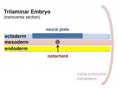
Trilaminar Embryo
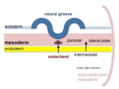
Paraxial and Lateral Plate
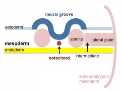
Somites
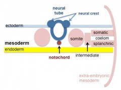
Somatic and Splanchnic


- Central Nervous System (CNS) - the neural plate undergoes morphological changes to form the primitive central nervous system. An epithelial layer of cells which contributes all neural (brain, spinal cord, peripheral nervous system) and the external epithelium (surface layer of the skin) of the embryo. Neurogenesis begins towards the end of week 3, when the neural tissues separate from this germ cell layer.
- Peripheral Nervous System (PNS) - the neural crest cells in the body region migrate and spread to different regions of the embryo forming the PNS and many other embryonic tissues. Neural crest cells in the head region form skeletal and other structures.
Human Neuralation - Early Stages

|

|
| Cranial Neural Pore (stage 11) | Caudal Neural Pore (stage 12) |
The stages below refer to specific Carneigie stages of development.
- stage 8 (about 18 postovulatory days) neural groove and folds are first seen
- stage 9 the three main divisions of the brain, which are not cerebral vesicles, can be distinguished while the neural groove is still completely open
- stage 10 (two days later) neural folds begin to fuse near the junction between brain and spinal cord, when neural crest cells are arising mainly from the neural ectoderm
- stage 11 (about 24 days) the cranial neuropore (rostral, cephalic or anterior) closes within a few hours; closure is bidirectional, it takes place from the dorsal and terminal lips and may occur in several areas simultaneously. The two lips, however, behave differently.
- stage 12 (about 26 days) The caudal (posterior) neuropore takes a day to close
- the level of final closure is approximately at future somitic pair 31
- corresponds to the level of sacral vertebra 2
- stage 13 (4 weeks) the neural tube is normally completely closed (More? neural tube defects)
![]()
Three primary brain vesicles develop initially due to the neural plate being broader at the cranial (brain) end than the narrower caudal (spinal cord) end. When the plate fuses to form a tube, these 3 initial expansions (vesicles) result.

Pharyngeal Arches and Placodes
In the head region, two main components of head development form the pharyngeal arches and sensory placodes.
- Pharyngeal arches form a series of ventral folds under the brain in a rostral to caudal sequence. These arches will form most of the head and neck structures of the embryo and contain all three germ layers (ectoderm, mesoderm and endoderm). The topic of head and sensory development is covered in detail in BGD cycle B.
- Small patches of ectoderm form pairs of specialised placodes that eventually contribute to specific sensory components, cranial ganglia and the anterior pituitary (adenohypophysis).
| Cranial End of Embryo (Carnegie stage 11) | |
|---|---|

|

|


- Links: Placodes
Cardiogenesis

|

|
| Human embryos (ventral view between 20 to 22 days (GA week 5 to 6). | Human Embryo (ventral view Week 4, Carnegie stage 10) |
As the tubular heart grows it develops dilations and constrictions which form the truncus arteriosus, bulbus cordis, primitive ventricle, primitive atrium and sinus venosus.
Embryo Stage 13
Movies - Embryo Carnegie stage 13 - These are rotating animations based upon reconstruction of individual serial slice images of the stage 13 embryo.
| Central Nervous System | Gastrointestinal | Cardiovascular |
Week 4 Dynamics
Note that many of the movies start in week 4 and continue on through later embryonic development.
Ectoderm
|
|
Mesoderm
|
|
|
|
Endoderm
| Amniotic Cavity |
| Page | Play |
Neural Tube Defects (NTD)
Maternal Diet - FolateResearch has demonstrated that that supplementation of maternal diet with folate reduces incidence of NTDs.
|


Week 4 Interactive Component
| Attempt the Quiz - Week 4 | |
|---|---|

Here are a few simple Quiz questions that relate to Week 4 (GA week 6) from the lecture and practical. See your Quiz Result - Answer all the questions, then click "submit" to complete. The page will reload and you can then reopen this table to see your result and feedback.
|
Additional Information
| Additional Information - Content shown under this heading is not part of the material covered in this class. It is provided for those students who would like to know about some concepts or current research in topics related to the current class page. |
Timeline
| Week 4 - Human Embryo Stages and Events (GA week 6) | ||
|---|---|---|
| Embryo Week: Week 1 | Week 2 | Week 3 | Week 4 | Week 5 | Week 6 | Week 7 | Week 8 | Week 9 | ||
| Event | ||
| Stage 10 |  
neural crest - differentiation at spinal cord level from day 22 until day 26 neural - neural folds begin to fuse near the junction between brain and spinal cord, when neural crest - cells are arising mainly from the neural ectoderm neural crest - trigeminal, facial, and postotic ganglia components visible[1] neural crest - migration of vagal level neural crest cells begins (7-10 somite stage) neural - Brain rostral neural tube forms 3 primary brain vesicles (week 4) respiratory - Week 4 laryngotracheal groove forms on floor foregut. | |
| heart - begins to beat in Humans by day 22-23, first functioning embryonic organ formed. | ||
| Stage 11 | 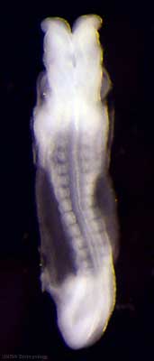
thyroid - thyroid median endodermal thickening in the floor of pharynx neural - rostral (or cephalic) neuropore closes within a few hours; closure is bidirectional, it takes place from the dorsal and terminal lips and may occur in two areas simultaneously. The two lips, however, behave differently. Optic ventricle appears | |
| Stage 12 | 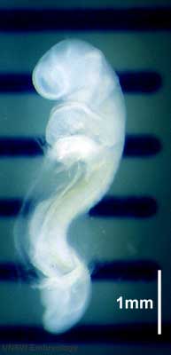
pituitary - Week 4 hypophysial pouch, Rathke's pouch, diverticulum from roof liver septum transversum forming liver stroma and hepatic diverticulum forming hepatic trabeculae[2] neural - caudal neuropore takes a day to close (closure is approximately at future somitic pair 31/sacral vertebra 2) neural - secondary neurulation begins neural crest - cardiac crest, neural crest from rhombomeres 6 and 7 that migrates to pharyngeal arch 3 and from there the truncus arteriosus[1] neural crest - vagal neural crest enter the foregut (20-25 somite stage) | |
| Stage 13 | 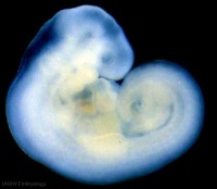 Neural - the neural tube is normally completely closed, ventricular system now separated from amniotic fluid. Neural crest at spinal level is segregating, and spinal ganglia are in series with the somites. Spinal cord ventral roots beginning to develop.[3] Neural - the neural tube is normally completely closed, ventricular system now separated from amniotic fluid. Neural crest at spinal level is segregating, and spinal ganglia are in series with the somites. Spinal cord ventral roots beginning to develop.[3]
telencephalon cavity appears liver epithelial cord proliferation enmeshing stromal capillaries[2] smell Crest comes from the nasal placodes[4] integumentary - 4 weeks simple ectoderm epithelium over mesenchyme integumentary - 1 to 3 months ectoderm - germinative (basal) cell repeated division of generates stratified epithelium; mesoderm - differentiates into connective tissue and blood vessels | |
| Note - the day timing of stages is only approximate, system names link to first page of that specific system, and events are based upon the literature cited below. | ||
References
| ||
Neural Development
| Neural Tube | Primary Vesicles | Secondary Vesicles | Adult Structures |
|---|---|---|---|
| week 3 | week 4 | week 5 | adult |
| prosencephalon (forebrain) | telencephalon | Rhinencephalon, Amygdala, hippocampus, cerebrum (cortex), hypothalamus, pituitary | Basal Ganglia, lateral ventricles | |
| diencephalon | epithalamus, thalamus, Subthalamus, pineal, posterior commissure, pretectum, third ventricle | ||
| mesencephalon (midbrain) | mesencephalon | tectum, Cerebral peduncle, cerebral aqueduct, pons | |
| rhombencephalon (hindbrain) | metencephalon | cerebellum | |
| myelencephalon | medulla oblongata, isthmus | ||
| spinal cord, pyramidal decussation, central canal | |||
Neural Tube Defects
Australian Statistics
- Women who have one infant with a neural tube defect have a significantly increased risk of recurrence
- 40-50 per thousand compared with 2 per thousand for all births.
- A randomised controlled trial conducted by the Medical Research Council of the United Kingdom demonstrated a 72% reduction in risk of recurrence by periconceptional (ie before and after conception) folic acid supplementation (4mg daily).
- Other epidemiological research, including work done in Australia, suggests that primary occurrences of neural tube defects may also be prevented by folic acid either as a supplement or in the diet.
- This has been confirmed in a randomised controlled trial from Hungary, which found that a multivitamin supplement containing 0.8mg folic acid was effective in reducing the occurrence of neural tube defects in first births.
Wheat flour has contained folic acid since September 2009.
Before mandatory folic acid fortification was introduced:
- mean dietary folic acid intakes for women aged 16–44 years (the target population) in Australia was 108 micrograms (μg) of folic acid per day and in New Zealand was 62 μg of folic acid per day, well below the recommended 400 μg per day.
- there were 149 pregnancies affected by NTDs in 2005 in Australia (rate of 13.3 per 10,000 births) in the three states that provide the most accurate baseline of NTD incidence (South Australia, Western Australia and Victoria), and 63 pregnancies affected by NTDs in 2003 in New Zealand (rate of 11.2 per 10,000 births).
- Food Standards (FSANZ) had allowed industry two years to prepare to add folic acid to wheat flour used in making bread.
- Links: Victoria - Folate information for health professionals | NHMRC - Nutrient Reference Values for Australia and New Zealand Including Recommended Dietary Intakes | NHMRC - Iodine supplementation for Pregnant and Breastfeeding Women
UK
A randomised controlled trial conducted by the Medical Research Council of the United Kingdom demonstrated a 72% reduction in risk of recurrence by periconceptional (ie before and after conception) folic acid supplementation (4mg daily).
USA
Women who have one infant with a neural tube defect have a significantly increased risk of recurrence (40-50 per thousand compared with 2 per thousand for all births)


- Food and Drug Administration (USA) in 1996 authorized that all enriched cereal grain products be fortified with folic acid, with optional fortification beginning in March 1996 and mandatory fortification in January 1998. The data in the above graphs show the subsequent changes in anencephaly and spina bifida rate over that period.
Embryo Stages and Events
The collapsible table shown below identifies Carnegie stages and events that occur during week 4 (GA week 6).
| Week 4 - Human Embryo Stages and Events (GA week 6) | ||
|---|---|---|
| Embryo Week: Week 1 | Week 2 | Week 3 | Week 4 | Week 5 | Week 6 | Week 7 | Week 8 | Week 9 | ||
| Event | ||
| Stage 10 |
neural crest - differentiation at spinal cord level from day 22 until day 26 neural - neural folds begin to fuse near the junction between brain and spinal cord, when neural crest - cells are arising mainly from the neural ectoderm neural crest - trigeminal, facial, and postotic ganglia components visible[1] neural crest - migration of vagal level neural crest cells begins (7-10 somite stage) neural - Brain rostral neural tube forms 3 primary brain vesicles (week 4) respiratory - Week 4 laryngotracheal groove forms on floor foregut. | |
| heart - begins to beat in Humans by day 22-23, first functioning embryonic organ formed. | ||
| Stage 11 |
thyroid - thyroid median endodermal thickening in the floor of pharynx neural - rostral (or cephalic) neuropore closes within a few hours; closure is bidirectional, it takes place from the dorsal and terminal lips and may occur in two areas simultaneously. The two lips, however, behave differently. Optic ventricle appears | |
| Stage 12 |
pituitary - Week 4 hypophysial pouch, Rathke's pouch, diverticulum from roof liver septum transversum forming liver stroma and hepatic diverticulum forming hepatic trabeculae[2] neural - caudal neuropore takes a day to close (closure is approximately at future somitic pair 31/sacral vertebra 2) neural - secondary neurulation begins neural crest - cardiac crest, neural crest from rhombomeres 6 and 7 that migrates to pharyngeal arch 3 and from there the truncus arteriosus[1] neural crest - vagal neural crest enter the foregut (20-25 somite stage) | |
| Stage 13 | Neural - the neural tube is normally completely closed, ventricular system now separated from amniotic fluid. Neural crest at spinal level is segregating, and spinal ganglia are in series with the somites. Spinal cord ventral roots beginning to develop.[3]
telencephalon cavity appears liver epithelial cord proliferation enmeshing stromal capillaries[2] smell Crest comes from the nasal placodes[4] integumentary - 4 weeks simple ectoderm epithelium over mesenchyme integumentary - 1 to 3 months ectoderm - germinative (basal) cell repeated division of generates stratified epithelium; mesoderm - differentiates into connective tissue and blood vessels | |
| Note - the day timing of stages is only approximate, system names link to first page of that specific system, and events are based upon the literature cited below. | ||
References
| ||