BGDA Practical 12 - Second Trimester
Introduction

- Early fetal - week 12
- placental changes and fetal membrane changes
- length changes
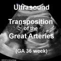
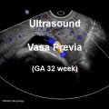
- clinical diagnosis (ultrasound)
- fetal endocrine
- sensory systems
- Genital male and female external genital differences observable
- Respiratory Month 3-6 - lungs appear glandular, end month 6 alveolar cells type 2 appear and begin to secrete surfactant
Week 12
<html5media height="420" width="550">File:Ultrasound_Fetus_02.mp4</html5media>
- Links: ultrasound
Size

- Crown Rump Length (CRL) 85 mm (week 10 CRL 40mm), femur length 15 mm, biparietal diameter 25 mm

- Biparietal diameter
- Head circumference
- Abdominal circumference
- Femur length
- Foot length (correlates with CRL)
- Placental parameters (covered in the next practical)
Head

|

|
| Fetal head lateral | Fetal head medial |

- Week 12 - tongue first differentiated epithelial cells.
Week 16+ (4 months)
Respiratory Development Overview

- week 4 - 5 embryonic
- week 5 - 17 pseudoglandular
- week 16 - 25 canalicular
- week 24 - 40 terminal sac
- late fetal - 8 years alveolar
Pituitary Development
Fetal endocrine systems become active during second semester.
- Week 4 - hypophysial pouch, Rathke’s pouch, diverticulum from roof
- Week 5 - elongation, contacts infundibulum, diverticulum of diencephalon
- Week 6 - connecting stalk between pouch and oral cavity degenerates
- Week 10 - growth hormone and ACTH detectable
- Week 16 - adenohypophysis fully differentiated
- Week 20 to 24 - growth hormone levels peak, then decline
Thyroid Development
- Maternal - Initial thyroid hormone supply across placenta.
- Fetal - Later thyroid required for neural development, stimulates metabolism (protein, carbohydrate, lipid), reduced/absence = cretinism (see abnormalities)
- Hormones - (amino acid derivatives) Thyroxine (T4), Triiodothyronine (T3)

- thyroid median endodermal thickening in the floor of pharynx, outpouch – thyroid diverticulum
- 24 days - thyroid median endodermal thickening in the floor of pharynx, outpouch – thyroid diverticulum
- Week 11 - colloid appearance in thyroid follicles, iodine and thyroid hormone (TH) synthesis
- Fetal Thyroid Hormone
- Initial secreted biologically inactivated by modification, late fetal secretion develops brown fat
- Iodine deficiency- during this period, leads to neurological defects (cretinism)
- Birth - TSH levels increase, thyroxine (T3) and T4 levels increase to 24 h, then 5-7 days postnatal decline to normal levels
Skin Development

- 4 weeks
- simple ectoderm epithelium over mesenchyme.
- 1 - 3 months
- ectoderm - germinative (basal) cell repeated division of generates stratified epithelium.
- mesoderm - somite dermatome spreads out under the epithelium, differentiates into connective tissue and blood vessels.
- 4 months
- basal cell - proliferation generates folds in basement membrane.
- neural crest cells - (melanocytes) migrate into epithelium. These are the pigment cell of the skin.
- embryonic connective tissue - differentiates into dermis, a loose ct layer over a dense ct layer. Beneath the dense ct layer is another loose ct layer that will form the subcutaneous layer.
- Ectoderm contributes to nails, hair follictles and glands.
- Nails form as thickening of ectoderm epidermis at the tips of fingers and toes. These form germinative cells of nail field.
- Cords of these cells extend into mesoderm forming epithelial columns.
- These form hair follicles, sebaceous and sweat glands.
- 5 months
- Hair growth initiated at base of cord, lateral outgrowths form associated sebaceous glands.
- Other cords elongate and coil to form sweat glands.
- Cords in mammary region branch as they elongate to form mammary glands.
- These glands will complete development in females at puberty. Functional maturity only occurs in late pregnancy.
- vernix caseosa begins to form and cover the skin (arms an intact visible coating in the third trimester)
- Links: integumentary
Genital Development
Genital male and female external genital differences now observable. In the ovary primary follicles begin to form and are characterized by the developing oocyte.
The animations below shows the development of external female and male genitalia from the indifferent external structure, during the period of week 9 to 12. Note - this content will be covered in detail in BGDB Genital development.
|
|
- Links: Genital animations
Neural

|

|
| Brain and Ventricular Development[1] | Brain Fissure Development[1] |
| Sylvian Fissure Development | |
|---|---|
| <html5media height="600" width="600">File:Neural_-_Sylvian_fissure.mp4</html5media> | |
At about 19 weeks (GA 21 weeks) neuronal migration ends and the radial glial cells, that aided this migration, now become transformed into astrocytes and astrocytic precursors.[2]
- Links: Glial Development
Sensory Development
- hearing - middle ear bones (ossicles) - not free to move until birth. Inner ear organ of Corti developing.
- vision - months 3 to 4 retina development, eyelids closed (until 5-7 months), lens grows through third trimester, cannot focus even postnatally.
Ossification
Ongoing Template:Endochondral ossification throughout skeleton and Template:Intramembranous in skull. Here are a few examples see bone timeline for specific details.
- Fetal Week 10 - osteogenic process begins in the humeral head. Primitive glenohumeral ligament present
- Fetal Week 11 - osteogenic process begins in the scapula
- Fetal Week 16-24 - Centres of ossification appear in remaining cartilage of otic capsule form petrous portion of temporal bone. Continues to ossify to form mastoid process of temporal bone.
- Links:bone timeline
Ultrasound
About Ultrasound
Different body tissues reflect sound waves differently. In ultrasound, a beam of sound waves (frequency 3 to 10 MHz) are passed through the body, the reflected waves are analysed by a computer, and an image is then generated on a display screen. The sound source is usually a transducer placed on the surface of the abdomen.
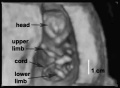
Ultrasound Measurements
There can be number of different parameters, depending on gestational age, that are commonly recorded during an ultrasound procedure. These measurements (and ratios) include embryonic/fetal size and key lengths and sizes of specific structures, fetal membrane sizes/volumes, placental location/size. In addition, general body movements, including heartbeat, can be observed.

|
| Ultrasound Fetal (GA 19 weeks)
Ultrasound scan through the fetal trunk to measure abdominal circumference (AC). |

Embryonic/Fetal Size: crown-rump length (CRL), biparietal diameter (BPD), abdominal circumference (AC)
Embryonic/Fetal Structures: femur length (FL), head circumference, nuchal translucency, heart size/rate
Fetal Membrane: gestational sac diameter (GS), yolk sac diameter (YS), meconium peritonitis, umbilical cord stricture
Placenta: location, size, umbilical cord stricture
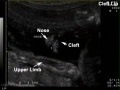
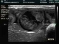
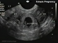
Ultrasound - Abnormalities

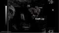
Cleft lip 1
Cleft lip 1 measurement
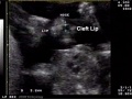
Cleft lip 2

Gastroschisis


See Ultrasound movie for Cleft lip 1
Second Trimester Interactive Component
| Attempt the Quiz - Second Trimester | ||
|---|---|---|

Here are a few simple Quiz questions that relate to Second Trimester development from the practical.
|
Additional Information
| Additional Information - Content shown under this heading is not part of the material covered in this class. It is provided for those students who would like to know about some concepts or current research in topics related to the current class page. |
| ||||||||||||||||||||||||||||||||||||||||||||||||||||||||||||||||||||||||||||||||
| ||||||||||||||||||||||||||||||||||||||||||||||||||||||||||||||||||||||||||||||||

Crown-Rump Length (CRL))
| Fertilization and Gestational Age - Crown-Rump Length (measured by ultrasound) | |||||||||||||||||||||||||||||||||||||||||||||||||||||||||||||||||||||||||||||||||||||||||||||||||||||||||||||||||||||||||||||||||||||||||||||||||||||||||||||||||||||||||||||||||||||||||||||||||||||||||||
| |||||||||||||||||||||||||||||||||||||||||||||||||||||||||||||||||||||||||||||||||||||||||||||||||||||||||||||||||||||||||||||||||||||||||||||||||||||||||||||||||||||||||||||||||||||||||||||||||||||||||||
Second Trimester Intestinal Growth
Both the small and large intestine grow in a linear manner during the second trimester.[3][4]


Second Trimester
| Event | ||
| Fertilisation Age FA | Gestational Age GA | |
| 14 |  Week 12 - CRL 85 mm, femur length 15 mm, biparietal diameter 25 mm Week 12 - CRL 85 mm, femur length 15 mm, biparietal diameter 25 mm
Hearing Week 12-16 - Capsule adjacent to membranous labrynth undegoes vacuolization to form a cavity (perilymphatic space) around membranous labrynth and fills with perilymph Genital male and female external genital differences observable Respiratory Month 3-6 - lungs appear glandular, end month 6 alveolar cells type 2 appear and begin to secrete surfactant Tongue Week 12 - first differentiated epithelial cells (Type II and III) Genital female genital canal (80 days) formed with absorption of the median septum | |
| 15 | Tongue Week 12 to 13 - maximum synapses between cells and afferent nerve fibers
Hearing - Outer Ear Development Week 13 - Meatal plug disc-like, innermost surface in contact with the primordial malleus, contributes to the formation of the tympanic membrane. | |
| 16 | Tongue Week 14 to 15 - taste pores develop, mucous
Ovary Development 100 days - primary follicles present Nail Development toenails appear Head Development facial skeleton remodelling begins | |
| 17 | Pancreas glucagon detectable in fetal plasma.
Spleen Week 15 -alpha-smooth muscle actin (alpha-SMA)-positive reticulum cells scattered around the arterioles. [5] | |
| 18 | 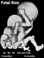 Hearing Week 16-24 - Centres of ossification appear in remaining cartilage of otic capsule form petrous portion of temporal bone. Continues to ossify to form mastoid process of temporal bone. Hearing Week 16-24 - Centres of ossification appear in remaining cartilage of otic capsule form petrous portion of temporal bone. Continues to ossify to form mastoid process of temporal bone.
Pituitary adenohypophysis fully differentiated Respiratory Week 16 to 25 lung histology - canalicular Hearing - Outer Ear Development Week 16.5 - External auditory meatus is fully patent throughout its length, lumen is still narrow and curved. Skin 4 months - basal cell- proliferation generates folds in basement membrane; neural crest cells- (melanocytes) migrate into epithelium; embryonic connective tissue- differentiates into dermis, a loose ct layer over a dense ct layer. Beneath the dense ct layer is another loose ct layer that will form the subcutaneous layer. Ectoderm contributes to nails, hair follictles and glands. Nails form as thickening of ectoderm epidermis at the tips of fingers and toes. These form germinative cells of nail field. Cords of these cells extend into mesoderm forming epithelial columns. These form hair follocles, sebaceous and sweat glands. primary follicles begin to form in the ovary and are characterized by an oocyte glandular urethra forms and skin folds present | |
| 19 |  Neural - Brain development histology week 17 Neural - Brain development histology week 17
| |
| 20 |  Tongue Week 18 - substance P detected in dermal papillae, not in taste bud primordia Tongue Week 18 - substance P detected in dermal papillae, not in taste bud primordia
Skin vernix caseosa covers skin Spleen Week 18 - alpha-SMA-positive reticulum cells increase in number and began to form a reticular framework. An accumulation of T and B lymphocytes occurred within the framework, and a primitive white pulp was observed around the arterioles. [5] Hearing - Outer Ear Development week 18 - External auditory meatus is already fully expanded to its complete form. | |
| 21 | ||
| 22 | Pituitary week 20 to 24 growth hormone levels peak, then decline
Skin lanugo, skin hair Skin 5 months - Hair growth initiated at base of cord, lateral outgrowths form associated sebaceous glands; Other cords elongate and coil to form sweat glands; Cords in mammary region branch as they elongate to form mammary glands. | |
| 23 | ||
| 24 |  Neural brain cortical sulcation - sylvian fissure, interhemispheric fissure, callosal sulcus, parietooccipital fissure, and hippocampic fissures present[6] Neural brain cortical sulcation - sylvian fissure, interhemispheric fissure, callosal sulcus, parietooccipital fissure, and hippocampic fissures present[6]
Spleen - Week 22 - antigenic diversity of the reticular framework was observed, and T and B lymphocytes were segregated in the framework. T lymphocytes were sorted into the alpha-smooth muscle actin-positive reticular framework, and the periarteriolar lymphoid sheath (PALS) was formed around the arteriole. B lymphocytes aggregated in eccentric portions to the PALS and formed the lymph follicle (LF). The reticular framework of the LF was alpha-SMA-negative. [5] | |
| 25 | ||
| 26 | Respiratory Week 24 to 40 lung histology - terminal sac
Spleen Week 24 - marginal zone appeared in the alpha-smooth muscle actin-positive reticular framework around the white pulp.[5] Earliest potential survival expected if born ovarian follicles can consist of growing oocytes surrounded by several layers of granulosa cells | |
| 27 | Respiratory end month 6 alveolar cells type 2 appear and begin to secrete surfactant | |
Endocrine - Adrenal and Placenta
The feto-placental unit, and potential roles of dehydroepiandrosterone (DHEA) in prenatal and postnatal brain development[7]
- "Synthesis of dehydroepiandrosterone (DHEA) by the fetal adrenal gland is important for placental oestrogen production, and may also be important for modulating the effects of glucocorticoids on the developing brain. ... Together, the studies outlined in this review indicate that the androgen DHEA is an important hormone of adrenal and Central Nervous System (CNS) origin in the fetal and postnatal spiny mouse. Disturbance of the development of these fetal tissues, and/or of the relationship between the fetal adrenal gland and placenta during pregnancy, may have significant consequences for fetal development, placental function, and maturation of the brain. It is proposed that such disturbances of normal adrenal function could account for some of the neuropathologies that arise in juvenile and adult offspring following illness and stress experienced by the mother during pregnancy."
References
- ↑ 1.0 1.1 Huang H, Xue R, Zhang J, Ren T, Richards LJ, Yarowsky P, Miller MI & Mori S. (2009). Anatomical characterization of human fetal brain development with diffusion tensor magnetic resonance imaging. J. Neurosci. , 29, 4263-73. PMID: 19339620 DOI.
- ↑ Kadhim HJ, Gadisseux JF & Evrard P. (1988). Topographical and cytological evolution of the glial phase during prenatal development of the human brain: histochemical and electron microscopic study. J. Neuropathol. Exp. Neurol. , 47, 166-88. PMID: 3339373
- ↑ FitzSimmons J, Chinn A & Shepard TH. (1988). Normal length of the human fetal gastrointestinal tract. Pediatr Pathol , 8, 633-41. PMID: 3244599
- ↑ Archie JG, Collins JS & Lebel RR. (2006). Quantitative standards for fetal and neonatal autopsy. Am. J. Clin. Pathol. , 126, 256-65. PMID: 16891202 DOI.
- ↑ 5.0 5.1 5.2 5.3 <pubmed>19255788</pubmed>
- ↑ <pubmed>11158907</pubmed>
- ↑ Quinn TA, Ratnayake U, Dickinson H, Castillo-Melendez M & Walker DW. (2016). The feto-placental unit, and potential roles of dehydroepiandrosterone (DHEA) in prenatal and postnatal brain development: A re-examination using the spiny mouse. J. Steroid Biochem. Mol. Biol. , 160, 204-13. PMID: 26485665 DOI.
BGDA: Lecture 1 | Lecture 2 | Practical 3 | Practical 6 | Practical 12 | Lecture Neural | Practical 14 | Histology Support - Female | Male | Tutorial
Glossary Links
- Glossary: A | B | C | D | E | F | G | H | I | J | K | L | M | N | O | P | Q | R | S | T | U | V | W | X | Y | Z | Numbers | Symbols | Term Link
Cite this page: Hill, M.A. (2026, July 26) Embryology BGDA Practical 12 - Second Trimester. Retrieved from https://embryology.med.unsw.edu.au/embryology/index.php/BGDA_Practical_12_-_Second_Trimester
- © Dr Mark Hill 2026, UNSW Embryology ISBN: 978 0 7334 2609 4 - UNSW CRICOS Provider Code No. 00098G