ANAT2241 Urinary System

| ANAT2241 This practical support page content is not part of the virtual science practical class and provides additional information for student self-directed learning purposes. All practical class pages are located on Moodle - ANAT2241 |
General Objective
To know the microanatomical structure of the kidney, ureter, urinary bladder and urethra.
Specific Objectives
- To describe the microanatomy of the kidney: cortex, medulla, pyramid, papillae, medullary rays, cortical columns, calyx, and pelvis.
- To know the structure and ultrastructure of the nephron including the glomerulus, Bowman’s capsule, proximal and distal tubules, loop of Henle, and juxtaglomerular apparatus.
- To identify collecting tubules in medullary rays of cortex, and in the inner and outer medulla.
- To know the vascular circulation in the kidney.
- To describe the structure of the ureter, urethra and urinary bladder and changes, which occur in the bladder during stretching.
Learning Activities
Examine the following virtual slides, and in course manual identify, draw and label the features listed and note their function.
Virtual Slides: Urinary System
- Renal Histology: Histology | Histology Stains | Renal Development
- Kidney - Nephron overview | Glomerulus | Vascular and renal poles | Medullary ray | tubules
- Ureter - Ureter labeled | Ureter epithelium
- Bladder - overview | wall 1 | wall 2 | transitional epithelium | Urinary Bladder Development
Kidney Anatomy

|

Adult nephron structure |
Nephron
|
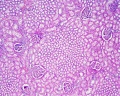
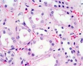
Nephron Histology
- Development - mean glomerular number shown to level at 36 weeks, increasing from about 15,000 at 15 weeks to 740,000 at 40 weeks.
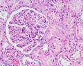
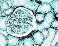
Glomerulus

|

|
| Glomerulus structure | Vascular and renal poles |
| Bowmans Capsule | |
|---|---|
Bowmans Capsule forms two layers with a space between these layers.
|

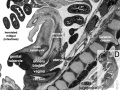
Within the glomerulus a high magnification view of a podocyte showing the interdigitated foot processes (pedicels) that are wrapped around the exterior of glomerular capillaries.[1] |

| |
Nephron Tubules

Nephron overview


- Renal System Histology: Nephron tubule overview | glomerulus structure | vascular and renal poles | Medullary rays | Nephron tubules
Large Images
medullary rays
glomerulus
distal tubule and collecting duct
proximal and distal tubule
distal and intermediate tubule
medullary ray
glomerulus
proximal tubule


Ureter Histology
|

|


Bladder Histology
Can be described anatomically by its 4 layers from inside outward:
Note that while the smooth muscle fibre layer organisation is described as longitudinal or circular, this is only a general organisation of fibre direction, and is better described as a "spiral" organisation. |

|


Fetal Kidney


- Fetal Renal Links: fetal kidney histology 01 | fetal kidney histology 02 | fetal kidney histology 03 | fetal kidney histology 04
Fetal Urethra Histology


Images
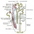
Nephron structure
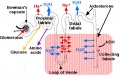
Nephron physiology
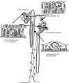
Nephrons - cortical and juxtamedullary
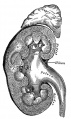
Kidney and adrenal gland (adult)
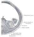
retroperitoneal
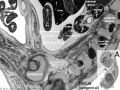
Fetal urogenital region most lateral right
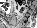
Fetal urogenital region lateral right
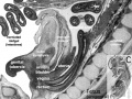
Fetal urogenital region medial
Fetal urogenital region midline
Fetal kidney (10 weeks)
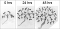
Mouse E12.5 kidney in vitro


Practical Overview
Normal Histology
- kidneys, ureters, urinary bladder, urethra
Anatomy
- retroperitoneal
- kidney - bean shaped
- rich blood supply
Kidney Function
- elimination of foreign substances
- regulation of the amount of water in the body
- control of the concentration of most compounds in the extracellular fluid
- filtration - glomeruli of the kidney
- selective resorption and excretion - tubular system of the kidney
Capsule
|
Cortex
|
Medulla
|
Blood Supply
- renal artery
- interlobar arteries (across medulla thru renal columns)
- arcuate arteries (cortico-medullary junction)
- interlobular arteries
- afferent glomerular arterioles
- glomerular capillary network
- efferent glomerular arterioles
Vasa Recta
- descending arterioles (arteriole rectae) + ascending venules (venulae rectae)
Glomerulus
- glomerulus - round (~0.2 mm in diameter) blind beginning of the nephron
- vascular pole - invaginated by a tuft of capillaries
- urinary pole - substances leave the capillaries enter the renal tubule
- Bowman's capsule - anatomical glomerulus is enclosed by two layers of epithelium.
- outer or parietal layer of Bowman's capsule form a simple squamous epithelium.
- inner layer, podocytes in the visceral layer, are extremely complex in shape.
- Mesangial cells in the glomerulus form the connective tissue that gives structural support to podocytes and vessels (Podocytes, mesangial cells, glomerular capillaries)
- Juxtaglomerular cells - smooth muscle cells afferent glomerular arteriole (epithelial-like cells)
- Macular Densa
- distal convoluted tubule near vascular pole (narrower and taller than rest of DCT)
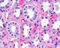
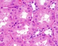
Tubules
Proximal Convoluted Tubules (PCT)
- brush border
- star-shaped
- larger outside diameter
Distal Convoluted Tubules (DCT)
- clean lumen surface
- apical nuclei
Collecting Tubules (CT)
- larger lumen than DCT (about size of PCT)
- cuboidal cells and smaller than DCT
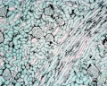
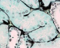
Renal Pyramids
- medullary straight tubules, ducts and vasa recta
- apical renal papilla - simple cuboidal/columnar epithelia
- calyx - lined by transitional epithelia
Note the urinary system transitional epithelium is also known as urothelium.
Ureters
- epithelium - transitional epithelia
- lamina propria - mainly of dense connective tissue, with many bundles of coarse collagenous fibres
- muscularis - consists of an inner longitudinal and outer circular layer of smooth muscle cells
- In lower parts of the ureter and the bladder an additional outer longitudinal layer of muscles is added to the first two.
Bladder
- epithelium - transitional epithelia
- apical plaques - thickened domain allows great changes in surface area.
- lamina propria - mainly of dense connective tissue, with many bundles of coarse collagenous fibres
- muscularis - consists of an inner longitudinal and outer circular layer of smooth muscle cells
- In the bladder (and lower parts of the ureter) an additional outer longitudinal layer of muscles is added to the first two.
Urethra
- penile urethra within corpus spongiosum
- pseudostratified columnar epithelia
- distal end - stratified squamous
- continuous with outer skin
Terms
| Renal Terms | ||
|---|---|---|
| ||
|
- arcuate artery -
- Bowman's capsule - Term describing the cup-shaped double epithelium surrounding the glomerulus of the nephron within the kidney. The outer parietal layer is a squamous simple epithelium. The inner visceral layer is formed by podocytes covering glomerular capillaries.
- collecting tubule -
- distal tubule -
- glomerulus -
- medullary ray -
- nephron - Term describing the functional unit of the kidney. Formed by blood vessels and specialised epithelia.
- podocyte - (glomerular podocyte) Kidney epithelial cell type in the nephron (kidney functional unit) located in the glomerulus. Podocytes form the visceral layer of Bowman's capsule and are at the filtration barrier between capillary blood and the nephron tubular system and function to ultrafiltrate blood, and support glomerular capillary pressures. The differentiation of podocytes involves the formation of cellular foot processes and then the slit membrane.
- proximal tubule -
- renal corpuscle -
- transitional epithelium - (urothelium)
References
<
External Links
External Links Notice - The dynamic nature of the internet may mean that some of these listed links may no longer function. If the link no longer works search the web with the link text or name. Links to any external commercial sites are provided for information purposes only and should never be considered an endorsement. UNSW Embryology is provided as an educational resource with no clinical information or commercial affiliation.
- Blue Histology Urinary System
- UNSW Virtual Slides Urinary Tract Histology
- UIOWA Virtual Slidebox of Histology Urinary tract
- Scott RP & Quaggin SE. (2015). Review series: The cell biology of renal filtration. J. Cell Biol. , 209, 199-210. PMID: 25918223 DOI.
Course Links
- Histology Glossary: A | B | C | D | E | F | G | H | I | J | K | L | M | N | O | P | Q | R | S | T | U | V | W | X | Y | Z | ANAT2241 Support | Histology | Histology Stains | Embryology Glossary
| Common Histology Stains | ||||||||||||||||||||||||||||||||||||||||||||||||||||||||||||||||||||||||||||||||||||||||||||||||||||||||||||||||||||||||||||||||||||||||||||||||
|---|---|---|---|---|---|---|---|---|---|---|---|---|---|---|---|---|---|---|---|---|---|---|---|---|---|---|---|---|---|---|---|---|---|---|---|---|---|---|---|---|---|---|---|---|---|---|---|---|---|---|---|---|---|---|---|---|---|---|---|---|---|---|---|---|---|---|---|---|---|---|---|---|---|---|---|---|---|---|---|---|---|---|---|---|---|---|---|---|---|---|---|---|---|---|---|---|---|---|---|---|---|---|---|---|---|---|---|---|---|---|---|---|---|---|---|---|---|---|---|---|---|---|---|---|---|---|---|---|---|---|---|---|---|---|---|---|---|---|---|---|---|---|---|---|
| ||||||||||||||||||||||||||||||||||||||||||||||||||||||||||||||||||||||||||||||||||||||||||||||||||||||||||||||||||||||||||||||||||||||||||||||||
| ||||||||||||||||||||||||||||||||||||||||||||||||||||||||||||||||||||||||||||||||||||||||||||||||||||||||||||||||||||||||||||||||||||||||||||||||
Practical Support
- Pages can be accessed from any internet connected computer.
ANAT2241 Support Links: The Virtual Microscope | Covering and Lining Epithelia | Glandular Epithelia | CT Components | CT Types | Bone, Bone Formation and Joints | Muscle | Nervous | Blood | Eye | Cardiovascular | Respiratory | Integumentary | Gastrointestinal | Gastrointestinal Organs | Lymphatic and Immune | Endocrine | Urinary | Female Reproductive | Male Reproductive | Histology Stains | Histology Drawings | Practicals Health and Safety 2013 | Moodle - 2019
ANAT2241 This practical support page content is not part of the science practical class and provides only background information for student self-directed learning purposes.
Cite this page: Hill, M.A. (2026, July 22) Embryology ANAT2241 Urinary System. Retrieved from https://embryology.med.unsw.edu.au/embryology/index.php/ANAT2241_Urinary_System
- © Dr Mark Hill 2026, UNSW Embryology ISBN: 978 0 7334 2609 4 - UNSW CRICOS Provider Code No. 00098G