2011 Lab 2 - Week 3
| 2011 Lab 2: Introduction | Week 1 | Week 2 | Week 3 | Online Assessment | Group Project |
Introduction

Approximate cross-section of an 18 day human conceptus. Identify the 3 layers of the trilaminar embryo: ectoderm (columnar cells), intraembryonic mesoderm (mesenchymal cells, endodermal cells (cuboidal single layer). Identify primitive groove with dense cluster of primitive streak cells below it. (Image: Nishimura etal., 1977) |
Key events of human development during the third week (week 3) following fertilization or Clinical week 5 (LMP). Note that during this time the conceptus cells not contributing to the embryo are contributing to placental membranes and the early placenta. During the third week of development conceptus implantion in the uterus wall is complete and trophoblast cells continue to invade uterine wall in the process of early placentation (villi formation). Within the conceptus, gastrulation converts the bilaminar embryo into the trilaminar embryo (ectoderm, mesoderm, endoderm). Morphological changes include an epithelial to mesenchymal cell transition and folding of the the embryonic disc. Carnegie stages 7 to 9 covers this period of human embryo development. Outside of the embryo, the extraembryonic spaces (chorionic, amniotic, yolk) and intraembryonic spaces (coeloms) continue to develop. Trophoblast cells continue to invade uterine wall and the emdometrium is being converted into the decidua, the process of villi formation and early placentation has begun. Gastrulation means "gut forming" and converts the inner cell mass which then formed the bilaminar embryo (epiblast, hypoblast) into the trilaminar embryo (ectoderm, mesoderm, endoderm). The process involves the migration of cells from the epiblast layer through the primitive streak to form first the endoderm layer and then a second intermediate layer the mesoderm layer. Once all cells have left the epiblast layer it now becomes the ectoderm layer. These three germ cell layers (ectoderm, mesoderm, endoderm) will form in a layer specific manner all the future tissues of the developing embryo. |
- Week 3 Links: Lecture - Week 3 | Gastrulation | Placenta | Coelomic Cavity
- Embryo Week: Week 1 | Week 2 | Week 3 | Week 4 | Week 5 | Week 6 | Week 7 | Week 8 | Week 9
Conceptus Cavities Week 2 and Week 3
| File:Chorion 001 icon.jpg</wikiflv> | Animation shows the events following implantation and focuses on changes in the the spaces surrounding the embryonic disc, the extraembryonic coelom. The blastoceol cavity is converted into two separate spaces: the yolk sac and the chorionic cavity. The third space lies above the epiblast, the amniotic cavity.
blue - epiblast layer yellow - hypoblast layer red cells - extraembryonic mesoderm layer green - trophoblast layer red spaces - blood-filled spaces, maternal lacunae white cells - (left) endometrial gland (right) endometrial epithelium |
Germ Layers and Dynamic Processes
- From 1 layer of cells to 3 layers that define all tissues of the entire embryo.
The conceptus is now fully implanted in the uterine wall. This time is now when a small cluster of cells (the inner cell mass) will differentiate into two simple cell layers (epiblast and hypoblast). From the single epiblast layer cells will migrate through a narrow region (primitive streak) to form firstly two layers epiblast and endoderm, then later the three layers ectoderm, mesoderm and endoderm. These final three layers (germ layers) are the trilaminar embryo and will form all the tissues of the body.
During implantation cells on the outside (trophoblast layer) of the conceptus are forming part of the early placenta. The cells on the inside (inner cell mass) now begin to form the three germ layers that will form the entire embryo. Note that as the conceptus also forms the fetal component of the placenta, not all cells from our blastocyst will form part of the developing embryo.
The images below show the embryo (week 3 between 15 to 17 days) as a disc 0.4 mm diameter in size.


Stage 7 Human Embryo (15 to 17 days)
Stage 10 Human Embryo (22 to 23 days)
Stage 7

|
|
| Features: embryonic disc, primitive node, primative streak, primitive groove, yolk sac
Facts: Week 3, 15 - 17 days, 0.4 mm View 1: embryonic disc, showing the epiblast viewed from the amniotic (dorsal) side. Events: Gastrulation is continuing as cells migrate from the epiblast, continuing to form mesoderm. Mesoderm lies between the ectoderm and endoderm as a continuous sheet except at the buccopharyngeal and cloacal membranes. These membranes have ectoderm and endoderm only and will lie at the rostral (head) and caudal (tail) of the gastrointestinal tract.
|
Gastrulation: Through the primitive streak cells migrate continuously through week 3 into week 4. Initial cells replace hypoblast as an epithelial layer the endoderm. Later migrating cells spread between the two epithelial layers to form mesoderm. |

|

|
| Axes: embryonic disc is shown rostral (head) to top and caudal (tail) to bottom. Left and right are the lateral margins of the disc as shown. |
Gastrulation
Carnegie Stage 7 and 8, gastrulation, migration of cells through the primitive streak to form endoderm and mesodermal layers of embryo.

 Scanning electron micrograph showing the early forming 3 layers: ectoderm, mesoderm and endoderm.
![]()
Timeline - Week 3
| Event | ||
| Stage 7 |  | |
| Stage 8 |  | |
 | ||
| Stage 9 | 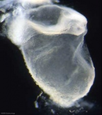 Musculoskeletal - somitogenesis, first somites form and continue to be added in sequence caudally Musculoskeletal - somitogenesis, first somites form and continue to be added in sequence caudally
Neural - the three main divisions of the brain, which are not cerebral vesicles, can be distinguished while the neural groove is still completely open Neural Crest - mesencephalic neural crest is visible[1] | |
| Heart - cardiogenesis, week 3 begins as paired heart tubes. |
- Carnegie Stages: 1 | 2 | 3 | 4 | 5 | 6 | 7 | 8 | 9 | 10 | 11 | 12 | 13 | 14 | 15 | 16 | 17 | 18 | 19 | 20 | 21 | 22 | 23 | About Stages | Timeline
Week 2 and 3 Movies
| Implantation | Mesoderm | Chorionic Cavity | Amniotic Cavity | Week 3 |
References
- ↑ <pubmed>17848161</pubmed>
| 2011 Lab 2: Introduction | Week 1 | Week 2 | Week 3 | Online Assessment | Group Project |
Glossary Links
- Glossary: A | B | C | D | E | F | G | H | I | J | K | L | M | N | O | P | Q | R | S | T | U | V | W | X | Y | Z | Numbers | Symbols | Term Link
Cite this page: Hill, M.A. (2026, July 31) Embryology 2011 Lab 2 - Week 3. Retrieved from https://embryology.med.unsw.edu.au/embryology/index.php/2011_Lab_2_-_Week_3
- © Dr Mark Hill 2026, UNSW Embryology ISBN: 978 0 7334 2609 4 - UNSW CRICOS Provider Code No. 00098G