User:Z5059996
| 2017 Project Groups | |||||
|---|---|---|---|---|---|
| Group 1 | Group 2 | Group 3 | Group 4 | Group 5 | Group 6 |
| Mark Hill - Lab 1 page | |||||
Student Page Here is the Student Page demonstration page I showed in the Practical class.
Use this page to practice editing and don't forget to add a topic to the 2017 Group Project 3 page.
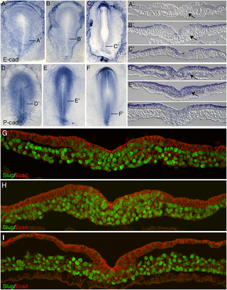
Chicken embryo E-cadherin and P-cadherin in gastrulation[1]
Project Contribution Diary
Week 4: Starting researching the topic of abnormal cardiovascular development - posted several promising links
Week 6: Decided upon several key examples of abnormal development, including ventricular septal defect (VSD), atrial septal defect (ASD), transposition of the great vessels, coarctation of the aorta, tetralogy of fallot, hypoplastic left heart syndrome, patent ductus arteriosus, pulmonary atresia.
Cardiac Septation
This stage of heart morphogenesis refers to the development of the four main cardiac chambers from the primitive atrium and ventricle. Cardiac septation is comprised of three main events:
- Division of the atrioventricular canal
- Atrial septation
- Ventricular septation
Division of the atrioventricular canal
Division of the atrioventricular canal (AVC) begins with the formation of the superior and inferior endocardial cushions, which are located on the dorsal and ventral aspects of the AVC respectively.[2] These cushions develop as mesenchymal cells invade and proliferate within swollen regions of cardiac jelly of the AVC; this mesenchyme is derived from endothelial cells that have transdifferentiated via the process of epithelial-mesenchymal transformation (EMT) [1,2]. Throughout the fifth week of development, the endocardial cushions project inwards and eventually fuse to partition the AVC into the left and right atrioventricular canals [1,2]. These canals will serve as the orifices in which the tricuspid and mitral valves are situated [1,2].
Atrial septation
As the AVC is undergoing division, a muscular outgrowth, referred to as the septum primum, extends inferiorly from the roof of the primordial atrium [3]. This septum partially divides the atrial chamber into left and right halves, leaving a temporary communication located between the inferior border of septum primum and the endocardial cushions known as the foramen primum [1]. As the size of the foramen primum diminishes, perforations in the superior portion of the septum primum develop as a result of apoptosis, forming a second communication between the atrial chambers called the foramen secundum [2]. Concurrently, an additional muscular septum, known as the septum secundum, projects inferiorly to the right of the septum primum [1]. Eventually, the septum secundum will extend beyond the length of the foramen secundum, generating a partial division of the atria that forms the upper boundary of the foramen ovale [3]. The development of the foramen ovale is critical as it allows oxygen-rich blood from the placenta to bypass pulmonary circulation of the embryo and directly enter systemic circulation [4]. Following birth, the foramen ovale is obliterated as the septum primum and septum secundum fuse, resulting in complete formation of the interatrial septum [1].
Ventricular septation
As with atrial septation, differentiation of the ventricles is initiated within week 4 of development [1]. A muscular ridge, referred to as the interventricular septum primordium, develops and extends superiorly from the caudal aspect of the primitive ventricular chamber [2]. Growth of the septum is attributed to the expansion of the ventricles, which involves the development of muscular trabeculae as cardiomyocytes proliferate within the chamber walls [3]. The end result is a partial division between the left and right ventricles, which forms the muscular portion of the interventricular septum [2]. A communication between the ventricles, known as the interventricular foramen, remains until approximately week 7 of development [5]. The foramen is obliterated by the fusion of the septum intermedium and the bulbar ridges of the bulbus cordis; this constitutes the membranous portion of the interventricular septum [1].
