User:Z5059996: Difference between revisions
No edit summary |
|||
| Line 225: | Line 225: | ||
===Group 4: Eye=== | ===Group 4: Eye=== | ||
{| border="1" align="left" | |||
|- | |||
|Criteria | |||
|Strengths | |||
|Weaknesses | |||
|- | |||
| 1. The choice of content shows a good understanding of the topic area | |||
| | |||
| | |||
|- | |||
|2. Content is correctly cited and referenced | |||
| | |||
| | |||
|- | |||
|3. The wiki has an element of teaching at a peer level | |||
| | |||
| | |||
|- | |||
|4. Relates the topic and content of the Wiki entry to learning aims of embryology | |||
| | |||
| | |||
|- | |||
|5. The content of the wiki should demonstrate to the reader that your group has researched adequately on this topic | |||
| | |||
| | |||
|} | |||
===Group 5: Lung=== | ===Group 5: Lung=== | ||
{| border="1" align="left" | |||
|- | |||
|Criteria | |||
|Strengths | |||
|Weaknesses | |||
|- | |||
| 1. The choice of content shows a good understanding of the topic area | |||
| | |||
| | |||
|- | |||
|2. Content is correctly cited and referenced | |||
| | |||
| | |||
|- | |||
|3. The wiki has an element of teaching at a peer level | |||
| | |||
| | |||
|- | |||
|4. Relates the topic and content of the Wiki entry to learning aims of embryology | |||
| | |||
| | |||
|- | |||
|5. The content of the wiki should demonstrate to the reader that your group has researched adequately on this topic | |||
| | |||
| | |||
|} | |||
===Group 6: Cerebellum=== | ===Group 6: Cerebellum=== | ||
{| border="1" align="left" | |||
|- | |||
|Criteria | |||
|Strengths | |||
|Weaknesses | |||
|- | |||
| 1. The choice of content shows a good understanding of the topic area | |||
| | |||
| | |||
|- | |||
|2. Content is correctly cited and referenced | |||
| | |||
| | |||
|- | |||
|3. The wiki has an element of teaching at a peer level | |||
| | |||
| | |||
|- | |||
|4. Relates the topic and content of the Wiki entry to learning aims of embryology | |||
| | |||
| | |||
|- | |||
|5. The content of the wiki should demonstrate to the reader that your group has researched adequately on this topic | |||
| | |||
| | |||
|} | |||
==Cardiac Septation== | ==Cardiac Septation== | ||
Revision as of 17:21, 11 October 2017
| 2017 Project Groups | |||||
|---|---|---|---|---|---|
| Group 1 | Group 2 | Group 3 | Group 4 | Group 5 | Group 6 |
| Mark Hill - Lab 1 page | |||||
Student Page Here is the Student Page demonstration page I showed in the Practical class.
Use this page to practice editing and don't forget to add a topic to the 2017 Group Project 3 page.
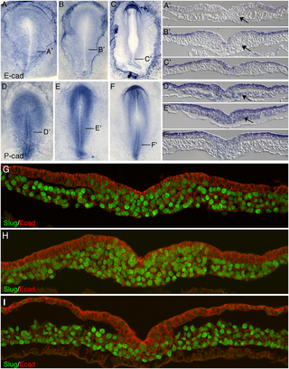
Chicken embryo E-cadherin and P-cadherin in gastrulation[1]
| Criteria | Strengths | Weaknesses |
| 1. The choice of content shows a good understanding of the topic area | ||
| 2. Content is correctly cited and referenced | ||
| 3. The wiki has an element of teaching at a peer level | ||
| 4. Relates the topic and content of the Wiki entry to learning aims of embryology | ||
| 5. The content of the wiki should demonstrate to the reader that your group has researched adequately on this topic |
Peer Reviews
Marking Criteria
Group Assessment Criteria (modified and condensed from the student page)
1. The choice of content shows a good understanding of the topic area
- The choice of content, headings and sub-headings, diagrams, tables, graphs show a good understanding of the topic area
- Project sub-heading structure is appropriate
- No key topic areas have been missed.
2. Content is correctly cited and referenced
- Text within the project page is correctly cited.
- Research articles and reviews can both be used, but are clearly identified.
- Images within the project page is correctly cited.
- Reference list does not contain multiple copies of the same article.
3. The wiki has an element of teaching at a peer level
- The wiki has an element of teaching at a peer level using the student's own innovative diagrams, tables or figures and/or using interesting examples or explanations.
- The content has been designed for an university undergraduate science student level.
- Acronyms and terms are well explained.
- Image summaries include a useful description in the context of the project page and not simply the original legends (though these can be included).
4. Relates the topic and content of the Wiki entry to learning aims of embryology
- Well-structured project page showing a broad understanding of embryological development.
- Includes relevant historic and current research.
- Molecular signaling mechanisms are included within the project page.
5. The content of the wiki should demonstrate to the reader that your group has researched adequately on this topic
- The content of the wiki should demonstrate to the reader that your group has researched adequately on this topic and covered the key areas necessary to inform your peers in their learning.
- Includes links to other UNSW Embryology related topic/content pages.
Suggested topic sub-headings (obtained from the student page)
- Developmental origin
- Developmental timeline
- Key discoveries
- Developmental signaling processes
- Identify review and research articles
- Current research
- Animal models
- Abnormal development
- Future questions
Group 1: Cerebral Cortex
| Criteria | Strengths | Weaknesses |
| 1. The choice of content shows a good understanding of the topic area | The developmental origin of the cerebral cortex is addressed well under the sub-heading ‘Early development of the brain’.
Abnormal development of the cerebral cortex and the associated conditions are covered in an immense amount of detail. The accompanying images and videos enhance the written information, as well as making it easier for the reader to comprehend. In addition, the sub-headings of this section compartmentalize the congenital diseases in a logical manner that highlights the link between abnormal development and specific diseases. |
There are several key topic areas missing from the page:
Some sections that have been included are somewhat irrelevant to the subject matter. For example, there is a large (unfinished) section on the anatomy and functions of the cerebral cortex. While it is important to provide a bit of an anatomical background on the subject, it shouldn’t be a major focus of this assignment. Focus more on the sections mentioned above, and keep the project focused on the embryology of the cerebral cortex. |
| 2. Content is correctly cited and referenced | There have been attempts at referencing throughout the assignment. A reference list has been produced and appears mostly correct. References have not been repeated throughout the list.
Peer-reviewed primary research articles have been used in this assignment. The student-drawn image has been cited correctly, as have most of the images used in the ‘abnormal development’ section. |
Overall, referencing in this assignment is very poor. Most of the content is completely devoid of any references (see ‘introduction’, ‘anatomy of the cortex’ and ‘abnormal development), and sections that have been referenced have been referenced “by paragraph” (see ‘timeline of corticogenesis’)
Many of the images have been cited incorrectly and used without permission. Remember to include the full reference, the original summary and the copyright license information for each image. |
| 3. The wiki has an element of teaching at a peer level | The information presented is mostly at a level appropriate for peers. Images and hand-drawn diagrams have been included to facilitate the readers understanding of the subject matter. Some of the images contain useful descriptions of the subject matter, and aid in understanding of the topic. | Many of the acronyms and terms used in this assignment are not well explained. Include a glossary of terms to make some of the content easier to follow and understand. |
| 4. Relates the topic and content of the Wiki entry to learning aims of embryology | The development of the cerebral cortex was covered extensively, which is a very important learning aim of embryology. | There are certain learning aims of embryology that have not been included in this assignment, such as developmental signaling processes (see criteria 1 for more information). There has been no discussion of relevant historical or current research (adding in the subheadings “key developments” and “current research” would help rectify this). |
| 5. The content of the wiki should demonstrate to the reader that your group has researched adequately on this topic | Certain aspects have been researched and presented well (such as embryological development).
Links to other pages of the UNSW embryology wiki have been included, however they have been used as references rather than just links. |
Information from the UNSW embryology wiki has been used as direct sources of information. Instead they should be included to relate this particular wiki page to other areas of learning.
The small number of sources cited in the reference list demonstrates a poor and narrow approach to researching this topic. A greater library of sources should be used to create this page (mainly primary research articles. |
Grade: FAIL
General Comment: While some aspects of the wiki page have been done well, the page is largely unfinished. Many sections still need to be added, and others are in need of improvement.
Group 2: Kidney
| Criteria | Strengths | Weaknesses |
| 1. The choice of content shows a good understanding of the topic area | The embryology timeline is well done and very informative – it gives the reader a general understanding of the process before each stage of development is done in detail.
The topic of ‘kidney development’ is described clearly and in detail. The well-structured subheadings make this section of the wiki page easier to follow. The chosen figures also enhance the information presented, and facilitate the readers understanding. The brief introduction to the anatomy of the kidney provides a nice introduction to the topic, and helps the reader understand the basics. |
The wiki page is missing several important areas of information:
Even though some information regarding signaling processes has been integrating throughout the 'kidney development’ section, the wiki page may benefit from a section entirely dedicated to signaling processes (this is one of Mark’s recommended sub-headings) There is currently very little information regarding current research on the wiki page – this section needs some work. Although the heading “future questions” has been added to the wiki page, there is no information associated with it. |
| 2. Content is correctly cited and referenced | There have been attempts at referencing throughout the assignment. A reference list has been produced and appears mostly correct. References have not been repeated throughout the list.
The reference list is comprised mainly of peer-reviewed primary research articles. Some images have been referenced correctly – see ‘figure 3’. |
Overall, referencing throughout the wiki page is poor. Some sections completely lack referencing (see ‘kidney structure’, ‘genes expressed’). Other sections have only 1 link attached to them (see ‘nephrogenesis’). Any information that is not original (in idea or structure) needs proper sentence-by-sentence citations. The most well-referenced section is the introduction to ‘developmental abnormalities’, and that still contains some uncited material.
Try not to rely on only one source of information per section (see ‘nephrogenesis’). Try to find a variety of research articles to source your material from. This will increase the quality and reliability of the information in the wiki page. Many of the images have been cited incorrectly and used without permission (see ‘figure 1’ and ‘figure 2’) Remember to include the full reference, the original summary and the copyright license information for each image. |
| 3. The wiki has an element of teaching at a peer level | The information presented is mostly at a level appropriate for peers. Some background information has been provided to aid in the reader’s understanding.
The chosen visual aids make some of the more complex ideas easier to comprehend (see ‘figure 3’). Some of the images also contain helpful descriptions that aid in understanding of the material (see ‘figure 3’ and ‘figure 8’) |
Many of the acronyms and terms used in this assignment are either poorly explained, or not explained at all. By including a glossary, the reader will be able to understand some of the more difficult subject areas.
No student-drawn diagrams have been included in the wiki page. Try to include some hand-drawn images, as well as other devices (e.g. tables, analogies) to aid the reader. |
| 4. Relates the topic and content of the Wiki entry to learning aims of embryology | The ‘Kidney development’ section was well-structured and was covered in great detail.
Developmental signaling processes were addressed, which is another important learning aim of embryology. Current research regarding kidney development has been mentioned. |
There has been no discussion of key discoveries regarding kidney development.
Although a thorough understanding of certain topics areas has been demonstrated, certain areas (such as current research) still need improvement. |
| 5. The content of the wiki should demonstrate to the reader that your group has researched adequately on this topic | Certain aspects have been well researched such as anatomy of the kidney, kidney development and developmental abnormalities. | No links to other pages on the UNSW embryology wiki have been included. Try linking this wiki page to other aspects of the embryology wiki.
The small number of sources cited in the reference list demonstrates a poor and narrow approach to researching this topic. A greater library of sources should be used to create this page (mainly primary research articles. |
Grade: CREDIT
General Comment: Most of the sections on the page have been done well, but some areas still need improvement.
Group 4: Eye
| Criteria | Strengths | Weaknesses |
| 1. The choice of content shows a good understanding of the topic area | ||
| 2. Content is correctly cited and referenced | ||
| 3. The wiki has an element of teaching at a peer level | ||
| 4. Relates the topic and content of the Wiki entry to learning aims of embryology | ||
| 5. The content of the wiki should demonstrate to the reader that your group has researched adequately on this topic |
Group 5: Lung
| Criteria | Strengths | Weaknesses |
| 1. The choice of content shows a good understanding of the topic area | ||
| 2. Content is correctly cited and referenced | ||
| 3. The wiki has an element of teaching at a peer level | ||
| 4. Relates the topic and content of the Wiki entry to learning aims of embryology | ||
| 5. The content of the wiki should demonstrate to the reader that your group has researched adequately on this topic |
Group 6: Cerebellum
| Criteria | Strengths | Weaknesses |
| 1. The choice of content shows a good understanding of the topic area | ||
| 2. Content is correctly cited and referenced | ||
| 3. The wiki has an element of teaching at a peer level | ||
| 4. Relates the topic and content of the Wiki entry to learning aims of embryology | ||
| 5. The content of the wiki should demonstrate to the reader that your group has researched adequately on this topic |
Cardiac Septation
This stage of heart morphogenesis refers to the development of the four main cardiac chambers from the primitive atrium and ventricle. Cardiac septation is comprised of three main events:
- Division of the atrioventricular canal
- Atrial septation
- Ventricular septation
Division of the atrioventricular canal
Division of the atrioventricular canal (AVC) begins with the formation of the superior and inferior endocardial cushions, which are located on the dorsal and ventral aspects of the AVC respectively.[2] These cushions develop as mesenchymal cells invade and proliferate within swollen regions of cardiac jelly of the AVC; this mesenchyme is derived from endothelial cells that have transdifferentiated via the process of epithelial-mesenchymal transformation (EMT). Throughout the fifth week of development, the endocardial cushions project inwards and eventually fuse to partition the AVC into the left and right atrioventricular canals [1,2]. These canals will serve as the orifices in which the tricuspid and mitral valves are situated [1,2].
Atrial septation
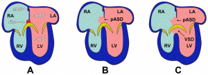
As the AVC is undergoing division, a muscular outgrowth, referred to as the septum primum, extends inferiorly from the roof of the primordial atrium [3]. This septum partially divides the atrial chamber into left and right halves, leaving a temporary communication located between the inferior border of septum primum and the endocardial cushions known as the foramen primum [1]. As the size of the foramen primum diminishes, perforations in the superior portion of the septum primum develop as a result of apoptosis, forming a second communication between the atrial chambers called the foramen secundum [2]. Concurrently, an additional muscular septum, known as the septum secundum, projects inferiorly to the right of the septum primum [1]. Eventually, the septum secundum will extend beyond the length of the foramen secundum, generating a partial division of the atria that forms the upper boundary of the foramen ovale [3]. The development of the foramen ovale is critical as it allows oxygen-rich blood from the placenta to bypass pulmonary circulation of the embryo and directly enter systemic circulation [4]. Following birth, the foramen ovale is obliterated as the septum primum and septum secundum fuse, resulting in complete formation of the interatrial septum [1].
Ventricular septation
As with atrial septation, differentiation of the ventricles is initiated within week 4 of development [1]. A muscular ridge, referred to as the interventricular septum primordium, develops and extends superiorly from the caudal aspect of the primitive ventricular chamber [2]. Growth of the septum is attributed to the expansion of the ventricles, which involves the development of muscular trabeculae as cardiomyocytes proliferate within the chamber walls [3]. The end result is a partial division between the left and right ventricles, which forms the muscular portion of the interventricular septum [2]. A communication between the ventricles, known as the interventricular foramen, remains until approximately week 7 of development [5]. The foramen is obliterated by the fusion of the septum intermedium and the bulbar ridges of the bulbus cordis; this constitutes the membranous portion of the interventricular septum [1].

