User:Z3491219
| Student Information (expand to read) | ||||||||||||||||||||||||||||||||||||||||||||||||||||||||||||||||
|---|---|---|---|---|---|---|---|---|---|---|---|---|---|---|---|---|---|---|---|---|---|---|---|---|---|---|---|---|---|---|---|---|---|---|---|---|---|---|---|---|---|---|---|---|---|---|---|---|---|---|---|---|---|---|---|---|---|---|---|---|---|---|---|---|
| Individual Assessments | ||||||||||||||||||||||||||||||||||||||||||||||||||||||||||||||||
|
Please leave this template on top of your student page as I will add your assessment items here. Beginning your online work - Working Online in this course
Click here to email Dr Mark Hill | ||||||||||||||||||||||||||||||||||||||||||||||||||||||||||||||||
| Lab 1 Assessment - Researching a Topic | ||||||||||||||||||||||||||||||||||||||||||||||||||||||||||||||||
In the lab I showed you how to find the PubMed reference database and search it using a topic word. Lab 1 assessment will be for you to use this to find a research reference on "fertilization" and write a brief summary of the main finding of the paper.
| ||||||||||||||||||||||||||||||||||||||||||||||||||||||||||||||||
| Lab 2 Assessment - Uploading an Image | ||||||||||||||||||||||||||||||||||||||||||||||||||||||||||||||||
OK you are now in a group
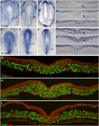
Initially the topic can be as specific or as broad as you want. Chicken embryo E-cad and P-cad gastrulation[1] References
| ||||||||||||||||||||||||||||||||||||||||||||||||||||||||||||||||
| Lab 4 Assessment - GIT Quiz | ||||||||||||||||||||||||||||||||||||||||||||||||||||||||||||||||
|
ANAT2341 Quiz Example | Category:Quiz | ANAT2341 Student 2015 Quiz Questions | Design 4 quiz questions based upon gastrointestinal tract. Add the quiz to your own page under Lab 4 assessment and provide a sub-sub-heading on the topic of the quiz. An example is shown below (open this page in view code or edit mode). Note that it is not just how you ask the question, but also how you explain the correct answer. | ||||||||||||||||||||||||||||||||||||||||||||||||||||||||||||||||
| Lab 5 Assessment - Course Review | ||||||||||||||||||||||||||||||||||||||||||||||||||||||||||||||||
| Complete the course review questionnaire and add the fact you have completed to your student page. | ||||||||||||||||||||||||||||||||||||||||||||||||||||||||||||||||
| Lab 6 Assessment - Cleft Lip and Palate | ||||||||||||||||||||||||||||||||||||||||||||||||||||||||||||||||
| ||||||||||||||||||||||||||||||||||||||||||||||||||||||||||||||||
| Lab 7 Assessment - Muscular Dystrophy | ||||||||||||||||||||||||||||||||||||||||||||||||||||||||||||||||
| ||||||||||||||||||||||||||||||||||||||||||||||||||||||||||||||||
| Lab 8 Assessment - Quiz | ||||||||||||||||||||||||||||||||||||||||||||||||||||||||||||||||
| A brief quiz was held in the practical class on urogenital development. | ||||||||||||||||||||||||||||||||||||||||||||||||||||||||||||||||
| Lab 9 Assessment - Peer Assessment | ||||||||||||||||||||||||||||||||||||||||||||||||||||||||||||||||
| ||||||||||||||||||||||||||||||||||||||||||||||||||||||||||||||||
| Lab 10 Assessment - Stem Cells | ||||||||||||||||||||||||||||||||||||||||||||||||||||||||||||||||
As part of the assessment for this course, you will give a 15 minutes journal club presentation in Lab 10. For this you will in your current student group discuss a recent (published after 2011) original research article (not a review!) on stem cell biology or technology.
| ||||||||||||||||||||||||||||||||||||||||||||||||||||||||||||||||
| Lab 11 Assessment - Heart Development | ||||||||||||||||||||||||||||||||||||||||||||||||||||||||||||||||
| Read the following recent review article on heart repair and from the reference list identify a cited research article and write a brief summary of the paper's main findings. Then describe how the original research result was used in the review article.
<pubmed>26932668</pubmed>Development | ||||||||||||||||||||||||||||||||||||||||||||||||||||||||||||||||
| ||||||||||||||||||||||||||||||||||||||||||||||||||||||||||||||||
lab attendance
Z3491219 (talk) 14:34, 5 August 2016 (AEST)
Z3491219 (talk) 13:13, 19 August 2016 (AEST)
Z3491219 (talk) 13:07, 26 August 2016 (AEST)
Z3491219 (talk) 13:08, 9 September 2016 (AEST)
Z3491219 (talk) 13:00, 23 September 2016 (AEST)
Z3491219 (talk) 14:37, 14 October 2016 (AEDT)
Z3491219 (talk) 13:12, 21 October 2016 (AEDT)
| Mark Hill 13 October 2016 - Lab attendance records and a number of online assessments are missing from your student page. Please be aware that individual assessments contribute 20% of your final mark for this course. |
Reference
PMID 27486480
Lab Assessment 1
Reference
<pubmed>22577141 </pubmed>
In human fertilisation, the spermatazoan attaches itself to the zona pellucida of the egg using sperm receptors and induces an acrosome reaction. This research article looks at the α7 nicotinic acetylcholine receptor (α7nAChR) as a plausible sperm receptor that is activated by the epidermal growth factor receptor (EGFR) to generate an acrosome reaction. The study involved using capacitated mouse sperm and mouse zona pellucidae that were isolated by Percoll gradient centrifugation of ovarian homogenates. These were then fertilized using IVF and various procedures like immunoblot anaylsis, immunocytochemistry and immunoprecipitation were used to determine an interesting interaction between α7nAChR and EGFR, which is a necessary step in the mechanism leading to the acrosome reaction.
The experiment showed that ZP-induced AR is enhanced via activation of α7nAChR and that α7nAChR mediates the AR by activating a pathway leading to Ca2+ influx into the sperm. The α7nAChR activates the EGFR through Src activation. In conclusion the experiment clearly indicates that α7nAChR and the EGFR are possibly new sperm receptors for ZP protein.
| Mark Hill 18 August 2016 - You have added the citation correctly and written a brief summary of the article findings. It was also good to include the methods employed in the study, hopefully you are familiar with these techniques? Is this the actual mouse sperm activation pathway, or just how Ca2+ influx can trigger the reaction. | Assessment 5/5 |
Lab 2 Assessment
| Mark Hill 29 August 2016 - All information Reference, Copyright and Student Image template correctly included with the file. Note that you could have used the PMID 21383844 for the reference section in the summary box.
|
Assessment 3/5 |
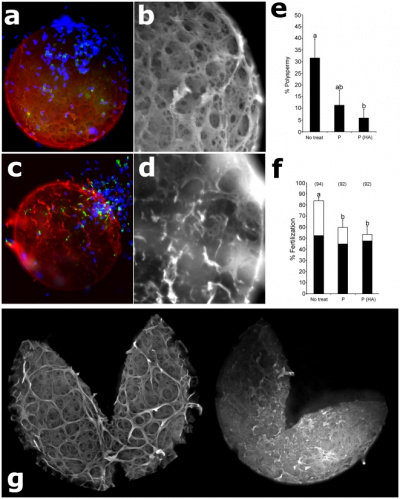
Degradation of Intact Oocyte Zonae by Isolated Sperm Proteasomes[1]
Lab 5 Assessment
Course Review Questionnaire completed
Lab 4 Assessment
Quiz
Lab 6 Assessment
Identify a known genetic mutation that is associated with cleft lip or palate
IRF6 is a known gene which when mutated is associated with non-syndromic cleft lip or palate.
Identify a recent research article on this gene
How does this mutation affect developmental signalling in normal development
The identification of cis-regulatory elements using sequence conservation across multiple species, analysis of animal models and biochemical analyses resulted in the identification of one specific sequence variant (rs642961) located within an enhancer ~10 kb upstream of the IRF6 transcription start site that is significantly over-transmitted in non-syndromic cleft lip only. [2] This mutated allele disrupts one of the binding sites for transcription factor AP-2α, which is mutated in the autosomal dominant CLP disorder branchio-oculo-facial syndrome therefore strongly suggesting it is a contributory variant. [3] A role of IRF6 in CLP is further supported by testing and analysis on animal models. Recent research has shown that Irf6 mutant mice exhibit a hyper-proliferative epidermis that fails to undergo terminal differentiation, which leads to multiple epithelial adhesions that can occlude the oral cavity and result in cleft palate. [4] These results showed that IRF6 is a key factor in the keratinocyte proliferation/differentiation switch and further research showed that IRF6 also plays a key role in the formation of oral periderm. [5]
Lab 7 Assessment
What is/are the dystrophin mutation(s)?
The DMD gene, encoding the dystrophin protein, is one of the longest human genes known, covering 2.3 megabases (0.08% of the human genome) at locus Xp21. [6] Mutations in the DMD gene that lead to the production of too little or a defective, internally shortened but partially functional dystrophin protein, result in a display of a much milder dystrophic phenotype in affected patients, like with Becker's muscular dystrophy (BMD). Duchenne muscular dystrophy (DMD), however, is caused by mutations in the gene that encodes the 427-kDa cytoskeletal protein dystrophin. [7]
In some cases the patient's phenotype is such that experts may decide differently on whether a patient should be diagnosed with DMD or BMD. The theory currently most commonly used to predict whether a variant will result in a DMD or BMD phenotype, is the reading frame rule. [8]
What is the function of dystrophin?
Dystrophin is a protein located between the sarcolemma and the outermost layer of myofilaments in the muscle fiber (myofiber). It is a cohesive protein, linking actin filaments to another support protein that resides on the inside surface of each muscle fiber’s plasma membrane (sarcolemma). This support protein on the inside surface of the sarcolemma in turn links to two other consecutive proteins for a total of three linking proteins. Dystrophin supports muscle fiber strength, and the absence of dystrophin reduces muscle stiffness, increases sarcolemmal deformability, and compromises the mechanical stability of costameres and their connections to nearby myofibrils; as shown in recent studies where biomechanical properties of the sarcolemma and its links through costameres to the contractile apparatus were measured, and helps to prevent muscle fiber injury.[9]
What other tissues/organs are affected by this disorder?
the heart has been known to be affects by this disorder resulting in complications like heart failure and arrhythmias. Weakness in respiratory muscles can severely shorten the lifespan of DMD patients.[10]
What therapies exist for DMD?
The long nature of the dystrophin gene puts forward many challenges like replacement, nature of viral vectors used in gene replacement. Various genetic approaches like gene product modifiers (exon skipping) and utrophin modulation strategies are being investigated. [11]
Administration of corticosteroids for suppression of inflammation of muscles is the main pharmacological treatment for DMD but it is known to have side effects. [12]
What animal models are available for muscular dystrophy?
The most commonly used animal is the mouse however golden retrievers and pigs have also been used. [9] [13] [14]
Lab 9 Assessment
These are great reviews of the project pages, with some good specific examples. You seem to understand how to provide a balanced critical assessment, tough a little more critical would also work, given the existing status of some of these pages. Please use a more professional language when providing feedback. 8/10
Group 1 peer assessment
Overall, you guys have done a great job on the research. Its really comprehensive and seems to cover all the important aspects of the topic. There are some incomplete topics which I’m sure when completed will prove to be a great source of information.
There are a few areas which can be improved. There isn’t much of an introduction to the topic. I think there should be some sort of introduction in layman terms on what your topic is about before diving into the semantics of the Wnt signalling pathway. Another aspect that can be touched upon is a little background on the discovery of the Wnt signalling pathway just for interest. You could also include a little detail on some of the important molecules involved, for example beta catenin, APC, etc. Some of the abbreviations have not been included for example: TCF/LEF family, SWI/SNF complex. The full forms should be written first and the abbreviated forms used thereafter. An easier, professional and more organised way to reference could be to include all the references in the end with footnoting and appropriate in text referencing. Normally, using bullet points is a lot clearer but in areas like the introduction, conclusion it would be ideal to use paragraphs instead. The use of diagrams to explain the Wnt signalling pathway is a good idea as well and there are some great images online. It makes it easier to understand the concept. Another thing I noticed was the formatting for each student was different. The formatting, use of headings, use of bullet points or use of paragraphs should be consistent throughout the page. For example: The Wnt-Calcium Ion pathway has paragraphs and is structure as all points under various articles, but the previous subheading is in bullet points and is quite vague with less detail. The non canonical pathway studies can be elaborated slightly more. This is quite an interesting topic and has a lot of potential for discussion as well as there isn’t much research done on it.
Overall, this topic seems very interesting and you guys are off to a great start. I’m sure taking all these and the other peer review points into consideration will help you to produce an enlightening resource for this topic.
Group 3 peer assessment
On browsing through your page for the first time, I was extremely impressed with the organisation and the headings and subheadings. It made it extremely easy to comprehend the information provided in an efficient manner. The hand drawn diagram was very informative and showed good understanding of the topic. The quiz at the end is a different and useful element to add to the page as well and helps to improve our understanding on the topic. Incorporating tables and diagrams is always a great idea so well done on that! The referencing has also been done in an organised and appropriate format. The flow of the page is great as well.
Some points of improvement include making sure all the abbreviations have been written in their full form when used for the first time on the page. For example, EWSR1. I think another thing that can be included is a small description on the important molecules of the pathway. For the quiz a link could be attached to the ‘submit’ option taking you to a page with the correct answers and explanations as well. The abnormalities could maybe include a sentence on the current treatment procedures for the same. Or this could be a separate heading all together. This could be included to get a wholesome idea of the abnormality from pathogenesis to treatment.
Overall, I think this is an amazing start to the project and you guys have done a great job covering all aspects of the assessment criteria. I’m sure this is going to be an awesome page!
Group 4 peer assessment
Group 4 is off to a great start with their project with well carried out research and reference to a lot of research papers mentioned as well. The referencing style used is easy to navigate and is appropriate in text referencing has been used as well.
However, there are a few points which can be improved upon to make this a great wiki page. The image included doesn’t have a description. The description is always necessary for relation to the text and to understand the figure. To get a wholesome idea of the pathway and also to educate the layman on the pathway there should be an introduction which covers the general aspects involved like where the pathway is used, the molecules involved, etc.
I also think for the mechanism heading, a general introduction or overview should be given and then you could delve into the mechanisms in the species. A table to bring out the difference between the mechanisms in the two species could also be included as a concise and clear manner to display the above information.
There were some formatting errors as well like the subheadings under mechanisms were in bold when ideally the heading should be in bold and the subheadings in normal font.
Under the abnormality heading, the sub headings seem comprehensive enough but I also think treatment could be included as it goes hand in hand with diagnosis and it seems incomplete without it.
There were a few complicated terms like organogenesis used which were not explained. Maybe a glossary could be included or just a simple definition can be included under the heading.
Overall, this group is off to a promising start with their page. I’m sure after incorporating the reviews given here the page will be fantastic!
Group 5 peer assessment
To be honest, this page is really good and already near completion in many aspects and there are very few negative points I could think of while going through the page. The first thing that appealed to many which was missing in a few of the other pages was the inclusion of an introduction. The page immediately stood out because of the inclusion of well formatted tables and images all related to the sub headings they were placed under. The research as well was extremely comprehensive and covered all topics required to gain a complete understanding of the T-box genes and their signalling. I particularly found the history aspect of it to be quite interesting. I also noticed that a wide variety of resources were used and the referencing for them was done with proper and consistent formatting making it easy to navigate through various articles and sources.
Some minor edits that can be done are that a few of a the PMID links remain under the headings which should be removed and included at the end with appropriate in text citation. Some of the abbreviations were not given in their full form when first used on the page.
This page is really great already and all the criteria have been carefully integrated and followed. Just a few changes here and there and you guys should have a top notch project!
Group 6 Peer Assessment
I think this group is off to a good start. An aspect that appealed to me is the plan to include an introduction which is always necessary to explain the broader aspects before going into details and the inclusion of history. The sub headings mentioned seem to cover most of the information important to understanding the topic. There is great room for improvement, however. The first aspect is the flow of the topics. The section on the history should be included in the introduction or after the introduction and then the mechanism, regulation and so on. The main idea of these projects is to look at the pathways in relation to embryogenic development and their abnormalities so the majority of the page should be dedicated to these aspects. I also noticed that there was a problem in the formatting. The main heading is the introduction with the other topics as sub headings of the introduction. There should be multiple headings and the respective sub headings under that. The inclusion of an image is good but the image title is missing. This is necessary to understand the relation to the text and to get a brief idea of what the image is about. There is only one image included and there is a large scope for other various visual aids like tables and figures with this topic. The referencing on this page is very poor. There has to be appropriate in text citation to easily navigate to the sources and the sources should be listed accordingly at the end of the page. Sources for images, table, figures, etc. should also be included. The main primary resource should be peer reviewed journal articles and not websites. Even if websites are include they should be referenced at the end appropriately and the referencing style should be consistent throughout.
I think this group is headed in the right direction but there are a lot of improvements that have to be made. It has the potential to be a great.
| Lab 10 - Stem Cell Presentations 2016 | |
|---|---|
| Group Mark | Assessor General Comments |
|
Group 1: 15/20 Group 2: 19/20 Group 3: 20/20 Group 4: 19/20 Group 5: 16/20 Group 6: 16/20 |
The students put great effort in their presentation and we heard a nice variety of studies in stem cell biology and regenerative medicine today. The interaction after the presentation was great.
As general feedback I would like to advise students to:
|
Lab 11 Assessment
Assessment - 5/5
<pubmed>22538609 </pubmed>
Summary
The authors of this article used multicolour clonal analysis to outline the contributions of cardiomyocytes as the zebrafish heart morphs from its primitive embryonic structure to its adult form. the findings show that the single cardiomyocyte thick wall of the juvenile ventricle forms by lateral expansion of several dozen cardiomyocytes into diverse muscle patches. Adult cortical muscle originates from approximately 8 cardiomyocytes which display clonal dominance reminiscent of stem cell populations. Cortical cardiomyocytes initially originate from internal myofibrils that in rare events breach the juvenile ventricular wall and expand over the surface. These results show the dynamic proliferative behaviours that generate adult cardiac structure, revealing clonal dominance as a key mechanism that shapes a vertebrate organ.
Integration into Review Article
This article was used while describing ventricular wall maturation. The authors compare the modest expansion of the ventricular wall development in zebrafish to the thick wall in other species like tuna fish and mammals. This article is used to highlight that cortical layer formation is a developmental response to biomechanical stress, and that the cardiomyocytes that build the final layer arise stochastically.
References
- ↑ <pubmed>21383844</pubmed>
- ↑ <pubmed>10934030</pubmed>
- ↑ <pubmed>18423521 </pubmed>
- ↑ <pubmed>17041603 </pubmed>
- ↑ <pubmed>19439425</pubmed>
- ↑ <pubmed>7719347</pubmed>
- ↑ <pubmed>15470384</pubmed>
- ↑ <pubmed>16770791</pubmed>
- ↑ 9.0 9.1 <pubmed>26140716</pubmed>
- ↑ <pubmed>27340611</pubmed>
- ↑ <pubmed>26140505</pubmed>
- ↑ <pubmed>27621596</pubmed>
- ↑ <pubmed>22968479</pubmed>
- ↑ <pubmed>27634466</pubmed>


