User:Z3439257
| Student Information (expand to read) | ||||||||||||||||||||||||||||||||||||||||||||||||||||||||||||||||
|---|---|---|---|---|---|---|---|---|---|---|---|---|---|---|---|---|---|---|---|---|---|---|---|---|---|---|---|---|---|---|---|---|---|---|---|---|---|---|---|---|---|---|---|---|---|---|---|---|---|---|---|---|---|---|---|---|---|---|---|---|---|---|---|---|
| Individual Assessments | ||||||||||||||||||||||||||||||||||||||||||||||||||||||||||||||||
|
Please leave this template on top of your student page as I will add your assessment items here. Beginning your online work - Working Online in this course
Click here to email Dr Mark Hill | ||||||||||||||||||||||||||||||||||||||||||||||||||||||||||||||||
| Lab 1 Assessment - Researching a Topic | ||||||||||||||||||||||||||||||||||||||||||||||||||||||||||||||||
In the lab I showed you how to find the PubMed reference database and search it using a topic word. Lab 1 assessment will be for you to use this to find a research reference on "fertilization" and write a brief summary of the main finding of the paper.
| ||||||||||||||||||||||||||||||||||||||||||||||||||||||||||||||||
| Lab 2 Assessment - Uploading an Image | ||||||||||||||||||||||||||||||||||||||||||||||||||||||||||||||||
OK you are now in a group
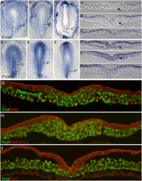
Initially the topic can be as specific or as broad as you want. Chicken embryo E-cad and P-cad gastrulation[1] References
| ||||||||||||||||||||||||||||||||||||||||||||||||||||||||||||||||
| Lab 4 Assessment - GIT Quiz | ||||||||||||||||||||||||||||||||||||||||||||||||||||||||||||||||
|
ANAT2341 Quiz Example | Category:Quiz | ANAT2341 Student 2015 Quiz Questions | Design 4 quiz questions based upon gastrointestinal tract. Add the quiz to your own page under Lab 4 assessment and provide a sub-sub-heading on the topic of the quiz. An example is shown below (open this page in view code or edit mode). Note that it is not just how you ask the question, but also how you explain the correct answer. | ||||||||||||||||||||||||||||||||||||||||||||||||||||||||||||||||
| Lab 5 Assessment - Course Review | ||||||||||||||||||||||||||||||||||||||||||||||||||||||||||||||||
| Complete the course review questionnaire and add the fact you have completed to your student page. | ||||||||||||||||||||||||||||||||||||||||||||||||||||||||||||||||
| Lab 6 Assessment - Cleft Lip and Palate | ||||||||||||||||||||||||||||||||||||||||||||||||||||||||||||||||
| ||||||||||||||||||||||||||||||||||||||||||||||||||||||||||||||||
| Lab 7 Assessment - Muscular Dystrophy | ||||||||||||||||||||||||||||||||||||||||||||||||||||||||||||||||
| ||||||||||||||||||||||||||||||||||||||||||||||||||||||||||||||||
| Lab 8 Assessment - Quiz | ||||||||||||||||||||||||||||||||||||||||||||||||||||||||||||||||
| A brief quiz was held in the practical class on urogenital development. | ||||||||||||||||||||||||||||||||||||||||||||||||||||||||||||||||
| Lab 9 Assessment - Peer Assessment | ||||||||||||||||||||||||||||||||||||||||||||||||||||||||||||||||
| ||||||||||||||||||||||||||||||||||||||||||||||||||||||||||||||||
| Lab 10 Assessment - Stem Cells | ||||||||||||||||||||||||||||||||||||||||||||||||||||||||||||||||
As part of the assessment for this course, you will give a 15 minutes journal club presentation in Lab 10. For this you will in your current student group discuss a recent (published after 2011) original research article (not a review!) on stem cell biology or technology.
| ||||||||||||||||||||||||||||||||||||||||||||||||||||||||||||||||
| Lab 11 Assessment - Heart Development | ||||||||||||||||||||||||||||||||||||||||||||||||||||||||||||||||
| Read the following recent review article on heart repair and from the reference list identify a cited research article and write a brief summary of the paper's main findings. Then describe how the original research result was used in the review article.
<pubmed>26932668</pubmed>Development | ||||||||||||||||||||||||||||||||||||||||||||||||||||||||||||||||
| ||||||||||||||||||||||||||||||||||||||||||||||||||||||||||||||||
Lab 1
ANAT2341 Lab 1 - Online Assessment
PMID 26043223
Paper Review: This article has firstly proved that the proteins (ZP-1, ZP-2 & ZP-3) in mouse zona pellucida form functional amyloid surrounding the oocytes. Firstly, several experiments such as staining and western blot were performed to confirm that the proteins are forming amyloid structure. Then, the amino acid sequence of the proteins from six different taxa were analysed and essential amyloidogenic sites were identified. It has been described that different ZP proteins will aggregate with each other or self-aggregate in different ways, depends on the amyloidogenic sites it possess. Specifically, ZP-N repeats, which is on of the amyloidogenic sites, has been stated to be related to the recognition of sperm. Therefore, the conclusion that ZP proteins exist in the ZP as functional amyloid and may get involved in prevention of polyspermy and cross-species fertilisation has been come up with. Moreover, since the amyloidogenic sites across species seem to be conserved, the amyloid formed are expected to possess similar functions, making this study on mouse model useful on human researches. Furthermore, some proteins exist in other parts of the body also have the conserved amyloidogenic sites, part or all of their functions may be carried out through their amyloid structure formed.
Amyloid used to be associated with several neurodegenerative and prion diseases in mammals. However, it is becoming increasingly obvious that functional amyloid does exist and possesses a physiological function rather than a pathological function. The further research can be focusing on the specificity of the amyloid structure formed and the functions related. Based on the data published, a question about diagram 2A is raised. It is known that buffer was used to incubate with the PAD beads as the negative control. However, a light band can be observed in the negative control, which is not supposed to happen.
| Mark Hill 18 August 2016 - You have added the citation correctly and written a good summary of the article. Quite an interesting paper on protein structure of the ZP, amyloid cross-β sheet fibrillar structure is an interesting association.
Unfortunately I have had to take marks off the final assessment as you have not added the reference correctly. Add a link to this reference using its PMID using this code <pubmed>XXXXX</pubmed> replacing the Xs with just the PMID number (no text). You have just added the link, here is the reference: <pubmed>26043223</pubmed> I also fixed your formatting that had a space at the beginning (no deduction for this). |
Assessment 3/5 |
Lab 2
ANAT2341 Lab 2 - Online Assessment
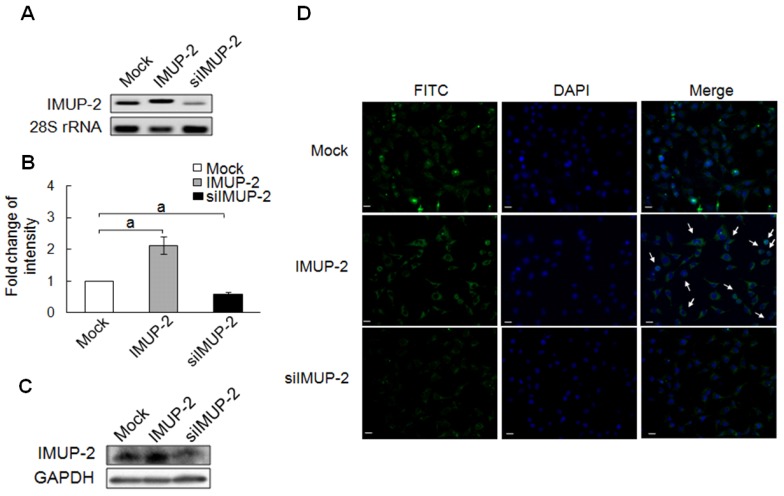
Human trophoblasts stained with different markers
| Mark Hill 29 August 2016 - All information Reference, Copyright and Student Image template correctly included with the file and referenced on your page here.
Your image requires a legend and the reference citation correctly with the legend. You need to include the ref name for a citation, as shown below: Code: <ref name="PMID25949126"><pubmed>25949126</pubmed></ref> and with the citation added to legend below, you lost marks for no citation. |
Assessment 4.5/5 |
Human trophoblasts stained with different markers[1]
Lab 3
ANAT2341 Lab 3 - Online Assessment
| Mark Hill 31 August 2016 - Lab 3 Assessment Quiz - Mesoderm and Ectoderm development. | Assessment 1.5/5 |
Lab 4
ANAT2341 Lab 4 - Online Assessment
| Mark Hill 11 October 2016 - GIT Quiz missing? | Assessment |
Lab 5
ANAT2341 Lab 5 - Online Assessment
Assessment: survey. Completed.
| Mark Hill 11 October 2016 - Questionnaire on course structure. | Assessment 5/5 |
Lab 6
ANAT2341 Lab 6 - Online Assessment
- Interferon Regulatory Factor 6 (IRF6) is a well established gene which mutations are related with non-syndromic Cleft lip and palate (NSCL/P).
- PMID 21331089 & PMID 22438645 were reviewed to achieve a better understanding about IRF6 mutations and NSCL/P.
- It seems that there are many types of mutations related with IRF6 gene, and different mutations are related with different outcomes. IRF6 has been identified to play a role in keratinocyte proliferation/differentiation switch. Further investigation has suggested that IRF6 is involved in the formation of oral periderm. The spatio-temporal regulation of the keratinocytes established by IRF6 protein is able to ensure the appropriate palatal adhesion. Moreover, p63 has been identified as an upstream regulator of IRF6, and mutations at p63 locus may cause similar pathological effects as the IRF6 mutations.
- Reference
<pubmed>21331089</pubmed> <pubmed>22438645</pubmed>
| Mark Hill 13 October 2016 - Interferon Regulatory Factor 6 IRF6 belongs to a family of transcription factors that share a highly conserved helix-turn-helix DNA-binding domain. This should have been included in your answer about signaling mechanism. | Assessment 4/5 |
Lab 7
Lab 8
ANAT2341 Lab 8 - Online Assessment
Paper based quiz.
Lab 9
For peer review, please see the section below: Group project.
Lab 10
Group presentation.
Lab 11
PMID 23302686 This article is referenced in the review article under the section cardiomyocyte turnover in adult mammals. It mainly demonstrates that cardiomyocyte proliferation, together with hypertrophy, may contribute to cardiac development during the first two decades. According to current knowledge on postnatal cardiac development, cardiomyocytes will only undergo hypertrophy. However, more and more evidences have revealed that in many animals, proliferation is also possible to support postnatal cardiac development. The research data in this article have firstly revealed that there is ongoing, though decreasing, myocyte cell cycle activity through out the life in human myocyte cells. However, increased cell cycle may lead to increased cell division, increased nuclei and increased ploidy. Therefore, experiments were also performed to quantify the effect of binucleation and polypoidization, so that the cardiomyocyte cytokinesis can be corrected with those data in order to obtain the number of cells produced from myocyte proliferation. Based on the results obtained, it seems that both hypertrophy and proliferation is taking place in the experimental models, and the developmental effect is continuous throughout the first two decades, although both the effects of hypertrophy and proliferation are decreased.
Back to the review article, the author seems to have made the same conclusion as the authors of the research article. The conclusion is used to support the recently proposed theory that: in adult heart, cell proliferation is taking place. Cardiac myocytes is under homeostatic regulation in adult cardiac tissue, although the ability of producing new cells is decreasing. Together with the results from other articles, a brief overview about cardiac myocyte turnover has been illustrated. Moreover, in the following paragraph, research findings have indicated that this repopulation could be induced by a group of hypoxic cardiac myocytes. Further research may shed light on this point and reveal the more detailed and in-depth mechanism of cardiomyocyte proliferation, which may make it possible for a patient to regenerate his heart.
The formatted reference for the review article
<pubmed> 26932668 </pubmed>
The formatted reference for the research article
<pubmed>23302686</pubmed>
Lab 12
Group Project
Peer Assessment
These are good brief reviews of project pages, with some specific examples. I think some projects required a more critical approach. 7/10
Group 2 Peer Review
Really well organised structures! Nice and clear. The history part is really good. It makes your project both educative and attractive. After the history, the molecular background has been well explained. You guys have really looked into this pathway in depth and understood it clearly. I'm really looking forward to reading the non-canonical and the regulation part of the molecular basis. As ANAT2341 is an anatomy course, you guys have perfectly caught the main point of this project. Most of your following paragraphs focus on the anatomical aspect related to Notch signalling pathway. Moreover, you have also identified some abnormalities associated with Notch which makes your page look really good (almost like a lecture note page)!
Just a few points about your page. Firstly, I think it might be better if you guys can find another or draw a picture yourselves showing important components of this pathway. The one that you guys have now looks a little bit massy. I think you guys can photoshop the picture a little bit, just to make the important components more obvious. Moreover, I believe that the differences between canonical pathway and non-canonical pathway are more than component differences. Maybe address more about what does the canonical pathway do and what does the non-canonical pathway do, so that those pathways can be differentiated better. However, since you haven't pasted you non-canonical part yet, it hard to judge whether or not my comment is appropriate.
Overall, I think this page is really good, for it has a lot of attractive contents and well structured which makes us easy to follow. What you guys need is just to complete it. Great job!
Group 3 Peer Review
Good Job Group 3! Very Well organised page with amazing contents. The signalling pathway is well illustrated with your hand drawing. A lot of articles were reviewed although some more citations may be required for some sentences on the page. The history section is really good making the page very interesting. Developmental effects and abnormalities are also described. Some sections need to be filled in. Seems that you guys are trying to make a few quiz questions in the end, that's a really good idea. Quizzes can definitely improve our understanding about the signalling pathway.
About the introduction part, it may be better if you can combine the introduction, history and overview together. Those three sections posses similar function--provide background information and attract the reader, therefore, i think it would be good to put them together, at least, make history and overview two subsections of introduction. Moreover, the format of the table for FGFR subtypes can be adjusted. Thirdly, I understand that some theories are well studied or well proved, however, it would be better if you can find more recent articles.
Overall, this web page is really good. It is well structured and only some sections need to be completed. I really recommend using of more recent articles because our understanding about the pathway will improve over time. Maybe read through some related articles, they will usually validate the previous results first before they start their own experiments.
Group 4 Peer Review
Another well organised web page and a lot of references were used. Excellent job Group 4! The picture below the title looks clear and educative. However, that may need a citation. Some sections were left blank. They need to be filled as well. The mechanism of the signalling is well explained.
First of all, both mechanisms about the signalling in mammals and other vertebrates were discussed, therefore, it would be better if the title can be changed. Secondly, it would be better if you can build up more connections between the word-version descriptions and the flow chart graph you used. Thirdly, it might be better if you can put more pictures about the phenotype in normal development and abnormalities. Moreover, I think more citations are necessary to support your story. And it would be good to add a section about the terms used on your page.
overall, this web page is really good. There are some problems about formats, but i think you can do it after you have filled all your sections in first.
Group 5 Peer Review
I have to say this page is the best across all groups. Very impressive work. Lots of references have been utilised and they are very supportive. The pictures cited are beautiful and are closely related to the contents. The molecular basis has been well explained and specific components of the pathway were pointed out in each signalling events related in embryo development. Moreover, you guys have also shed light on the ancient origin (evolutionary aspect) of this pathway which is really interesting.
Only a few points to mention. More pictures will make your page look better. Maybe one picture per section? Secondly, the origin and the ancient origin are a little bit confusing, maybe change their names and combine all the background things like those together? A few sections need to be completed and there are some structural problems need to be adjusted.
This is a really impressive web page. Good job Group 5!
Group 6 Peer Review
The structure of this page is really good. But it needs more contents. The pictures about the mechanism of this pathway is really good, but it would be better if it can has some colour. Or maybe draw one yourself. The pathway is well studied and well explained. Good job!
A few points about the down side. Since this is an anatomy course, it would be better if you can focus more on the anatomical aspect of this pathway. Secondly, animal models can also be an interesting section to be included. Thirdly, the order of the titles needs to be reconsidered. The history part can be put at the very first or the very end. The reference part should also be structured.
Overall, this page is still under constructing. But it has already got a very good structure. Good luck Group 6!
Reference
Jung R, Choi JH, Lee HJ, Kim JK, Kim GJ. Effect of Immortalization-Upregulated Protein-2 (IMUP-2) on Cell Death of Trophoblast. Development & Reproduction. 2013;17(2):99-109. doi:10.12717/DR.2013.17.2.099.
| Lab 10 - Stem Cell Presentations 2016 | |
|---|---|
| Group Mark | Assessor General Comments |
|
Group 1: 15/20 Group 2: 19/20 Group 3: 20/20 Group 4: 19/20 Group 5: 16/20 Group 6: 16/20 |
The students put great effort in their presentation and we heard a nice variety of studies in stem cell biology and regenerative medicine today. The interaction after the presentation was great.
As general feedback I would like to advise students to:
|
Lab Attendance
Z3439257 (talk) 14:35, 5 August 2016 (AEST) Z3439257 (talk) 14:40, 12 August 2016 (AEST) Z3439257 (talk) Z3439257 (talk) Z3439257 (talk) 13:20, 9 September 2016 (AEST) Z3439257 (talk) 13:25, 23 September 2016 (AEST) Z3439257 (talk) 13:11, 21 October 2016 (AEDT) Z3439257 (talk) 13:38, 28 October 2016 (AEDT)
referencing
PMID 24614230
- ↑ <pubmed>25949126</pubmed>


