Renal System Histology: Difference between revisions
No edit summary |
mNo edit summary |
||
| (84 intermediate revisions by 2 users not shown) | |||
| Line 1: | Line 1: | ||
{{Header}} | |||
==Introduction== | ==Introduction== | ||
[[File:Nephron histology.jpg|thumb|Nephron histology]] | [[File:Nephron histology.jpg|thumb|Nephron histology]] | ||
This section of notes gives an overview mainly of adult renal histology, see also [[Renal System Development]] notes. A key structure of the kidney functional unit, the nephron, is the glomerulus (renal corpuscle), which represents the initial vascular/renal interface. | This section of notes gives an overview mainly of adult renal histology, see also [[Renal System Development]] notes. A key structure of the kidney functional unit, the nephron, is the glomerulus (renal corpuscle), which represents the initial vascular/renal interface. | ||
[[File:HMsmall.jpg|left]] Page also provides further background information for Medicine phase 1 Health Maintenance B Urinary Tract Histology Practical [ | [[File:HMsmall.jpg|left]] Page also provides further background information for Medicine phase 1 Health Maintenance B Urinary Tract Histology Practical - [https://moodle.telt.unsw.edu.au/mod/book/view.php?id=856230 Moodle - Virtual Slides]. | ||
'''This support page content is not part of the HMB practical class.''' | |||
:{{ | ::'''HMB:''' [[Gastrointestinal Tract - Pancreas Histology|Pancreas Histology]] | [[Gastrointestinal_Tract_-_Liver_Histology|Liver Histology]] | [[Gastrointestinal_Tract_-_Gall_Bladder_Histology|Gall Bladder Histology]] | [[Renal System Histology]] | [[Histology Stains]] | ||
===Renal Virtual Slides=== | |||
[https://moodle.telt.unsw.edu.au/mod/book/view.php?id=856230 Moodle - Virtual Slides] (requires Moodle log in) | |||
* [https://www.best.edu.au/s/share?url=1atsbric%2Faibw4p9h&data=8%400!9%4063360!10%40-30743&version=1 Kidney LS and TS] (rat, stain - PAS, Tartrazine) | |||
* [https://www.best.edu.au/s/zgd1j4w3/kbyi69ht/invite/wcivg3rk?data=7%40%5B%5D!8%401!9%408758!10%40-8762.5&version=1 Kidney Pelvis] | |||
* [https://www.best.edu.au/s/p7srbd8z/gvgr4b9z/invite/2suh26aq?data=7%40%5B%5D!8%400!9%4043680!10%40-30539.5&version=1 Renal vascular structures] | |||
* [https://www.best.edu.au/s/gzb56l8f/8ecp3bsd/invite/dlnyuazz?data=8%402!9%4011846!10%40-2991&version=1 Ureter TS] | |||
* [https://www.best.edu.au/s/i2eayqlq/4tzcinmy/invite/s7vrspjx?data=8%404!9%401212!10%40-1585&version=1 Ureter TS] | |||
* [https://www.best.edu.au/s/e6jweivj/d7bya6gc/invite/689ai5az?data=8%401!9%4024960!10%40-21414&version=1 Urinary bladder] | |||
{{Renal Links}} | |||
{{Renal Histology Links}} | |||
==Kidney Anatomy== | ==Kidney Anatomy== | ||
{| | {| | ||
| | | [[File:Kidney cartoon.jpg]] | ||
* | # Parenchyma | ||
# Cortex | |||
# Medulla | |||
# Perirenal fat | |||
# Capsule | |||
# Ureter | |||
# Pelvis of kidney | |||
# Renal vessels | |||
# Hilum | |||
# Calyx | |||
| [[File:Gray1128.jpg|400px|]] | |||
Adult nephron structure | |||
| '''Nephron''' | |||
* Functional unit of kidney | |||
* Humans up to 1 million | * Humans up to 1 million | ||
* Filtration of waste from blood | * Filtration of waste from blood | ||
* Endocrine | * Endocrine | ||
* Blood pressure regulation | * Blood pressure regulation | ||
|} | |} | ||
== Nephron == | == Nephron Histology== | ||
* Development - mean glomerular number shown to level at 36 weeks, increasing from about 15,000 at 15 weeks to 740,000 at 40 weeks. | |||
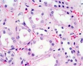
===Glomerulus=== | |||
{| | {| | ||
| [[File:Nephron_histology_01.jpg]] | | [[File:Nephron_histology_01.jpg]] | ||
| Line 32: | Line 60: | ||
| Glomerulus structure | | Glomerulus structure | ||
| Vascular and renal poles | | Vascular and renal poles | ||
|} | |||
{| | |||
| width=605px|[[File:Mouse renal podocyte EM02.jpg|600px]] | |||
Mouse renal podocyte (EM)<ref name="PMID28283912"><pubmed>28283912</pubmed></ref> | |||
| Transmission electron micrscopy of a mouse glomerular capillary revealing the structural composition of the glomerulus. | |||
The primary filtrate drains into Bowman’s space ('''BS''') and, via the urinary pole, reaches the proximal tubule. | |||
On the outside of the capillary, the foot processes ('''FP''') cover a major part of the glomerular basement membrane ('''GBM''') circumference. | |||
The slit-diaphram can be discerned in between the foot processs. | |||
The endothelial cell coats the inner surface of the capillary wall and is followed by the three layers of the glomerular basement membrane ('''GBM'''). | |||
|} | |||
:'''Links:''' [[:File:Nephron_histology 01.jpg|glomerulus structure image]] | [[:File:Nephron_histology 02.jpg|vascular and renal poles image]] | |||
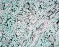
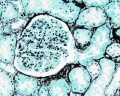
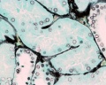
===Nephron Tubules=== | |||
[[File:Nephron histology.jpg]] | |||
Nephron overview | |||
[[File:Nephron histology 03.jpg]] [[File:Nephron histology 04.jpg]] | |||
{{Renal Histology}} | |||
===Large Images=== | |||
<gallery> | |||
File:Renal_histology_01.jpg|medullary rays | |||
File:Renal_histology_02.jpg|glomerulus | |||
File:Renal_histology_03.jpg|distal tubule and collecting duct | |||
File:Renal_histology_04.jpg|proximal and distal tubule | |||
File:Renal_histology_05.jpg|distal and intermediate tubule | |||
File:Renal_histology_06.jpg|medullary ray | |||
File:Renal_histology_07.jpg|glomerulus | |||
File:Renal_histology_08.jpg|proximal tubule | |||
</gallery> | </gallery> | ||
==Ureter Histology== | |||
{| | |||
| | |||
* The adult ureter is a thick-walled muscular tube, 25 - 30 cm in length, running from the kidney to the urinary bladder. | |||
* Anatomically can be described in two parts the abdominal part (''pars abdominalis'') and pelvic part (''pars pelvina''). | |||
* The ureter is composed of three layers: outer fibrous layer (''tunica adventitia''), muscular layer (''tunica muscularis'') and mucous layer (''tunica mucosa''). | |||
* The muscular layer has also been described as being subdivided into 3 fibre layers: | |||
# an external longitudinal | |||
# a middle circular | |||
# an internal longitudinal | |||
| [[File:Adult_bladder.jpg|400px]] | |||
|} | |||
[[File:Ureter histology 001.jpg]] [[File:Ureter histology 002.jpg]] | |||
==Bladder Histology== | |||
{| | |||
| Can be described anatomically by its 4 layers from inside outward: | |||
* '''Mucosa''' - (mucus layer) a transitional epithelium layer formed into folds (rugae). | |||
* '''Submucosa''' - connects the muscular layer with the mucous layer. | |||
* '''Muscular''' - the detrusor muscle is the muscle of the urinary bladder wall. | |||
* '''Serous''' - the superior or abdominal surfaces and the lateral" surfaces of the bladder are covered by visceral peritoneum, the serous membrane (serosa) of the abdominal cavity, consisting of mesthelium and elastic fibrous connective tissue. | |||
'''Detrusor Muscle''' | |||
* The adult detrusor muscle consists of three layers of smooth (involuntary) muscle fibres. | * The adult detrusor muscle consists of three layers of smooth (involuntary) muscle fibres. | ||
** internal layer - fibres arranged longitudinally | |||
** middle layer - fibres arranged circularly | |||
** external layer - fibres arranged longitudinally | ** external layer - fibres arranged longitudinally | ||
Note that while the smooth muscle fibre layer organisation is described as longitudinal or circular, this is only a general organisation of fibre direction, and is better described as a "spiral" organisation. | |||
| [[File:Bladder histology.jpg|400px]] | |||
|} | |||
<gallery> | |||
File:Bladder histology 001.jpg | |||
File:Bladder histology 002.jpg | |||
File:Bladder histology 003.jpg | |||
File:Bladder histology 004.jpg | |||
</gallery> | |||
==Fetal Kidney== | |||
<gallery> | |||
File:Human fetal kidney histology 01.jpg| | |||
File:Human fetal kidney histology 02.jpg| | |||
File:Human fetal kidney histology 03.jpg| | |||
File:Human fetal kidney histology 04.jpg| | |||
</gallery> | |||
:'''[[Renal System Development|Fetal Renal Links]]''': [[:File:Human fetal kidney histology 01.jpg|fetal kidney histology 01]] | [[:File:Human fetal kidney histology 02.jpg|fetal kidney histology 02]] | [[:File:Human fetal kidney histology 03.jpg|fetal kidney histology 03]] | [[:File:Human fetal kidney histology 04.jpg|fetal kidney histology 04]] | |||
==Fetal Urethra Histology== | |||
<gallery> | |||
File:Male_histology_001.jpg| | |||
File:Male_histology_002.jpg| | |||
File:Male_histology_003.jpg| | |||
File:Male_histology_004.jpg| | |||
</gallery> | |||
==Images== | ==Images== | ||
| Line 73: | Line 180: | ||
File:Fetal_10wk_urogenital_1.jpg|Fetal kidney (10 weeks) | File:Fetal_10wk_urogenital_1.jpg|Fetal kidney (10 weeks) | ||
File:Mouse-kidney in vitro.jpg|Mouse E12.5 kidney in vitro | File:Mouse-kidney in vitro.jpg|Mouse E12.5 kidney in vitro | ||
File:Mouse renal podocyte EM01.jpg|Mouse renal podocyte EM<ref name="PMID28283912"><pubmed>28283912</pubmed></ref> | |||
</gallery> | </gallery> | ||
==External Links== | ==External Links== | ||
{{External Links}} | {{External Links}} | ||
[http://vslides.unsw.edu.au/VirtualSlideV2.nsf/id/7CCC0D Virtual | * '''Blue Histology''' [http://www.lab.anhb.uwa.edu.au/mb140/CorePages/Urinary/urinary.htm Urinary System] | ||
* '''UNSW Virtual Slides''' [http://vslides.unsw.edu.au/VirtualSlideV2.nsf/id/7CCC0D Urinary Tract Histology] | |||
* '''UIOWA Virtual Slidebox of Histology''' [http://www.path.uiowa.edu/cgi-bin-pub/vs/fpx_browse.cgi?cat=o_urinary&div=nlm Urinary tract] | |||
== | ==Practical Overview== | ||
''' | [[File:Gray1126.png|thumb|retroperitoneal kidney]] | ||
'''Normal Histology''' | |||
* kidneys, ureters, urinary bladder, urethra | |||
''' | '''Anatomy''' | ||
* retroperitoneal | |||
* kidney - bean shaped | |||
* rich blood supply | |||
''' | '''Kidney Function''' | ||
* elimination of foreign substances | |||
* regulation of the amount of water in the body | |||
* control of the concentration of most compounds in the extracellular fluid | |||
* '''filtration''' - glomeruli of the kidney | |||
* '''selective resorption and excretion''' - tubular system of the kidney | |||
''' | {| | ||
| valign="top"|'''Capsule''' | |||
''' | * outer layer - dense CT (fibroblasts and collagen | ||
* inner layer - myofibroblasts | |||
| valign="top"|'''Cortex''' | |||
* outer renal corpuscles | |||
* medullary rays | |||
** only straight tubules + straight collecting tubules | |||
''' | ** 400-500 project medulla to cortex | ||
* between medullary rays - convoluted tubules of nephrons | |||
| valign="top"|'''Medulla''' | |||
* medullary pyramids (together with associated cortical region = '''renal lobe''') | |||
** base at cortioco-medullary border | |||
** apex at renal papilla (surrounded by '''minor calyx''') | |||
''' | * minor calyces converge to form '''major calyces''' then '''renal pelvis''' | ||
|} | |||
''' | |||
===Blood Supply=== | |||
* renal artery | |||
* interlobar arteries (across medulla thru renal columns) | |||
* arcuate arteries (cortico-medullary junction) | |||
* interlobular arteries | |||
*afferent glomerular arterioles | |||
* glomerular capillary network | |||
* efferent glomerular arterioles | |||
''' | '''Vasa Recta''' | ||
* descending arterioles (arteriole rectae) + ascending venules (venulae rectae) | |||
===Glomerulus=== | |||
* glomerulus - round (~0.2 mm in diameter) blind beginning of the nephron | |||
* ''' vascular pole''' - invaginated by a tuft of capillaries | |||
* '''urinary pole''' - substances leave the capillaries enter the renal tubule | |||
* '''Bowman's capsule''' - anatomical glomerulus is enclosed by two layers of epithelium. | |||
** outer or parietal layer of Bowman's capsule form a simple squamous epithelium. | |||
** inner layer, '''podocytes''' in the visceral layer, are extremely complex in shape. | |||
* '''Mesangial cells''' in the glomerulus form the connective tissue that gives structural support to podocytes and vessels (Podocytes, mesangial cells, glomerular capillaries) | |||
* Juxtaglomerular cells - smooth muscle cells afferent glomerular arteriole (epithelial-like cells) | |||
* '''Macular Densa''' | |||
** distal convoluted tubule near vascular pole (narrower and taller than rest of DCT) | |||
===Tubules=== | |||
'''Proximal Convoluted Tubules (PCT)''' | |||
* brush border | |||
* star-shaped | |||
* larger outside diameter | |||
''' | '''Distal Convoluted Tubules (DCT)''' | ||
* clean lumen surface | |||
* apical nuclei | |||
''' | '''Collecting Tubules (CT)''' | ||
* larger lumen than DCT (about size of PCT) | |||
* cuboidal cells and smaller than DCT | |||
===Renal Pyramids=== | |||
* medullary straight tubules, ducts and vasa recta | |||
* apical renal papilla - simple cuboidal/columnar epithelia | |||
* calyx - lined by transitional epithelia | |||
''' | Note the urinary system transitional epithelium is also known as '''urothelium'''. | ||
'''ureter''' | ==Ureters== | ||
* epithelium - transitional epithelia | |||
* lamina propria - mainly of dense connective tissue, with many bundles of coarse collagenous fibres | |||
* muscularis - consists of an '''inner longitudinal''' and '''outer circular layer''' of smooth muscle cells | |||
** In lower parts of the ureter and the bladder an '''additional outer longitudinal layer''' of muscles is added to the first two. | |||
''' | ==Bladder== | ||
* '''epithelium''' - transitional epithelia | |||
** apical plaques - thickened domain allows great changes in surface area. | |||
* '''lamina propria''' - mainly of dense connective tissue, with many bundles of coarse collagenous fibres | |||
* '''muscularis''' - consists of an '''inner longitudinal''' and '''outer circular layer''' of smooth muscle cells | |||
** In the bladder (and lower parts of the ureter) an '''additional outer longitudinal layer''' of muscles is added to the first two. | |||
''' | ==Urethra== | ||
* penile urethra within '''corpus spongiosum''' | |||
* '''pseudostratified columnar epithelia''' | |||
* distal end - '''stratified squamous''' | |||
* continuous with outer skin | |||
==References== | |||
<references/> | |||
==Terms== | |||
{{Renal terms}} | |||
{{Glossary}} | |||
{{ | {{Footer}} | ||
[[Category:Histology]][[Category:Renal]] | [[Category:Histology]][[Category:Renal]] | ||
Latest revision as of 10:35, 30 August 2017
| Embryology - 11 May 2024 |
|---|
| Google Translate - select your language from the list shown below (this will open a new external page) |
|
العربية | català | 中文 | 中國傳統的 | français | Deutsche | עִברִית | हिंदी | bahasa Indonesia | italiano | 日本語 | 한국어 | မြန်မာ | Pilipino | Polskie | português | ਪੰਜਾਬੀ ਦੇ | Română | русский | Español | Swahili | Svensk | ไทย | Türkçe | اردو | ייִדיש | Tiếng Việt These external translations are automated and may not be accurate. (More? About Translations) |
Introduction
This section of notes gives an overview mainly of adult renal histology, see also Renal System Development notes. A key structure of the kidney functional unit, the nephron, is the glomerulus (renal corpuscle), which represents the initial vascular/renal interface.

Page also provides further background information for Medicine phase 1 Health Maintenance B Urinary Tract Histology Practical - Moodle - Virtual Slides.
This support page content is not part of the HMB practical class.
Renal Virtual Slides
Moodle - Virtual Slides (requires Moodle log in)
- Kidney LS and TS (rat, stain - PAS, Tartrazine)
- Kidney Pelvis
- Renal vascular structures
- Ureter TS
- Ureter TS
- Urinary bladder
- Renal Histology: Histology | Histology Stains | Renal Development
- Kidney - Nephron overview | Glomerulus | Vascular and renal poles | Medullary ray | tubules
- Ureter - Ureter labeled | Ureter epithelium
- Bladder - overview | wall 1 | wall 2 | transitional epithelium | Urinary Bladder Development
Kidney Anatomy

|

Adult nephron structure |
Nephron
|
Nephron Histology
- Development - mean glomerular number shown to level at 36 weeks, increasing from about 15,000 at 15 weeks to 740,000 at 40 weeks.
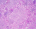
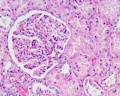
Glomerulus

|

|
| Glomerulus structure | Vascular and renal poles |

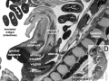
Mouse renal podocyte (EM)[1] |
Transmission electron micrscopy of a mouse glomerular capillary revealing the structural composition of the glomerulus.
|
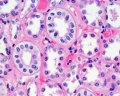
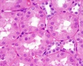
Nephron Tubules

Nephron overview


- Renal System Histology: Nephron tubule overview | glomerulus structure | vascular and renal poles | Medullary rays | Nephron tubules
Large Images
medullary rays
glomerulus
distal tubule and collecting duct
proximal and distal tubule
distal and intermediate tubule
medullary ray
glomerulus
proximal tubule


Ureter Histology
|

|


Bladder Histology
Can be described anatomically by its 4 layers from inside outward:
Note that while the smooth muscle fibre layer organisation is described as longitudinal or circular, this is only a general organisation of fibre direction, and is better described as a "spiral" organisation. |

|


Fetal Kidney


- Fetal Renal Links: fetal kidney histology 01 | fetal kidney histology 02 | fetal kidney histology 03 | fetal kidney histology 04
Fetal Urethra Histology


Images
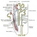
Nephron structure
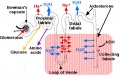
Nephron physiology
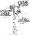
Nephrons - cortical and juxtamedullary
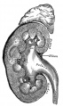
Kidney and adrenal gland (adult)
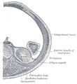
retroperitoneal
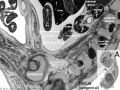
Fetal urogenital region most lateral right
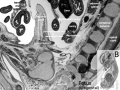
Fetal urogenital region lateral right
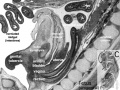
Fetal urogenital region medial
Fetal urogenital region midline
Fetal kidney (10 weeks)
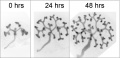
Mouse E12.5 kidney in vitro
![Mouse renal podocyte EM[1]](/embryology/images/thumb/d/d0/Mouse_renal_podocyte_EM01.jpg/90px-Mouse_renal_podocyte_EM01.jpg)
Mouse renal podocyte EM[1]


![Mouse renal podocyte EM[1]](/embryology/index.php?title=File:Mouse_renal_podocyte_EM01.jpg)
External Links
External Links Notice - The dynamic nature of the internet may mean that some of these listed links may no longer function. If the link no longer works search the web with the link text or name. Links to any external commercial sites are provided for information purposes only and should never be considered an endorsement. UNSW Embryology is provided as an educational resource with no clinical information or commercial affiliation.
- Blue Histology Urinary System
- UNSW Virtual Slides Urinary Tract Histology
- UIOWA Virtual Slidebox of Histology Urinary tract
Practical Overview
Normal Histology
- kidneys, ureters, urinary bladder, urethra
Anatomy
- retroperitoneal
- kidney - bean shaped
- rich blood supply
Kidney Function
- elimination of foreign substances
- regulation of the amount of water in the body
- control of the concentration of most compounds in the extracellular fluid
- filtration - glomeruli of the kidney
- selective resorption and excretion - tubular system of the kidney
Capsule
|
Cortex
|
Medulla
|
Blood Supply
- renal artery
- interlobar arteries (across medulla thru renal columns)
- arcuate arteries (cortico-medullary junction)
- interlobular arteries
- afferent glomerular arterioles
- glomerular capillary network
- efferent glomerular arterioles
Vasa Recta
- descending arterioles (arteriole rectae) + ascending venules (venulae rectae)
Glomerulus
- glomerulus - round (~0.2 mm in diameter) blind beginning of the nephron
- vascular pole - invaginated by a tuft of capillaries
- urinary pole - substances leave the capillaries enter the renal tubule
- Bowman's capsule - anatomical glomerulus is enclosed by two layers of epithelium.
- outer or parietal layer of Bowman's capsule form a simple squamous epithelium.
- inner layer, podocytes in the visceral layer, are extremely complex in shape.
- Mesangial cells in the glomerulus form the connective tissue that gives structural support to podocytes and vessels (Podocytes, mesangial cells, glomerular capillaries)
- Juxtaglomerular cells - smooth muscle cells afferent glomerular arteriole (epithelial-like cells)
- Macular Densa
- distal convoluted tubule near vascular pole (narrower and taller than rest of DCT)
Tubules
Proximal Convoluted Tubules (PCT)
- brush border
- star-shaped
- larger outside diameter
Distal Convoluted Tubules (DCT)
- clean lumen surface
- apical nuclei
Collecting Tubules (CT)
- larger lumen than DCT (about size of PCT)
- cuboidal cells and smaller than DCT
Renal Pyramids
- medullary straight tubules, ducts and vasa recta
- apical renal papilla - simple cuboidal/columnar epithelia
- calyx - lined by transitional epithelia
Note the urinary system transitional epithelium is also known as urothelium.
Ureters
- epithelium - transitional epithelia
- lamina propria - mainly of dense connective tissue, with many bundles of coarse collagenous fibres
- muscularis - consists of an inner longitudinal and outer circular layer of smooth muscle cells
- In lower parts of the ureter and the bladder an additional outer longitudinal layer of muscles is added to the first two.
Bladder
- epithelium - transitional epithelia
- apical plaques - thickened domain allows great changes in surface area.
- lamina propria - mainly of dense connective tissue, with many bundles of coarse collagenous fibres
- muscularis - consists of an inner longitudinal and outer circular layer of smooth muscle cells
- In the bladder (and lower parts of the ureter) an additional outer longitudinal layer of muscles is added to the first two.
Urethra
- penile urethra within corpus spongiosum
- pseudostratified columnar epithelia
- distal end - stratified squamous
- continuous with outer skin
References
Terms
| Renal Terms | ||
|---|---|---|
| ||
|
Glossary Links
- Glossary: A | B | C | D | E | F | G | H | I | J | K | L | M | N | O | P | Q | R | S | T | U | V | W | X | Y | Z | Numbers | Symbols | Term Link
Cite this page: Hill, M.A. (2024, May 11) Embryology Renal System Histology. Retrieved from https://embryology.med.unsw.edu.au/embryology/index.php/Renal_System_Histology
- © Dr Mark Hill 2024, UNSW Embryology ISBN: 978 0 7334 2609 4 - UNSW CRICOS Provider Code No. 00098G