|
|
| (55 intermediate revisions by the same user not shown) |
| Line 4: |
Line 4: |
| ! Online Editor | | ! Online Editor |
| |- | | |- |
| |[[File:Mark_Hill.jpg|50px|left]] This 1908 historic paper by Mall describes abnormal human development. Note the term "monsters" is historic terminology not currently used. | | | [[File:Mark_Hill.jpg|50px|left]] This 1908 historic paper by Mall describes abnormal human development. Note the term "monsters" is historic terminology not currently used. |
|
| |
|
| See also {{Mall Links}} | | See also the later papers by Mall: {{Ref-Mall1917}} |
|
| |
|
| | {{Ref-Mall1921}} |
| | <br> |
| | {{Mall Links}} |
| | <br><br> |
| '''Modern Notes:''' | | '''Modern Notes:''' |
| | <br> |
| {{Abnormality Links}} | | {{Abnormality Links}} |
| |} | | |} |
| | {{Mall1908a TOC}} |
| {{Historic Disclaimer}} | | {{Historic Disclaimer}} |
| =A Study of the Causes Underlying the Origin of Human Monsters= | | =A Study of the Causes Underlying the Origin of Human Monsters= |
| Volume Xix. February, 1908. Number I.
| |
|
| |
|
| Journal
| | (Third Contribution To The Study Of The Pathology Of Human Embryos.) |
|
| |
|
| Of
| | By [[Embryology_History_-_Franklin_Mall|Franklin P. Mall]] |
|
| |
|
| Morphology.
| | Professor of Anatomy. Johns Hopkins University. Baltimore. Mn. |
|
| |
|
| A Study Of The Causes Underlying The
| |
| Origin Of Human Monsters. (Third
| |
| Contribution To The Study
| |
| Of The Pathology Of
| |
| Human Embryos.)
| |
|
| |
|
| By Franklin P. Mall,
| | ''{{Online Editor}} - Please note the term "monster" is the historic term for gross developmental abnormalities and is both no longer used and also offensive. It has been left within this text as this is how such major abnormalities were medically referred at that time, and I apologise for any offence this term may cause.'' |
|
| |
|
| Pknrasson Or Anatomy. Johns Hopkins University. Baltimore. Mn.
| | ==Table of Contents== |
| | Introduction |
|
| |
|
| Table Of Contents.
| | ===PART I=== |
| | [[File:Mall1908a title page.jpg|thumb|Title page]] |
| | * [[Paper - A study of the causes underlying the origin of human monsters 1|Historical]] |
|
| |
|
| FAG!
| | Experimental |
| Introduction . . . . . . . . . . . . . . . . . . . . . . . . . . . . . . . . . . . . . . . . . . . . . . . . . . . . . . 3
| |
| PART I.
| |
| Historical . . . . . . . . . . . . . . . . . . . . . . . . . . . . . . . . . . . . . . . . . . . - . . . . . . . . . . . . . 9
| |
| Experimental : | |
| (a) Double Monster . . . . . . . . . . . . . . . . . . . . . . . . . . . . . . .1 . . . . . . . . .. 17
| |
| (b) Lithium embryos and nodul_ar forms in chicks and man. .. .. . 21
| |
| (c) Experiments with salts of potassium and changes especially
| |
| marked in the heart . . . . . . . . . . . . . . . . . . . . . . . . . . . . . . . . .. 25
| |
| (41) Sodium monsters--spina bifida and anencephaly . . . . . . . . . . .. 33
| |
| (2) The destruction of tissues in magnesium monsters, cyclopia and club-foot . . . . . . . . . . . . . . . . . . . . . . . . . . . . . . . . . .. 43
| |
| Pathological ova . . . . . . . . . . . . . . . . . . . . . . . . . . . . - . . . . . . . . . . . . . . . . . . . .. 48
| |
|
| |
|
| Twin pregnancies . . . . . . . . . . . . . . . . . . . . . . . . . . . . . . . . . . . . . . . . . . . . . . . .. 58
| | * [[Paper - A study of the causes underlying the origin of human monsters 2|(a) Double Monster]] |
| I
| |
| 2 MALL. [VoL. XIX. | |
|
| |
|
| PAGE
| | * [[Paper - A study of the causes underlying the origin of human monsters 3|(b) Lithium embryos and nodular forms in chicks and man]] |
| Unruptured tubal pregnancies . . . . . . . . . . . . . . . . . . . . . . . . . . . . . . . . . . . . . . .. 60
| |
| Ruptured tubal pregnancies . . . . . . . . . . . . . . . . . . . . . . . . . . . . . . . . . . . . . . .. 65
| |
|
| |
|
| Partial or complete destruction of the amnion, leaving only the
| | * [[Paper - A study of the causes underlying the origin of human monsters 4|(c) Experiments with salts of potassium and changes especially marked in the heart]] |
| umbilical vesicle . . . . . . . . . . . . . . . . . . . . . . . . . . . . .. . . . . . . . . . . . . . 69
| |
|
| |
|
| Ova in which the embryo and amnion have been destroyed, including
| | * [[Paper - A study of the causes underlying the origin of human monsters 5|Sodium monsters - spina bifida and anencephaly]] |
| certain moles . . . . . . . . . . . . . . . . . . . . . . . . . . . . . . . . . . . . . . . . . .. 82
| |
|
| |
|
| Pathological ova in which the embryo has been destroyed, leaving a
| | * [[Paper - A study of the causes underlying the origin of human monsters 6|The destruction of tissues in magnesium monsters, cyclopia and club-foot]] |
| portion of the umbilical cord and the amnion . . . . . . . . . . . . .. 90
| |
|
| |
|
| Pathological embryos of the second w-eek . . . . . . . . . . . . . . . . . . . . . . . . .. 97
| | * [[Paper - A study of the causes underlying the origin of human monsters 7|Pathological ova]] |
|
| |
|
| Embryos of the third week . . . . . . . . . . . . . . . . . . . . . . . . . . . . , . . . . . . . . .. 102
| | * [[Paper - A study of the causes underlying the origin of human monsters 8|Twin pregnancies]] |
|
| |
|
| Embryos of the fourth week . . . . . . . . . . . . . . . . . . . . . . . . . . . . . . . . . . . . .. 107
| | * [[Paper - A study of the causes underlying the origin of human monsters 9|Unruptured tubal pregnancies]] |
|
| |
|
| Embryos of the fifth week . . . . . . . . . . . . . . . . . . . . . . . . . . . . . . . . . . . . . . .. I13
| | * [[Paper - A study of the causes underlying the origin of human monsters 10|Ruptured tubal pregnancies]] |
|
| |
|
| Embryos of the sixth week . . . . . . . . . . . . . . . . . . . . . . . . . . . . . . . . . . . . . . .. 118
| | * [[Paper - A study of the causes underlying the origin of human monsters 11|Partial or complete destruction of the amnion, leaving only the umbilical vesicle]] |
|
| |
|
| Embryos of the seventh week . . . . . . . . . . . . . . . . . . . . . . . . . . . . . . . . . . . .. I27
| | * [[Paper - A study of the causes underlying the origin of human monsters 12|Ova in which the embryo and amnion have been destroyed, including certain moles]] |
|
| |
|
| Embryos of the eighth week and older . . . . . . . . . . . . . . . . . . . . . . . . . . . .. I33
| | * [[Paper - A study of the causes underlying the origin of human monsters 13|Pathological ova in which the embryo has been destroyed, leaving a portion of the umbilical cord and the amnion]] |
| PART 11.
| |
| Description of the specimens and figures . . . . . . . . . . . . . . . . . . . . . . . . . .. 139
| |
|
| |
|
| Description of the plates . . . . . . . . . . . . . . . . . . . . . . . . . . . . . . . . . . . . . . . . .. 305
| | * [[Paper - A study of the causes underlying the origin of human monsters 14|Pathological embryos of the second week]] |
| INTRODUCTION.
| |
|
| |
|
| The present communication is the outcome of a study of
| | * [[Paper - A study of the causes underlying the origin of human monsters 15|Embryos of the third week]] |
| I63 pathological human embryos which I have collected
| |
| during the past fifteen years. The first contribution‘ which
| |
| I made to this subject included a report of 53, and the second”
| |
| of 20 of these embryos. These two studies are rather
| |
| anatomical in nature and do not consider the causes which
| |
| produce pathological embryos, nor their relation to ordinary
| |
| human monsters. A more careful study of my specimens, | |
| which have more than doubled in number during the past five
| |
| years, establishes beyond doubt (I) the identity of pathological embryos and small monsters, that is, many of them
| |
| would have developed into real monsters if they had not been
| |
| aborted, and (2) that all of them are developed from normal
| |
| ova due to external influences,—in man to a condition which
| |
| I shall term faulty implantation.
| |
|
| |
|
| From the earliest ages in the world’s history the study of
| | * [[Paper - A study of the causes underlying the origin of human monsters 16|Embryos of the fourth week]] |
| monsters, and the causes which produce them, has been one
| |
| of the capital problems in anatomy, medicine and natural | |
| history. Supernatural causes for the production of monsters,
| |
| like the influence of the gods, celestial or diabolical, or lunar
| |
| influences, as expressed in their name, monster or moon—calf,
| |
| held for a long time universal sway and are still believed in
| |
| by many ignorant people. However, the Greek naturalists
| |
| and physicians were inclined to ascribe them to natural causes,
| |
| which belief was gradually displaced in the Dark Ages by the
| |
| one that monsters were hybrids of bestial origin, a theory
| |
| which was finally overthrown in the eighteenth century.
| |
| Equally general has been the theory that maternal impressions
| |
| affect the offspring and convert them into monsters. This
| |
|
| |
|
| ‘Mall, Welch Festschrift, Johns Hopkins Hospital Reports, IX, 1900.
| | * [[Paper - A study of the causes underlying the origin of human monsters 17|Embryos of the fifth week]] |
| ‘Mall, Vaughan Festschrift, Contributions to Medical Research, Ann
| |
|
| |
|
| Arbor, 1903.
| | * [[Paper - A study of the causes underlying the origin of human monsters 18|Embryos of the sixth week]] |
| 1 3
| |
| 4 MALL. [VoL. XIX.
| |
|
| |
|
| belief is of great antiquity, and is at present of world-wide
| | * [[Paper - A study of the causes underlying the origin of human monsters 19|Embryos of the seventh week]] |
| distribution. It also was attacked in the eighteenth century,
| |
| first by Blondel from a philosophical and then by Haller from
| |
| a scientific standpoint.
| |
|
| |
|
| All the theories can be resolved into the simple question:
| | * [[Paper - A study of the causes underlying the origin of human monsters 20|Embryos of the eighth week and older]] |
| “Are the conditions which produce a monster germinal and
| |
| therefore hereditary, or are they produced from normal
| |
| germs by external influences?” The discussion on both sides
| |
| of the question has been a long one, conducted during many
| |
| years by the ablest masters, among whom are always included the leading anatomists of the time. However, the
| |
| theory of external influences gradually gained ground during
| |
| the nineteenth century, as the science of embryology was | |
| cultivated more and more. But here again we have two
| |
| schools, the one believing that monsters are formed from
| |
| normal germs due to maternal impressions, the other that
| |
| they are due to mechanical influences. It may be noted here
| |
| that the obstetricians and gynecologists of America as a class
| |
| advocate strongly the theory of maternal impressions, due
| |
| largely, no doubt, to their insufficient scientific education.
| |
| On the other hand, we may pride ourselves over the masterful
| |
| strokes of American teratologists against this theory; the
| |
| experimental teratologists have produced double monsters,
| |
| spina bifida and cyclopia, under the very noses of these practitioners, but they continue their futile speculations over mere
| |
| coincidences.
| |
|
| |
|
| With Meckel, who laid the embryological foundation for a
| | ===PART II=== |
| scientific teratology, and the Saint-Hilaires, who made the
| |
| first teratological experiments, we have the beginning of the
| |
| development of the mechanical theory. It appears in a
| |
| variety of forms, as, for example, that monsters are due to
| |
| tight lacing which causes pressure upon the embryo, or to the
| |
| contracting uterus which naturally might have the same effect.
| |
| However, this theory was gradually transformed by teratologists so that now it rests upon the idea that amniotic bands
| |
| constrict or compress the embryo, thus bringing about its
| |
| deformity. Occasionally it has been found that monsters are
| |
| No. 1.] ORIGIN OF HUMAN MONSTERS. 5
| |
|
| |
|
| attached to the chorion through newly-formed bands of tissue,
| | * [[Paper - A study of the causes underlying the origin of human monsters 21|Description of the specimens and figures]] |
| and such bands, whether present or not, are held responsible | | * [[Paper - A study of the causes underlying the origin of human monsters 22|Description of the plates]] |
| for all terata. This coincidence, as I term it, cannot be of
| |
| frequent occurrence, for usually there is present an hydramnios. Nor can even the “coincidence” occur in anamniotic
| |
| animals. Furthermore, no amniotic bands were ever found in
| |
| any of the 169 specimens which I have studied.
| |
|
| |
|
| In place of these theories it is my purpose to demonstrate
| | ==Introduction== |
| that all monsters are produced by external influences upon
| | [[File:Franklin Mall 01.jpg|thumb|link=Embryology_History_-_Franklin_Mall|Franklin Mall (1911)]] |
| normal ova which affect the nutrition of the embryos due to
| | The present communication is the outcome of a study of 163 pathological human embryos which I have collected during the past fifteen years. The first contribution<ref>Mall, Welch Festschrift, Johns Hopkins Hospital Reports, IX, 1900.</ref> which I made to this subject included a report of 53, and the second<ref>Mall, Vaughan Festschrift, Contributions to Medical Research, Ann Arbor, 1903.</ref> of 20 of these embryos. These two studies are rather anatomical in nature and do not consider the causes which produce pathological embryos, nor their relation to ordinary human monsters. A more careful study of my specimens, which have more than doubled in number during the past five years, establishes beyond doubt (1) the identity of pathological embryos and small monsters, that is, many of them would have developed into real monsters if they had not been aborted, and (2) that all of them are developed from normal ova due to external influences,—in man to a condition which I shall term faulty implantation. |
| faulty implantation of the ovum. That the power to become
| |
| a monster is present in every ovum is fully demonstrated by
| |
| experiments upon a variety of vertebrates as well as by all
| |
| of my pathological ova, especially those obtained from tubal | |
| pregnancies.
| |
|
| |
|
| The changes found repeatedly in the chorion are no doubt
| |
| primary; they are usually of an hemorrhagic nature, often
| |
| indicating inflammatory changes in the uterus. I shall only
| |
| hint at the cause for the changes in the uterus, which may
| |
| interfere with the formation of the decidua, and recommend
| |
| this field as a very fertile one for gynecologists and obstetricians to investigate. At any rate, the change interferes much
| |
| with the attachment of the ovum, and this condition and what
| |
| results from it I have termed faulty implantation.
| |
|
| |
|
| It has been impossible, in fact it is not desirable, to discuss
| | From the earliest ages in the world’s history the study of monsters, and the causes which produce them, has been one of the capital problems in anatomy, medicine and natural history. Supernatural causes for the production of monsters, like the influence of the gods, celestial or diabolical, or lunar influences, as expressed in their name, monster or moon—calf, held for a long time universal sway and are still believed in by many ignorant people. However, the Greek naturalists and physicians were inclined to ascribe them to natural causes, which belief was gradually displaced in the Dark Ages by the one that monsters were hybrids of bestial origin, a theory which was finally overthrown in the eighteenth century. Equally general has been the theory that maternal impressions affect the offspring and convert them into monsters. This belief is of great antiquity, and is at present of world-wide distribution. It also was attacked in the eighteenth century, first by Blondel from a philosophical and then by Haller from a scientific standpoint. |
| extensively the immense amount of excellent literature upon
| |
| teratology. The whole makes one of the best chapters in
| |
| medical literature to which the greatest minds of medicine
| |
| have contributed their best efforts. One cannot go through
| |
| these writings, many of which are comprehensive, without
| |
| laying them aside with profound respect. In this study I
| |
| have used many of them freely, but make, however, very few
| |
| references.“ I have also avoided technical terms as much as
| |
|
| |
|
| ‘J’. F. Meckel, Handbuch d. pathol. Anatomic, Leipzig, I812.
| |
| Ffirster, Die Missbildungen des Menschen, Jena, 1865.
| |
|
| |
|
| Bischoff, Wagner’s Handwfirterbuch, I842.
| | All the theories can be resolved into the simple question: “Are the conditions which produce a monster germinal and therefore hereditary, or are they produced from normal germs by external influences?” The discussion on both sides of the question has been a long one, conducted during many years by the ablest masters, among whom are always included the leading anatomists of the time. However, the theory of external influences gradually gained ground during the nineteenth century, as the science of embryology was cultivated more and more. But here again we have two schools, the one believing that monsters are formed from normal germs due to maternal impressions, the other that they are due to mechanical influences. It may be noted here that the obstetricians and gynecologists of America as a class advocate strongly the theory of maternal impressions, due largely, no doubt, to their insufficient scientific education. On the other hand, we may pride ourselves over the masterful strokes of American teratologists against this theory; the experimental teratologists have produced double monsters, spina bifida and cyclopia, under the very noses of these practitioners, but they continue their futile speculations over mere coincidences. |
|
| |
|
| Ahlfeld, Die Missbildungen des Menschen, Leipzig, 1880-1882. Part
| |
| 6 MALL. [VoI.. XIX.
| |
|
| |
|
| possible, and in general have adopted Ballantyne’s classification, which, in turn, is based‘ upon Tarufii’s.
| | With Meckel, who laid the embryological foundation for a scientific teratology, and the Saint-Hilaires, who made the first teratological experiments, we have the beginning of the development of the mechanical theory. It appears in a variety of forms, as, for example, that monsters are due to tight lacing which causes pressure upon the embryo, or to the contracting uterus which naturally might have the same effect. However, this theory was gradually transformed by teratologists so that now it rests upon the idea that amniotic bands constrict or compress the embryo, thus bringing about its deformity. Occasionally it has been found that monsters are attached to the chorion through newly-formed bands of tissue, and such bands, whether present or not, are held responsible for all terata. This coincidence, as I term it, cannot be of frequent occurrence, for usually there is present an hydramnios. Nor can even the “coincidence” occur in anamniotic animals. Furthermore, no amniotic bands were ever found in any of the 169 specimens which I have studied. |
|
| |
|
| I wish it were possible to thank adequately the many
| |
| physicians who have contributed the specimens and who have
| |
| responded so generously to my many inquiries. All data
| |
| obtained from them are given in quotations under the description of the specimens, which are also properly credited to the
| |
| donors. Some names will be seen repeatedly, as Miller,
| |
| Boldt, Lamb, Brodel, Ballard, West and Minot. In addition,
| |
| I wish also to thank my colleagues at the Johns Hopkins,
| |
| who have aided me in every possible way to bring together
| |
| the gynecological, obstetrical, pathological and experimental
| |
| embryological evidence.
| |
|
| |
|
| In this paper all of the embryos mentioned in the two previous ones are discussed, and brought together in Part II. | | In place of these theories it is my purpose to demonstrate that all monsters are produced by external influences upon normal ova which affect the nutrition of the embryos due to faulty implantation of the ovum. That the power to become a monster is present in every ovum is fully demonstrated by experiments upon a variety of vertebrates as well as by all of my pathological ova, especially those obtained from tubal pregnancies. |
| The essence of the first contribution is given, and all of the
| |
| second contribution is incorporated in this publication, so in
| |
| a measure it may be viewed as a study of the whole collection. However, the va_rious steps by which I came to arrange | |
| the specimens, as I have, can be understood only by consulting
| |
| the two previous publications.
| |
|
| |
|
| III, which was to be the important part, never appeared. Fortunately,
| |
| however, Ah1fe1d’s library, which is very complete, was presented to the
| |
| Johns Hopkins University. This I have consulted freely and in a way
| |
| it makes up for the missing Part III.
| |
|
| |
|
| Marchand, Missbildungen, Eu1enburg’s Real-Encyclopedia, 3d edition,
| | The changes found repeatedly in the chorion are no doubt primary; they are usually of an hemorrhagic nature, often indicating inflammatory changes in the uterus. I shall only hint at the cause for the changes in the uterus, which may interfere with the formation of the decidua, and recommend this field as a very fertile one for gynecologists and obstetricians to investigate. At any rate, the change interferes much with the attachment of the ovum, and this condition and what results from it I have termed faulty implantation. |
| I897, Vol. XV. _
| |
|
| |
|
| Taruffi, 'Storia delli Teratologia, 8 volumes, Bologna, I881-I895.
| |
|
| |
|
| Hirst and Piersol, Human Monsters, Philadelphia. ,
| | It has been impossible, in fact it is not desirable, to discuss extensively the immense amount of excellent literature upon teratology. The whole makes one of the best chapters in medical literature to which the greatest minds of medicine have contributed their best efforts. One cannot go through these writings, many of which are comprehensive, without laying them aside with profound respect. In this study I have used many of them freely, but make, however, very few references.<ref>J. F. Meckel, Handbuch d. pathol. Anatomic, Leipzig, I812. Ffirster, Die Missbildungen des Menschen, Jena, 1865.; Bischoff, Wagner’s Handwfirterbuch, 1842.; Ahlfeld, Die Missbildungen des Menschen, Leipzig, 1880-1882.</ref> I have also avoided technical terms as much as possible, and in general have adopted Ballantyne’s classification, which, in turn, is based upon Tarufii’s. |
|
| |
|
| Piersol, Teratology, Ref. Hndbk. Med. Sci., new edition, Vol. VII,
| |
| I .
| |
| 9o4Ballantyne, Antenatal Pathology, 2 volumes, Edinburgh, 1904.
| |
| E. Schwalbe, Die Morphologie d. Missbildungen des Menschen und
| |
| der Thiere, Jena, Pt.- I, 1906, Pt. II, I907.
| |
| PART 1.
| |
| HISTORICAL AND GENERAL.
| |
| HISTORICAL.‘
| |
|
| |
|
| The changes found in the pathological embryos to be
| | I wish it were possible to thank adequately the many physicians who have contributed the specimens and who have responded so generously to my many inquiries. All data obtained from them are given in quotations under the description of the specimens, which are also properly credited to the donors. Some names will be seen repeatedly, as Miller, Boldt, Lamb, Brodel, Ballard, West and Minot. In addition, I wish also to thank my colleagues at the Johns Hopkins, who have aided me in every possible way to bring together the gynecological, obstetrical, pathological and experimental embryological evidence. |
| described in this memoir are so radical in nearly all specimens
| |
| that it is almost useless to speculate regarding the fate of the
| |
| embryos had they continued to grow to the end of a normal
| |
| pregnancy. Could the circulation be maintained these specimens
| |
| might have developed into amorphous monsters, a condition
| |
| which is probable only when there is a normal twin foetus to | |
| supply the nutrition. In only one of my specimens (No. 87)
| |
| are the possibilities for such a termination present. Here on
| |
| one side of the chorion there is a normal embryo of the third
| |
| week and on the other side a highly developed umbilical
| |
| vesicle with but a rudimentary amnion, but no real body of
| |
| an embryo. In all of the other twin specimens the changes
| |
| in both embryos are radical and identical, so that we could not | |
| hope to have had the one embryo dependent upon the other
| |
| for its circulation and nutrition.
| |
|
| |
|
| In general then the changes in the embryo and its membrane, due to the inflammatory action in the uterus, are so
| |
| great that if the ovum is not aborted at an early date (as it
| |
| usually is) it is converted into a solid mole which in the
| |
| course of time is likewise expelled. A few specimens, how~
| |
| ever, are but slightly changed, and these would probably have
| |
| grown into some sort of merosomatous monsters had they
| |
| been retained in the uterus. From my experience I am convinced that in the study of specimens like these we have the
| |
| key by which we can unlock lhany of the mysteries of
| |
| teratology.
| |
|
| |
|
| In my first two communications I carefully avoided | | In this paper all of the embryos mentioned in the two previous ones are discussed, and brought together in Part II. The essence of the first contribution is given, and all of the second contribution is incorporated in this publication, so in a measure it may be viewed as a study of the whole collection. However, the various steps by which I came to arrange the specimens, as I have, can be understood only by consulting the two previous publications. |
| all speculations on this subject, for I was well aware of the | |
| sad state this subject is in, and mere speculations would not
| |
|
| |
|
| ‘The data here recorded are taken largely from Ballantyne’s Antenatal Pathology.
| |
|
| |
|
| 9
| | III, which was to be the important part, never appeared. Fortunately, however, Ah1fe1d’s library, which is very complete, was presented to the Johns Hopkins University. This I have consulted freely and in a way it makes up for the missing Part III. |
| I0 MALL. [VoL. XIX.
| |
|
| |
|
| help teratology out of its difficult position. However, what
| | Marchand, Missbildungen, Eu1enburg’s Real-Encyclopedia, 3d edition, I897, Vol. XV. |
| little progress has been made in the study of terata has been
| |
| made by the embryologist and we naturally still have confidence in him. The course to be followed, therefore, is the
| |
| study of early abortions, and this I have done diligently.
| |
| I can, therefore, subscribe fully to what Ballantyne has
| |
| recently said in his able and scholarly treatise on antenatal
| |
| pathology. He says, page 77: “Now, in reference to the
| |
| inquiry into the problems of teratology or embryonic pathology, let me emphasize the importance of a thorough
| |
| scrutiny of the foetal membranes and of the routine examination, microscopic as well as macroscopic, of all abortion
| |
| sacs and their contents thrown off in- the early months of
| |
| pregnancy. What is most wanted at present are careful
| |
| descriptions of monstrous embryos from abortion sacs’, observations upon teratological conditions while the organism is
| |
| still in the embryonic period of antenatal life. These are
| |
| essential for the further progress of a knowledge of human
| |
| teratogenesis, and they are at the present time the desiderata
| |
| of embryonic pathology. Microscopic human monstrosities
| |
| are, as a matter of fact, almost unknown.”
| |
|
| |
|
| The last sentence is hardly justifiable, for a pretty large
| | Taruffi, 'Storia delli Teratologia, 8 volumes, Bologna, I881-I895. |
| number of young pathological embryos have been described
| |
| by His, Giacomini and myself, but these do not resemble
| |
| monsters at full term any more than an embryo of the fourth
| |
| week resembles a new-born child. Whether the early pathological embryos are young monsters, or young monsters of
| |
| so extreme a degree that they will not continue to grow, is
| |
| now the most important question of the capital problem in
| |
| teratology. I think that the specimens that are reported in
| |
| this publication contribute to the answer of this question, but
| |
| many more observations are required before the answer will
| |
| be accepted by all teratologists.
| |
|
| |
|
| The history of teratology co-exists with that of medicine
| | Hirst and Piersol, Human Monsters, Philadelphia. , |
| and includes mythology, the vilest superstitions and scientific | |
| embryology. The medical profession have abandoned the
| |
| idea of supernatural causes in the production of monsters
| |
| No. 1.] ORIGIN OF HUMAN MONSTERS. II
| |
| | |
| and have gladly exchanged the hybridity theory (cohabitation with lower animals) for the more innocent one of
| |
| maternal impressions. The last notion is of great antiquity,
| |
| is of world-wide distribution and is intimately related to
| |
| witchcraft. It is gratifying to note that these superstitions,
| |
| based upon coincidences, have been raised from medicine by
| |
| the study of scientific anatomy, and the more recent work by
| |
| J. F. Meckel in this direction can be ranked with that of
| |
| Morgagni and Virchow. Morgagni gave the first blow to
| |
| humoral pathology by giving medicine an anatomical basis,
| |
| Meckel cast out devils, witches and mother’s marks by placing
| |
| teratology on an embryological basis, and Virchow won the
| |
| third great victory for anatomy, probably the greatest contribution ever made to medicine, by giving it an histological
| |
| basis. It would be inappropriate to enter any further into a
| |
| discussion of teratogenesis in this publication, for in general
| |
| the superstitious notions are abandoned by scientific physicians, although they may still be entertained by a few practitioners of some eminence. It is humiliating to state that
| |
| these practitioners seem to reside exclusively in America,
| |
| but we have every reason to hope that when scientific medical
| |
| education becomes general with us they will also disappear.
| |
| | |
| Most of the great men who have contributed to the progress of medicine, from Hippocrates and Aristotle to the
| |
| modern scientists, tried to ascribe the production of monsters to natural and not to supernatural processes. From the
| |
| first the explanations were as satisfactory as they are to-day,
| |
| for even now we barely do better than Aristotle did. However, the spread of the scientific spirit beginning with the
| |
| study and practice of anatomy by all medical students has
| |
| driven medical superstitions pretty well out of the medical
| |
| profession. In this respect we differ from-the ancients. The
| |
| first scientific explanations were of a crude mechanical nature,
| |
| like those due to excessive lacing, malformations of the uterus
| |
| or a twin foetus, which might injure the embryo. This notion
| |
| was superseded in part by the theory of Morgagni, who
| |
| maintained that monsters were due to foetal disease. This
| |
| I2 MALL. [Von XIX.
| |
| | |
| again received its death-blow from J. F. Meckel, who pointed
| |
| out the well known fact that many structural anomalies are
| |
| hereditary. This observation naturally divided terata into
| |
| two groups: those which are hereditary and germinal, and
| |
| those which are not hereditary but due to mechanical injury
| |
| or disease. I think this line of division should be drawn
| |
| much sharper than it is, but until our data can be arranged
| |
| better than is now possible we are still quite uncertain regarding a large number of terata. It seems to me that many
| |
| merosomatous terata (all kinds of anatomical anomalies and
| |
| variations of the extremities, like polydactyly and possibly
| |
| some cases of arrested development like ectrodactyly and
| |
| hare—lip) are germinal and cannot be produced experimentally. Other monsters in which more or less of the
| |
| foetus is destroyed, as in iniencephaly, spina bifida, aneucephaly, cyclopia, club-foot and many varieties of arrested development, are not germinal but are produced in some mechanical way which usually interferes with the nutrition of
| |
| the embryo. In my notes I have been in the habit of calling
| |
| those belonging to the first group as being abnormal and
| |
| those to the second group as pathological. The one is
| |
| germinal with a hereditary tendency, and the other is acquired
| |
| and therefore not hereditary; polydactyly is inherited, cyclopia is not, although there seems to be a tendency for it to
| |
| occur more than once in abortions from the same woman.
| |
| However, if this is true, it may be due to the same cause in the
| |
| uterus of the mother affecting the nutrition of successive ova,
| |
| thus producing similar deformities in the embryos. Usually
| |
| a woman who gives birth to several monsters has the varieties mixed up pretty well, the first may have hydrocephalus,
| |
| the next hare-lip and the third cyclopia. Reducing it to a
| |
| matter of chance, a woman who has given birth to one
| |
| monster is more likely to give birth to a second one, which,
| |
| however, is rarely like the first. In experimental teratology
| |
| in birds and amphibia the result is the same. Here monsters
| |
| may be produced experimentally with a variety of agents,
| |
| even by treating the semen of toads with X—rays, but the
| |
| No. 1.] ORIGIN OF HUMAN MONSTERS. I3
| |
| | |
| variety of monster can never be predicted, and if there are
| |
| a number of them they are usually of -mixed types.”
| |
| | |
| What I have to say in this publication of monsters applies
| |
| only merosomatous terata which are not of an hereditary
| |
| nature and are no doubt produced by agents which interfere
| |
| with the nutrition of the embryo. Having taken only those
| |
| monsters from which the germinal factor is excluded, it
| |
| makes it necessary once more to consider some minor mechanical agents as their cause, which may be termed a modified
| |
| mechanical theory.
| |
| | |
| The advocates of the mechanical theory gradually lost
| |
| ground, for they had to combat the germinal theory on the one
| |
| hand, and on the other they were compelled to state that mechanical influences, generally those due to lacing, caused the
| |
| foetus to become monstrous by the pressure that was exerted
| |
| upon it. The theory was then modified to include primarily
| |
| intra-abdominal influences like tumors, malformations of the
| |
| pelvis and uterus, as well as those within the ovum itself.
| |
| Gradually we see less and less weight placed upon any of these
| |
| specific causes, and finally the modern advocates of the theory
| |
| believe that amniotic bands and adhesions are the main influences in the production of monstrosities. It is needless to
| |
| state that each advocate had his own combination of circumstances, and when all of them are taken together, with modifications and exceptions, it is practically impossible to make
| |
| general statements. Suffice to say that the objections to each
| |
| form of the theory appear to be sufficient to explode the
| |
| whole theory, and to the bulk of physicians maternal influences seem to be as rational a cause in the production of
| |
| monsters as mechanical influences, for the data of experience
| |
| are about as good in the former as in the latter.
| |
| | |
| There are some rare cases of spontaneous amputation of
| |
| the extremities which are said to be due to pressure of the
| |
| umbilical cord. However, these cases can be separated into
| |
| two marked groups, one in which there is an actual amputa
| |
| 'Bardeen, Iour. of Experimental Zool., 1907.
| |
| I4 MALL. [VoL. XIX.
| |
| | |
| tion and the other in which there is an atrophic or rudimentary hand or foot attached. In the latter instance it
| |
| seems to me that it is very irrational to hold the umbilical cord
| |
| responsible for the amputation. Furthermore, the cause is
| |
| possibly germinal, as may be the case in sympodia, syndactyly
| |
| and ectrodactyly. The rare cases in which there is actual
| |
| amputation of the extremity are more likely to have been
| |
| produced by mechanical injuries during labor than by having
| |
| the amputated limb caught in a loop of the umbilical cord.
| |
| In fact, we must admit that we are unable to explain by any
| |
| satisfactory hypothesis either congenital amputations or dislocations.
| |
| | |
| It has been noticed occasionally in merosomatous monsters
| |
| that the diseased or malformed part is tied by means of
| |
| bands of tissue either to the amnion or to adjacent parts "of
| |
| the body of the foetus. These observations, relatively few in
| |
| number, have led to the theory that the bands caused the
| |
| deformity. It seems to me that, in view of the idea that
| |
| many monsters are due simply to an arrest of development
| |
| of some part of the embryo, that hydramnios is usually present, and that all kinds of monstrosities may be produced in
| |
| lower animals (including amphibia which have no amnion),
| |
| it is highly probable that amniotic bands and the like are
| |
| secondary in their formation and have nothing whatever to
| |
| do with the production of monsters. The more the embryo»
| |
| logical theory is tested by experimental methods the more
| |
| all simple mechanical explanations suffer, and it seems to me
| |
| that all of them will have to be abandoned.
| |
| | |
| It is not especially remarkable to find that when the head or
| |
| face is malformed the diseased part occasionally forms a secondary attachment with the amnion; or that, as in exomphalos,
| |
| where the umbilical cord is “dilated,” the extruded viscera
| |
| come in direct contact with the placenta, as they should, and
| |
| the blood-vessels are scattered and run along the amnion to the
| |
| placenta, as should also be the case when the subject is
| |
| viewed from the standpoint of embryology. Furthermore, deformities of the extremities are of frequent occurrence, but
| |
| No. 1.] ORIGIN OF HUMAN MONSTERS. I5
| |
| | |
| amniotic bands are rarely found, and when they are present
| |
| they are often attached to the body of the embryo and not
| |
| to the deformed extremity. It seems to me, therefore, that as
| |
| facts accumulate it becomes clearer and clearer that the occasional amniotic adhesions found are due to the presence of
| |
| the monster and are not causal in nature.-3
| |
| | |
| Possibly I have devoted too much space to the discussion
| |
| of mechanical theories in teratogenesis. What has been said
| |
| is no doubt acceptable to all embryologists, and my apology
| |
| is due to the fact that the influence of maternal impressions
| |
| upon the offspring is still believed in by so large a number of
| |
| American medical writers of note and that mechanical notions
| |
| regarding embryology are entertained by physicians in general.
| |
| | |
| The great embryologists from Harvey onward explained
| |
| the conditions found in monsters as due to an arrest of
| |
| development, for they saw in these distorted individuals conditions which are normally found in the embryo. The
| |
| embryological theory was first well formulated by J. F.
| |
| Meckel, who explained the beast—1ike appearance of some
| |
| monsters by the fact that in his development man passes successively through stages found in lower animals. To those
| |
| who have accepted the doctrine of evolution this is all clear,
| |
| but it remains to be shown what are the factors in development, and the effect of changes in the embryo upon the
| |
| growth of the foetus.
| |
| | |
| As has been pointed out above, we must divide monsters into
| |
| two groups, those in which the proper conditions to produce
| |
| them are already in the germ (are therefore inherited), and
| |
| those due to certain external influences which act upon
| |
| the egg after it is fertilized. It is obvious that only the
| |
| second group can be considered in any experiments made
| |
| upon the embryo. So, if the pathological ova I have studied
| |
| | |
| 'Ballantyne says: “The reader may feel (and he is justified in so
| |
| feeling) that, after all, experimental teratogeny has not done much for
| |
| the understanding of the mode of origin of monstrosities, if it has
| |
| weakened a belief in the influence of the amnion.” I may add that this
| |
| argument can be applied to maternal impressions as a cause equally as well.
| |
| I6 MALL. [VoL. XIX.
| |
| | |
| are all due to a diseased chorion, which in turn is dependent
| |
| upon endometritis, then we should find embryos tending towards club-foot, anencephaly, iniencephaly, spina bifida and
| |
| cyclopia, which in fact proves to be the case. However, a
| |
| large group of new monsters, known only to embryologists,
| |
| make their appearance and from the very nature of the abnormality found but few of them could develop beyond the first
| |
| months of pregnancy. In their study comparisons have been
| |
| constantly made with normal embryos of the same size, and
| |
| in this way, to a certain degree, it is possible to picture the
| |
| order of events. It is found that in these specimens some
| |
| tissues are more susceptible than others, and when the nutrition of the ovum is impaired it is these that are affected first.
| |
| In very early stages the amnion and embryo are equally
| |
| susceptible and the umbilical vesicle and chorion are the
| |
| most resistant. Later it is the embryo alone, and still later
| |
| the head, central nervous system and extremities. It follows then that the parts most susceptible are those most
| |
| frequently found changed, or wanting, in merosomatous nongerminal monsters. In general the varieties found in my collection of young embryos correspond with those obtained
| |
| experimentally by others in birds and appear much like the
| |
| most common human monsters.
| |
| | |
| The Saint-‘Hilaires, who contributed so very much to our
| |
| knowledge of teratology, were the first to study the subject
| |
| experimentally. By a variety of experiments made upon the
| |
| shell of the egg (e. g., pricking and varnishing) the older
| |
| Saint-Hilaire produced a large number of anomalies in which
| |
| there were defective heads and spina bifida. His experiments
| |
| were made upon eggs after development was well under way,
| |
| and his results were pronounced enough to allow of comparison with human monsters. The younger Saint-Hilaire
| |
| extended the experiments to include the earliest days of
| |
| incubation, and found that the embryos which developed were
| |
| dwarfed or were wanting altogether. In no instance were
| |
| polysomatous monsters produced. At any rate, the experiments of the Saint—Hilaires show that a change in the external
| |
| No. L] ORIGIN OF HUMAN MONSTERS. 17
| |
| | |
| physical conditions may influence and modify normal development and thereby produce a variety of merosomatous terata.
| |
| | |
| During the following seventy-five years a great amount of
| |
| experimental work was done upon chicks by numerous investigators, which showed that the varieties of monsters produced were quite constant, no matter what agent is used, but
| |
| no single variety could be produced with certainty. It was
| |
| found impossible to experiment with precision, for a certain
| |
| | |
| per cent of eggs would produce one or more varieties of
| |
| deformed embryos.
| |
| | |
| EXPERIMENTAL.
| |
| | |
| RECENT WORK UPON THE PRODUCTION on POLYSOMATOUS
| |
| MONSTERS.
| |
| | |
| The various theories regarding teratogenesis which had
| |
| troubled mankind for so many centuries were finally exploded
| |
| by naturalists, whose speculations gradually led them to experiment upon this subject. Anatomists and zoologists had
| |
| deduced that the primary change lay in the egg about the
| |
| time of fertilization, and we read in J. Muller,’ Valentin’ and
| |
| Leuckart3 that a double monster is due to division of the
| |
| embryo—forming substance in the earliest stage of development. The experiments subsequently made by Gerlachf
| |
| Panum‘ and Dareste° upon chicks were negative in this
| |
| respect, but the tradition has come down to us that polysomatous monsters are produced by a process of splitting of the
| |
| primitive streak. The more recent anatomists—Fol, Rauber,
| |
| | |
| ‘Muller, Meckel’s Archiv, I828.
| |
| | |
| ‘Valentin, Handwiirterbuch d. Physiologic, I, 1842.
| |
| | |
| ‘Leuckart, De Monstris, Giittingen, I845.
| |
| | |
| ‘Gerlach, Doppeln-iissbildungen, I882.
| |
| | |
| ‘Panum, Entstehung der Missbildungen, 1860.
| |
| | |
| ‘Dareste, Recherches sur la production dc monstrosités, Paris, 1891..
| |
| 18 MALL. [VoL. XIX.
| |
| | |
| Born and O. Hertwig—who observed the developing egg and
| |
| experimented upon it, were at first inclined to the theory that
| |
| the first cause in the production of monsters is due to polyspermy, but this has not been substantiated.
| |
| | |
| The first reliable and valuable observations upon the production of double monsters were made by Vejdovskyf’ who
| |
| noticed that the eggs of Lumbricus produce more monsters in
| |
| warm than in cool weather, and he expressed the suspicion
| |
| that they were produced by the change in temperature.
| |
| Driesch“ seized upon this idea, experimented upon sea-urchins’
| |
| eggs, and found by subjecting them to high temperatures that
| |
| the cells, in the two—cell stage, separated, each growing into
| |
| an individual, but, however, remaining connected with each
| |
| other. Driesch had already shown that when the blastomeres
| |
| of these eggs are fully separated by shaking, each grows into
| |
| a whole embryo, and it was now clear to him that double
| |
| monsters are produced by separating the blastomeres slightly,
| |
| but still keeping them close enough together so that the independent embryos grow into each other’s bodies to form a
| |
| double individual.
| |
| | |
| By a very different method double monsters were also produced by Loeb.° He subjected sea—urchin eggs to an equal
| |
| mixture of sea-water and distilled water shortly after they
| |
| had been fertilized. The rapid absorption of water caused
| |
| many of the cell membranes to burst, and part of their protoplasm escaped, which, however, remained connected with that
| |
| inside of the membrane. All this took place before the nucleus
| |
| had divided. Upon returning the eggs to normal sea-water
| |
| cleavage began, and one of the first two nuclei wandered into
| |
| the extruded protoplasm. Each nucleus with its protoplasm
| |
| then became an embryo, and in case the embryo within the egg
| |
| was not separated from the extra-ovate embryo by its active
| |
| movements in the blastula and gastrula stage a double monster
| |
| | |
| ’Veidovsky, Entwicldsg. Untersuchungen, Prag, I890.
| |
| | |
| ‘Driesch, Zeit. f. wiss Zool., LV, 1892.
| |
| | |
| 'Loeb, Biological Lectures at Woods Hell, 1393: Pfliigex-'s Archiv,
| |
| LV, 1894; Roux’: Archiv, I, 1895; and Studies in General Physiology.
| |
| | |
| Chapter X, Chicago, 1905.
| |
| No. I.] ORIGIN OF HUMAN MONSTERS. 19
| |
| | |
| or “Siamese” twins were formed. Frequently they became
| |
| separated and independent animals developed. It often happened that the outflow of protoplasm was multiple, and then
| |
| the three, or even more, protoplasmic drops which were
| |
| formed developed respectively into triple or quadruple
| |
| monsters.
| |
| | |
| These important discoveries were soon extended to the
| |
| vertebrates by E. B. Wilson,1° who experimented upon the
| |
| eggs of Amphioxus, and by O. Schultze,“ who experimented
| |
| upon those of the frog. Wilson partly separated the blastomeres of Amphionus in the two-celled stage and produced a
| |
| variety of double monsters which developed until the first
| |
| gill-slits were formed. In the gastrula stage almost every
| |
| possible transition occurred between forms slightly expanded
| |
| laterally to those in which the two bodies were joined only by
| |
| a slender bridge of tissue. Incomplete separation of the blastomeres in the four-celled stage gave rise sometimes to double
| |
| embryos of equal size, triple embryos, one being as large as
| |
| the other two, or rarely to quadruple monsters. Wi1son’s
| |
| studies prove, he believes, “that the unity of the normal
| |
| embryo is not caused by a mere juxtaposition of the cells, but
| |
| they indicate that this unity is not mechanical but physiological, and point toward the conclusion that there must be a
| |
| structural continuity from cell to cell that is a medium of coordination, and that is broken by the mechanical displacement
| |
| of the blastomeres.”
| |
| | |
| Oskar Schultze produced monsters in frogs by fixing the
| |
| eggs between two glass plates, and after they had developed
| |
| to the morula stage the plates were inverted. A number of
| |
| the eggs righted themselves, but others grew into double
| |
| embryos. Wetzel” extended the observations of Schultze
| |
| and showed that there was a flow of protoplasm in each of
| |
| the blastomeres into their upper hemispheres, which may
| |
| | |
| "Wilson, Iour. of Morph., 1893.
| |
| “O. Schultze, Verhandl. d. anat. Gese11sch., 1894, and Roux’s Archiv.
| |
| | |
| I, 1895.
| |
| "Wetze1, Arch. f. mik. Anat., XLVI, I895.
| |
| 20 MALL. [VoL. XIX.
| |
| | |
| account for the separation of the primary cells, thus laying
| |
| the foundation for two embryos instead of one.
| |
| | |
| Spemann” has also produced polysomatous monsters from
| |
| the frog’s egg by tying a ligature loosely around it in the twocell stage between the two blastomeres. Specimens in which
| |
| the ligature struck the median plane of the embryo produced
| |
| two-headed monsters of all grades, their development depending somewhat upon the degree of the mechanical constriction. He performed similar experiments upon Triton
| |
| eggs, and in some instances found cyclopia in one or both of
| |
| the heads. By broadening the anlage of the tail through
| |
| splitting, a double tail may be formed, or in case limb-buds
| |
| are divided one or more times two or even a cluster of limbs
| |
| may be produced where but one develops normally.“
| |
| | |
| The experiments enumerated above, although not quite to
| |
| the point in the present study, are reviewed because they show
| |
| that teratogenetic problems are solved by experimental embryology and because they are very striking. If it is clear
| |
| that polysomatous monsters are produced experimentally
| |
| with such precision, the great variety of merosomatous terata
| |
| of the experimenter must be admitted worthy of careful study.
| |
| It is necessary to state this because only a small per cent of
| |
| them live for some length of time, but they show a great similarity with early stages of human terata with which we are
| |
| familiar. A glance at the great works of Dareste and Panum
| |
| makes it clear that the deformed embryos they obtained are not
| |
| easily interpreted and they could easily be pushed aside as not
| |
| bearing upon the subject in question. The same criticism may
| |
| be made against early pathological human embryos. That it is
| |
| difficult to see any marked relation between them and monsters
| |
| at the end of pregnancy caused me much confusion for a
| |
| long time, but after studying a large number of deformed
| |
| embryos I am finally convinced that the pathological embryos
| |
| are nothing but young monsters. This conclusion is sup
| |
| ”Spemann, Stizungsber. d. phys.—med. Gesellsch., Wfirzburg, I900;
| |
| Zool. Jahrbuch, VII Supplementband, I904.
| |
| “Tornier, Roux’s Archiv, XX, I905.
| |
| No. 1.] ORIGIN OF HUMAN MONSTERS. 2!
| |
| | |
| ported especially by the numerous investigations in experimental embryology, many of which are also at the same time
| |
| investigations in experimental teratology. For this reason I
| |
| shall consider briefly the recent work upon the production of
| |
| merosomatous monsters.
| |
| | |
| LITHIUM EMBRYOS AND NODULAR FORMS
| |
| IN CHICKS AND IN MAN.
| |
| | |
| Comparative teratology gives ample testimony to explain
| |
| the production of double monsters, and it is now clear how
| |
| they may be produced in man. But when the study is extended to merosomatous terata great difliculties arise in making
| |
| comparisons between the large number of experimental monsters in lower animals, the pathological embryos which I have
| |
| studied, and finished monsters at the end of pregnancy. The
| |
| endless literature upon these subjects is very difficult to blend
| |
| :into a continuous story on account of the various terminologies used by the different writers. However, I hope to draw
| |
| some satisfactory lines through it, being guided by comparative anatomy and embryology.
| |
| | |
| The immense number of experiments performed upon the
| |
| eggs of different species of animals has given the greatest
| |
| "variety of monsters of very irregular form, and extremely
| |
| difficult to interpret properly in the light of our present knowledge of embryology. Quite recently our distinguished tera.tologist, Morgan, has presented this problem in a new light
| |
| in his series of studies on the relation between normal and
| |
| abnormal development of the embryo of the frog.‘ No doubt
| |
| scientific investigations like these of Morgan will soon clear
| |
| up many of the questions in teratology which have perplexed
| |
| us so long. It may be noted that new kinds of monsters are
| |
| constantly being produced by the experimental teratologist,
| |
| one of the most interesting being that known as the lithium
| |
| larva.
| |
| | |
| ‘Morgan, Ten Studies in Roux’s Archiv, Vols. XV-XIX, 1902-I905.
| |
| 22 MALL. [Von XIX.
| |
| | |
| In 1893, Herbst,2 while studying sea-urchins’ eggs, observed
| |
| that the action of lithium salts upon them caused the layers
| |
| of the blastoderm to invert in development. The lithium experiments were repeated by others, including Morgan, upon
| |
| frogs’ eggs, who found that the upper protoplasmic contents
| |
| of the egg fails to move downward, which is followed by a
| |
| complete inversion of the germ layers. The entire upper part
| |
| of the egg sinks into its interior and forms a medullary plate
| |
| which is bent back upon itself. This change in development
| |
| is due to the physical and chemical action of lithium salts, for
| |
| it cannot be brought about by any other means.
| |
| | |
| Very recently Stockard3 experimented upon Fundulus with
| |
| solutions of lithium chloride and produced monsters which
| |
| developed into quite decent fishes, but at present it is impossible to compare them with lithium larvae of sea-urchins and
| |
| frogs. In these embryos the blastoderm is usually prevented
| |
| from growing downward over the yolk, as is also the case in
| |
| the frog, and therefore bulges as a cap upon the upper part
| |
| of the egg. In stronger solutions of lithium this cap often
| |
| constricts at its borders and finally pinches itself off from the
| |
| yolk and dies. In embryos which survive, the heart beats
| |
| slowly, the eyes often fail to develop, the blood is colorless and
| |
| therefore appears to lack hemoglobin. These characteristics,
| |
| taken with the inability to recover from the lithium effect,
| |
| seem to prove that they are due to chemical causes.
| |
| | |
| At present it is difficult to compare the great variety of
| |
| monsters produced in anamniotic with those obtained in
| |
| amniotic animals, for the number of the latter is relatively
| |
| small and their description meager. We have a series of
| |
| excellent papers on the production of monsters in the hen’s
| |
| egg by Panum,‘ Dareste5 and Féré.° These authors, however, devoted their main discussion to the teratogenic agents,
| |
| | |
| 'Herbst, Mitth. Zool. Sta., Neapel, 1893, and Roux’s Archiv, 1896.
| |
| ‘Joni’. Ex. Zool., Baltimore, 1906.
| |
| | |
| ‘Panum, Entstehung d. Missbildungen, Berlin, 1880.
| |
| ’Dareste, Recherches sur la production de monstrosités, Paris, 1891.
| |
| 'Féré, Cinquantenaire de la Société de Biologie, Paris, 1899.
| |
| No. 1.] ORIGIN OF HUMAN MONSTERS. 23
| |
| | |
| of which they used a great variety. In general, they employed
| |
| variations in temperature during incubation and, although
| |
| they produced many kinds of monsters, they never could
| |
| predict which kind they were to obtain from a given batch of
| |
| eggs. It is, therefore, very apparent why experimental teratologists did not make much headway as long as they experimented upon the chick. However, they did establish two facts:
| |
| first, that monsters are produced from the hen’s egg by all
| |
| kinds of external influences, as varnishing the shell, placing
| |
| the egg in the vertical position, change of temperature, traumatic means, shaking, magnetic and electrical influences, by
| |
| gases which penetrate the shell and by a great variety of
| |
| chemical poisons ‘and toxines injected into the white of the
| |
| egg. In general, any substance which either interferes with
| |
| the nutrition of the egg or poisons it, causes the embryo to
| |
| become abnormal, but a special kind of monster is never
| |
| produced by a given teratogenic agent. In this respect the
| |
| experiments upon anamniotic eggs are far more satisfactory.
| |
| | |
| Panum classified the monsters he produced into two great
| |
| groups: (I) Those in which the whole embryo is involved,
| |
| (2) those in which but part of it is abnormal. Under the
| |
| first group there are (a) flattened forms, that is, the germinal
| |
| area is not much changed in shape; (b) flattened forms with
| |
| the production of red blood, i. e., only the embryo is affected;
| |
| (c) cylindrical forms, the embryo becomes abnormal in a more
| |
| advanced stage; and (d) amorphous forms. This first group,
| |
| with its four subdivisions, may possibly be compared with the
| |
| great variety of irregular monsters of which the lithium larva
| |
| may be considered the type. At any rate, we may say that
| |
| there is an analogy. Certainly there is a similarity between
| |
| the cylindrical forms, which do not live long, and the deformed fishes obtained from Fundulus by means of lithium
| |
| chloride solutions. The great change which has taken place
| |
| in both varieties makes it impossible for either of them to
| |
| exist for a longer time. A similar form of monster is also
| |
| often found in mammals, and His, in adopting Panum’s terminology, has classed it with cylindrical monsters. The
| |
| 24 MALL. [VoL. XIX.
| |
| | |
| pathological changes in them are so radical that their lives
| |
| are also short. Amorphous forms are analogous with lithium
| |
| larvae and identical with the nodular form in man. In general, only part of the embryo continues to develop in an
| |
| irregular fashion and finally the whole embryo dies. The
| |
| flattened forms, with and without blood—vessels, are identical
| |
| in man with the vesicular forms, that is, ova containing
| |
| umbilical vesicles only, and to ova without embryos, respectively.
| |
| | |
| The merosomatous monsters in which the whole embryo
| |
| is affected, total monsters as they are also called, are not
| |
| likely to live long, but they are of great interest to those
| |
| studying teratological problems. VVhile they are being formed
| |
| a certain number of eggs develop into partial monsters, and
| |
| in man some of them grow into foetuses which may go on to
| |
| full term, and a very small number of them live on to maturity
| |
| after birth.
| |
| | |
| The recent total monsters produced by Bardeen7 in subjecting the sperm of toads to X-rays before fertilization can
| |
| be explained on the same ground as are lithium embryos. The
| |
| tadpoles which develop from such eggs are entirely diseased,
| |
| continue to grow in an irregular manner and appear much like
| |
| lithium larvae in Fundulus or as ordinary pathological embryos
| |
| in man. In all three cases the primary radical change involves
| |
| the whole embryo; in Bardeen’s experiment the cause affected the sperm before fertilization, in Fundulus shortly
| |
| after fertilization, and in human pathological embryos somewhat later. ‘ Although the methods employed are very different, the principle involved and the results obtained are much
| |
| the same. V
| |
| | |
| A very large number of monsters are to be classed as total
| |
| monsters. They are probably brought into existence by‘ a
| |
| variety of circumstances, all of which interfere with the
| |
| nutrition and growth of the whole embryo and the changes
| |
| | |
| in them are so radical that their lives are very short. In man
| |
| | |
| 'Bardeen, Jour. Ex. Zbo1., IV, 1907.
| |
| No. 1.] ORIGIN OF HUMAN MONSTERS. 25
| |
| | |
| the primary trouble cannot be due to the presence of poisons
| |
| in the blood of the mother, to correspond with chemicals used
| |
| by teratologists in producing lithium larvae, for instance, nor
| |
| to a fever, to correspond with the changes in temperature
| |
| used to produce chick monsters. The process in man is quite
| |
| different, being due probably to faulty implantation of the
| |
| ovum, which naturally affects the growth of the embryo.
| |
| This will be discussed under a subsequent heading.
| |
| | |
| EXPERIMENTS WITH SALTS or POTASSIUM AND CHANGES or
| |
| THE EMBRYO ESPECIALLY MARKED IN THE HEART.
| |
| | |
| In case interference in the nutrition of the embryo is not too
| |
| great the growth of part of the embryo, instead of all of it, may
| |
| be retarded, with an additional destruction of tissue, thus producing the partial monsters in man, which, frequently develop to
| |
| full term. However, there is every kind of gradation between
| |
| total and partial monsters, the former often showing many
| |
| indications of the latter and the latter are frequently multiple.
| |
| A total monster may have spina bifida, hare-lip, anencephaly,
| |
| club-foot, cyclopia, etc., and a partial monster may include
| |
| several of these types. The primary affection which is to
| |
| produce a pure spina bifida in man must be slight, must come
| |
| at the right time, and subsequently the faulty implantation
| |
| must be remedied so that the embryo may continue to grow.
| |
| In order to make my standpoint clear I shall first give
| |
| some data regarding the frequency of some types of monsters
| |
| as well as some experiments which show that the heart is
| |
| extremely susceptible, its growth being easily retarded and
| |
| often arrested after the embryo is well formed.
| |
| | |
| Von Winckell has given us some data regarding 87 monsters obtained from 12,378 births in Dresden, that is, there
| |
| | |
| ‘Von Winckel, Ueber die menschl. Missbildungen, Samml. klin.
| |
| Vortréige, Leipzig, 1904.
| |
| 26 MALL. [VoL. XIX.
| |
| | |
| was one monster in every 142 children born. He also states
| |
| that there were 105 monsters in 20,000 births in Munich, or
| |
| I to 190. The most marked deformity in each of the 87
| |
| Dresden cases number as follows:
| |
| | |
| Deformities of the head . . . . . . . . . . . . . . . . . . .. 23
| |
| | |
| Deformities of the face . . . . . . . . . . . . . . . . . . . . I2
| |
| | |
| Deformities of the neck . . . . . . . . . . . . . . . . . . . . 4
| |
| | |
| Deformities of the abdomen . . . . . . . . . . . . . . .. 7
| |
| | |
| Deformities of the back . . . . . . . . . . . . . . . . . . . . 3
| |
| | |
| Deformities of the upper extremity . . . . . . . . . . 9
| |
| | |
| Deformities of the lower extremity . . . . . . . . .. I7
| |
| | |
| Deformities of the skin . . . . . . . . . . . . . . . . . . . . II
| |
| | |
| Deformities of other organs . . . . . . . . . . . . . . .. I
| |
| | |
| I have compiled a similar table from Panum,2 who gives
| |
| data obtained from Otto and from Meckel. It is as follows:
| |
| | |
| Total number Number of
| |
| | |
| of monsters. cases.
| |
| | |
| Anencephalus . . . . . . . . . . . . . . . . .. 618 II9
| |
| Anencephalus (according to Bal
| |
| lantyne . . . . . . . . . . . . . . . . . . . . . 325 46
| |
| | |
| Hydrocephalus . . . . . . . . . . . . . . . . .. 618 26
| |
| | |
| Hydrocephalocele . . . . . . . . . . . . . .. 618 93
| |
| | |
| Hare-lip . . . . . . . . . . . . . . . . . . . . . . . 618 77
| |
| | |
| Cyclopia . . . . . . . . . . . . . . . . . . . . . .. 618 I6
| |
| | |
| Eyes missing . . . . . . . . . . . . . . . . . .. 618 9
| |
| | |
| Deformed upper jaw . . . . . . . . . . . .. 618 3
| |
| | |
| Deformed extremities . . . . . . . . . . .. 618 II 5
| |
| | |
| Spina bifida . . . . . . . . . . . . . . . . . . . . 404 38
| |
| | |
| In my collections of I63 pathological embryos there are 48
| |
| which show deformities which can easily be recognized as
| |
| similar or as being forerunners to foetal monsters. In 27 of
| |
| the embryos this deformity is limited to a single part of the
| |
| embryo, as indicated in the table, but in 21 of them two or
| |
| | |
| 'Panum, L c.
| |
| No. 1.] ORIGIN OF HUMAN MONSTERS. 27
| |
| | |
| more malformations are present. They are as follows. Total
| |
| number of cases, 48.3
| |
| | |
| Atrophichead.......... . . . . . . .
| |
| | |
| Malformed face and neck . . . . . . . . . . . . . . . . . . . I7
| |
| | |
| Displaced eyes . . . . . . . . . . . . . . . . . . . . . . . . . . . . 3
| |
| | |
| Deformed extremities . . . . . . . . . . . . . . . . . . . . . . 18
| |
| | |
| Spinabifida...... . . . . . . . . . . . . . . . . . . ......I2
| |
| | |
| Exomphaly . . . . . . . . . . . . . . . . . . . . . . . . . . . . . . . 5
| |
| | |
| In a general way the deformities in these 48 malformed
| |
| embryos correspond pretty well in per cent with the type
| |
| of monsters as recorded by Von Winckel and Panum. As
| |
| shown in the table on page 43, seven pregnancies out of every
| |
| | |
| ‘In order to make it possible to look up the histories of the embryos
| |
| | |
| given in this table I add the numbers of "the specimens which are included
| |
| under each heading:
| |
| | |
| Aggggic Ffiqcgcind Eye. Extremities. Spina bifida. Exomphaly.
| |
| 12 no 135 81 6 115
| |
| 60 122 201 94 I2 162
| |
| 69 124 285 122 54 166
| |
| 81 132 124 94 244
| |
| | |
| 104 201 I32 135 364
| |
| I32 212 13 5 182
| |
| 135 226 142 189
| |
| I37 232 177 226
| |
| 177 246 200 251
| |
| 182 251 230 293
| |
| 189 276 232 364
| |
| 200 297 251 365
| |
| 201 330 316
| |
| | |
| 207 343 325
| |
| | |
| 212 357 343
| |
| | |
| 226 364 344
| |
| | |
| 276 365 357
| |
| | |
| 295 366
| |
| | |
| 309
| |
| | |
| 335
| |
| | |
| 341
| |
| | |
| 343
| |
| | |
| 364
| |
| 28 MALL. [VoL. XIX.
| |
| | |
| IOO give pathological ova of which but one-third give well
| |
| formed embryo monsters, or two per cent of all pregnancies.
| |
| The number of monsters which go on to full term is about .6
| |
| per cent, and it is just this group which has escaped me. The
| |
| embryo and foetal monsters form, therefore, 2.6 per cent of all
| |
| pregnancies, or, in other words, three well-formed monsters
| |
| are aborted in the early months of pregnancy for every one
| |
| which goes on to the end of pregnancy.
| |
| | |
| It is clear that those cases in which the embryo is markedly
| |
| deformed or is absent altogether, as is the case in about 100 of
| |
| my specimens, cannot possibly develop for any great length
| |
| of time, for without either heart or form they cannot exist.
| |
| However, the second group of forty—eight embryo monsters
| |
| show within themselves such radical changes that they also
| |
| could not have existed much longer. In nearly all of them the
| |
| heart is markedly changed, is atrophic or is wanting altogether.
| |
| There are also many other changes, especially in the central
| |
| nervous system, which makes it probable that they have lived
| |
| as long as they could and were then finally aborted. In
| |
| general, I think that the form of the monsters and their classification show clearly that they are practically identical
| |
| with those that grow into foetuses and then to full term, differing only in the degree of their changes. These are so radical
| |
| in the embryo monsters that their lives are destroyed.
| |
| | |
| Teratologists have long ago observed that the heart must
| |
| be affected more or less in monsters on account of the frequent oedematous condition of the tissues and of the excessive
| |
| accumulation of fluids in the serous cavities and in the
| |
| amnion. In fact, a large per cent of monsters have hydrocephalus and hydramnios. These conditions are seen in
| |
| many of my specimens and have been observed by experimental teratologists like Panum and Dareste. However,
| |
| we now have some good experiments which throw some light
| |
| upon this subject.
| |
| | |
| In 1893 Loeb‘ made the brilliant discovery that the heart
| |
| beat could be arrested in Fundulus by placing the eggs in a
| |
| | |
| ‘Loeb, Pfli.iger’s Archiv, LIV, 1893.
| |
| No. 1.] ORIGIN OF HUMAN MONSTERS. 29
| |
| | |
| I. 5 per cent aqueous solution of potassium chloride shortly
| |
| after fertilization. He found that the eggs develop in a pretty
| |
| normal fashion with the exception that though the heart develops it does not beat at all. The blood-vessels develop properly as regards their course and division, but their lumina are
| |
| irregular, like a chain of beads, which Loeb believes to be
| |
| due to a lack of normal blood-pressure. Similar pictures
| |
| may be seen in pathological human embryos, that is, only part
| |
| of the vascular system is present, or the heart is atrophic but
| |
| some of the blood-vessels are present, or the whole vascular
| |
| system of the body is absent, with remnants of vessels in
| |
| the yolk sac and in the chorion. In the last instance the
| |
| vessels of the body may have been present at one time, for
| |
| they should not have reached the chorion without passing
| |
| first through the body. The embryos in Loeb’s experiments
| |
| rarely hatched, and all of them died before the sixteenth day,
| |
| due to heart poisoning. Loeb thought that all of the organs-—
| |
| brain, eye, ear and myotomes—developed without any marked
| |
| anomalies, but I do not think that his experiments were extensive enough to test this point thoroughly. However, he
| |
| states that the pigmentation of the yolk sac was aifected
| |
| decidedly by the absence of the circulation. Under normal
| |
| conditions the pigment, which is at first evenly scattered over
| |
| the yolk, wanders to the blood-vessels as soon as the circulation begins and stays there, forming pictures which correspond with the branching vessels. In potassium embryos where
| |
| the blood does no.t circulate the pigment cells are not attracted
| |
| to the blood-vessels, but remain scattered evenly over the
| |
| yolk.
| |
| | |
| By extirpating the heart anlage from very young frog em-bryos Knower5 obtained a similar arrest in the development,
| |
| but his experiments show that absence of the heart or early
| |
| defects in its action produces marked abnormalities in the development of the embryos. While the earlier stages of the
| |
| development of these frog embryos is normal, the later changes
| |
| | |
| ‘Knower, Anatomical Record, Amer. Jour. Anat., VII, I907.
| |
| 3o MALL. [Von XIX.
| |
| | |
| are arrested and strikingly abnormal. These embryos grow in
| |
| an irregular fashion, become oedematous and the lymph vessels,
| |
| blood-vessels and serous cavities are distended. Especially is
| |
| the pronephros thus affected and the glomerulus distorted.
| |
| The vascular system is much distended and very irregular, the
| |
| chief vessels being laid down, though incomplete. The aorta
| |
| and pronephric sinuses open into a mesenteric sac. The capillary system is imperfect or absent, blood corpuscles are relatively few and apt to be collected in the enlarged sinuses. Remarkable also is the role the lymph hearts play in such specimens. They continue to beat and pump the lymph containing
| |
| some blood into the veins, and from their periphery it must
| |
| pass again into the tissues. In connection with arrest in the development of organs the coiling of the intestines is limited to :1
| |
| single loop, the pancreas and liver are not normal, the subdivisions of the brain do not acquire their specific size and
| |
| shape, the eyes are much aborted and the musculature is vacuolated. Knower obtained similar results from frog embryos
| |
| developed in acetone chloroform, which inhibits the heart
| |
| action from its earliest stages.
| |
| | |
| The recent experiment of Bardeen, in which he produced
| |
| toad monsters by subjecting the sperm to the influence of
| |
| X-rays before fertilization, may also bear upon the question
| |
| of the importance of the heart in early development. “The
| |
| eggs develop at first apparently normally or even better than
| |
| the control, but beyond the gastrula stage the development
| |
| begins to become retarded, and at the time of hatching, as
| |
| the tail begins to grow out, marked deformities begin to
| |
| appear in the larvae.” The change takes place at the time
| |
| the heart begins to function, for the vascular system was
| |
| barely developed in any of the embryos experimented upon.
| |
| The heart is rudimentary and may have no continuous lumen.
| |
| The chief arteries and veins are incompletely developed.
| |
| There are but few blood corpuscles in any of the embryos,
| |
| and Bardeen‘ states that it is uncertain whether the blood had
| |
| | |
| ‘Bardeen, Jour. Ex. Zoolf, Baltimore, 1907.
| |
| No. 1.] ORIGIN OF HUMAN MONSTERS. 31
| |
| | |
| circulated at all in any of the embryos. In all of ‘them the
| |
| spaces in the tissues indicate that there is a marked oedema.
| |
| The mesenchyme is increased in amount, the cells being spread
| |
| apart by the fluids between them. Unfortunately for my
| |
| purpose, Bardeen has not examined early stages of his monsters, nor has Loeb examined with sufficient care the later
| |
| stages. If we keep Knower’s experiments in mind, we may
| |
| think for the present that the changes in circulation in Bardeen’s experiments are primary, and the other changes, like
| |
| irregular development of the nervous system, dropsy and
| |
| hydrocephalus, are secondary, that is, due to the absence of
| |
| the circulation. At any rate, Bardeen’s description of his
| |
| “X—ray toads” corresponds in many respects with those I
| |
| have given of pathological human embryos in earlier publications and in Part III of this publication.
| |
| | |
| It is also clear that the necrotic changes in the central
| |
| nervous system of these larvae, as well as those in pathological
| |
| human embryos, can be due to deficient nutrition, but in order
| |
| to produce a finished monster the nutrition must not be impaired too much. The heart may be poisoned somewhat,
| |
| which in turn may afiect the central nervous system, causing
| |
| histolysis and dissociation there, and the general development
| |
| may be retarded and the embryo deformed, but as soon as the
| |
| heart ceases to function all growth ceases and the embryos
| |
| gradually disintegrate, as has been the case in about Ioo specimens out of my 169.
| |
| | |
| In very rare instances human embryos continue to develop
| |
| to the end of pregnancy without a heart. Such a specimen
| |
| must be one embryo of duplicate twins, with a common
| |
| umbilical cord through which it may receive nourishment
| |
| from its healthy brother. Quite early in development its
| |
| circulation must be reversed in the descending aorta, for as
| |
| soon as its heart stops blood enters the body through the
| |
| umbilical arteries, which passes in a reversed direction up
| |
| the descending aorta. Under these conditions all kinds of
| |
| curious monsters develop, ranging from a single head without a body to a kind of teratoma known sometimes as a
| |
| 32 MALL. [VoL. XIX.
| |
| | |
| placental parasite. Of course, a “rescue” like this is out of
| |
| the question in nearly all cases, and the life of the embryo is
| |
| of short duration after its heart has ceased to beat.
| |
| | |
| Specimens similar to Bardeen’s and mine have often been
| |
| found in chicks by Dareste and by Panum. Panum describes
| |
| quite extensively the changes which take place in chick monsters. He recognizes in his classification that flattened monsters are of two kinds: (1) Anaemic ones, in which no blood
| |
| is found, and (2) those in which red blood had been found
| |
| but most of the vessels are retained in the area vasculosa. In
| |
| these probably the vascular anlage was destroyed very early
| |
| or it began in the area vasculosa and did not develop into
| |
| the embryo. The result is similar to that obtained by Bardeen, Knower and myself.
| |
| | |
| It is clear, I hope, that certain parts of the embryo are more
| |
| susceptible to insults than others, and thismust be admitted in
| |
| order to explain why potassium stops the heart, lithium affects
| |
| in a peculiar way the movement of the protoplasm in the
| |
| blastomeres, and sodium produces spina bifida by arresting
| |
| the movements which close the spinal canal. In order to
| |
| analyze the situation we must concern ourselves mostly with
| |
| simple reactions in their early stages, for they are not under
| |
| way long before they become very complex and beyond
| |
| our reach. Mesenchyme is less susceptible than nerve tissue,
| |
| and the brain is more susceptible than the spinal cord; and
| |
| so on. A susceptible tissue when affected undergoes certain
| |
| morphological changes well recognized by Panum. Panum
| |
| pictured to himself a kind of inflammation of the tissues,
| |
| | |
| tparenchymatous, due to disturbances in the nutrition of the
| |
| | |
| part, but it is by no means clear that anything like inflammation in the healing of wounds takes place in embryos or in
| |
| the various tissues after they have become partly necrotic.
| |
| Often it is noticed that cells accumulate in portions of the
| |
| embryo, and His thought that they wandered out from the
| |
| blood-vessels. In my former publications I spoke of these
| |
| cells as the wandering cells of His. Hertwig and Bardeen
| |
| speak of necrosis, and there is every evidence that destructive
| |
| No. 1.] ORIGIN OF HUMAN MONSTERS. 33
| |
| | |
| and not constructive processes are present in parts of the
| |
| embryo which are becoming deformed. As this question is
| |
| being investigated more and more it is clear that we must
| |
| build up a pathology of our own based upon observations
| |
| upon normal healing of wounds} etc., in embryos, just as
| |
| Morgan was compelled to study anew the development of the
| |
| frog in order to interpret properly the various malformed
| |
| eggs he had under consideration.
| |
| | |
| I am quite certain that in the human embryo the cells may
| |
| spread from the blood-vessels into the surrounding tissues
| |
| after the heart has stopped, and I am also certain that mesenchyme cells may separate and segregate, and that the cells of
| |
| the central nervous system may become necrotic in part, dissolve their connections and gradually fill the central canal.
| |
| It matters little what we call this process, it probably includes
| |
| a series of processes, but for the present I shall apply to it
| |
| the term dissociation. In doing this I do not commit myself
| |
| as to the origin of a group of cells. In order to describe my
| |
| human embryos properly I must also use the term maceration, and by it I mean that the process has taken place after
| |
| the death of the part. When cells dissociate they are still
| |
| alive, but they are on the way to meet their fate. As the
| |
| tissues continue to grow their sharp borders are broken and
| |
| they gradually become hopelessly confused. For the present
| |
| the terms dissociation and maceration will do; in a short
| |
| time it will be necessary to displace them, for experimental
| |
| teratology will continue to be a fruitful field of research.
| |
| | |
| SODIUM IVIONSTERS-——-SPINA BIFIDA AND ANENCEPHALY.
| |
| | |
| Probably the most satisfactory chapter at present in experimental teratology is the subject of spina bifida. Under this
| |
| heading, of course, is meant that kind of spina bifida which
| |
| is due to a lack of closure of the neural canal and not the
| |
| kind that may be produced in older embryos after the cord is
| |
| | |
| ‘See, for instance, Eyclesliymer, Amer. Iour. Anat., VII, 1907.
| |
| 34 MALL. [VoL. XIX.
| |
| | |
| formed perfectly and the vertebral canal remains open. However, it is easy to conceive of the second kind as a variety of
| |
| the first, for in it the development went on normally, but the
| |
| vertebral arches did not meet in the dorsal mid-line to produce the vertebral canal.
| |
| | |
| In 1892 O. Hertwig‘ published his remarkable article on
| |
| open blastopore in frogs’ eggs and its relation to spina bifida.
| |
| Hertwig was experimenting upon eggs to produce polyspermy, after they had been kept for some time after maturation and found that many of these eggs developed abnormally,
| |
| due to polyspermy, he believed. A part of the eggs segmented
| |
| irregularly and developed in a peculiarly pathological fashion,
| |
| that is the blastopore remained open much longer than it
| |
| should. The further study of these specimens showed that
| |
| they grew into embryo monsters with all kinds of deformity
| |
| of the spinal canal, often producing quite typical specimens
| |
| of spina bifida. Hertwig saw clearly the bearing of these
| |
| experiments upon the general explanation of spina bifida and
| |
| its relation to the blastopore. This he discusses at great
| |
| length and with much ability. He was able to show the relation of his work with that obtained by Lereboullet on the
| |
| pike, by Oellacher on the salmon, and by Rauber on the
| |
| trout. These investigations had shown that an open spinal
| |
| canal or even a total fissure of the body may result when the
| |
| germ ring does not unite properly to form the body of the
| |
| embryo.
| |
| | |
| It now became possible for the first time to follow spina
| |
| bifida from its very earliest stages in amphibian and fish
| |
| embryos up to a time when it is clear that the process is
| |
| identical with that found in birds and mammals. To be sure,
| |
| in the latter cases we must take the specimens as they occasionally come to us, for it is impossible at present to experiment upon mammals successfully, and in chicks the experiments are not very satisfactory. This comparison was
| |
| | |
| made by Hertwig with great acumen, using the excellent
| |
| | |
| ‘Hertwig, Arch. f. mik. Anat., XXXIX, I892.
| |
| No. L] ORIGIN OF HUMAN MONSTERS. 35
| |
| | |
| article of Von Recklinghausenz upon spina bifida as a representative one for man.
| |
| | |
| About the same time Morgan and Tsuda? in working upon
| |
| the orientation of the frogs’ eggs, subjected them to a great
| |
| variety of solutions, and found that a .6 per cent solution of
| |
| sodium chloride prevented closure of the blastopore. By them
| |
| the nail was hit upon the head; the other investigators only
| |
| obtained monsters occasionally (Hertwig incorrectly believed
| |
| them to be due to polyspermy), but Morgan and Tsuda obtained them in great number. It was found that less than .6
| |
| per cent of salt did not affect the embryo and a stronger
| |
| solution killed it. Successful specimens, and there were many
| |
| of them, were examined from stage to stage in their development and the exact steps by which the blastopore is closed
| |
| was followed. This gave them a decided advantage in study
| |
| over the hap-hazard one in finding embryos already formed.
| |
| The experiments were used mainly to study the orientation
| |
| of the embryo in its relation to the lips of the blastopore.
| |
| | |
| The crucial experiment of Morgan and Tsuda was immediately seized upon by Hertwig‘ and employed in his experiments on spina bifida. Spina bifida could now be studied
| |
| experimentally. Hertwig also found that a .6 per cent solution of common salt delayed the development of frogs’ eggs,
| |
| the intestines, chorda, myotomes and nervous system developing normally, but gastrulation was postponed for from twelve
| |
| to twenty-four hours. As a result of this the spinal cord
| |
| does not close posteriorly as rapidly as it should and permanent spina bifida follows. Often the walls of the spinal tube
| |
| are thin and its lumen is small, showing that there is a
| |
| general arrest of its development.
| |
| | |
| In general, Hertwig did not continue the experiments
| |
| beyond the sixth day, for the salt caused marked changes in
| |
| the exposed spinal cord. It seemed to be less resistant, inas
| |
| ‘Von Recklinghausen, Virch, Arch., 105, 1886.
| |
| ‘Morgan and Tsuda, Quart. Jour. Micr. Sei., N. S. 35, 1894. Also,
| |
| Morgan, Roux’s Archiv, pp. 266, 269 and 293, 19:2.
| |
| ‘Hertwig, Arch. f. mik. Anat., XLIV, I905.
| |
| 36 MALL. [VoL. XIX.
| |
| | |
| much as it underwent histolysis and cytolysis. The epidermis also showed changes; instead of being smooth on the
| |
| outside it became rough, grew up into numerous papillomata,
| |
| as Bardeen found in his X-ray larvae, and as I have often
| |
| found in pathological human embryos.
| |
| | |
| Hertwig explained Morgan’s remarkable experimental
| |
| production of spina bifida by assuming that the concentration
| |
| of the salt retarded the growth of the cells of the egg and
| |
| that the reduction of energy is unequal in different portions
| |
| of the egg. Through this change differences in the rate of
| |
| growth are established unlike those in the normal embryo,
| |
| which naturally ended in the production of an abnormal
| |
| embryo, that is, a monster. In this instance Morgan’s sodium
| |
| larva is the typical embryonic stage of spina bifida.
| |
| | |
| The spina bifida, although complete at first, rarely remains
| |
| so, for the neural tube closes more or less, remaining open
| |
| usually behind and often in front, giving quite typical specimens of anencephaly. It is clear, therefore, that this variety
| |
| of spina bifida is also due to an arrest of development
| |
| which could easily undergo secondary changes and produce
| |
| a condition which is often found in foetuses at full term.
| |
| Hertwig concludes, properly so, I think, that every human
| |
| ovum has within it the power to develop into a monster,
| |
| either anencephalic or otherwise, and that it is not due to any
| |
| abnormal condition of the germ, but to external influences
| |
| which affect the growth of the egg. A monster is due to the
| |
| influence of external substances which retard the growth of
| |
| the embryo, usually one portion more than the other. For a
| |
| long time teratologists have practically stated the same in
| |
| recognizing that monsters usually represent arrestments of
| |
| normal development. Not only is this true regarding merosomatous monsters, but every egg has within it the power
| |
| to develop into a polysomatous monster, or into duplicate
| |
| twins.
| |
| | |
| Later Hertwig‘ extended Morgan's experiments to Axolotl,
| |
| thus making it applicable to at least six species of animals.
| |
| | |
| ‘Hertwig, Gegenbaur’s Festschrift, II, 1896.
| |
| No. 1.] ORIGIN OF HUMAN MONSTERS. 37
| |
| | |
| In this animal the monster lives much longer than the larvae
| |
| of frogs and toads do, and for this reason terata with spina
| |
| bifida. or anencephaly are obtained that resemble very much
| |
| those found in man. It was found that a .5 per cent solution
| |
| of NaCl produced no perceptible effect on Axolotl, that a
| |
| .6 per cent solution made half of them grow into monsters,
| |
| and in a .7 per cent solution all of them had spina bifida. In
| |
| them it was found that the neural tube did not close regularly, and often several dorsal openings remained, some until
| |
| the embryos were quite large. In frogs gastrulation was
| |
| affected decidedly by the .6 per cent solution of salt; in
| |
| Axolotl gastrulation remained normal in the .7 per cent solution, the change being confined to the brain and cord, but did
| |
| not extend to its caudal end. The exposed cord underwent
| |
| a certain amount of histolysis and cytolysis with more or
| |
| less scar formation, thus resembling very much the condition
| |
| found in spina bifida in man. At the conclusion of Hertwig’s
| |
| paper he rightly asks whether it is not possible for chemical
| |
| substances in the blood, alcohol, toxines or doses of medicine,
| |
| to pass from the uterus to the ovum in man and produce
| |
| monsters. It is clear that he believes that monsters are not
| |
| germinal and hereditary, but that they may be produced from
| |
| every normal ovum through influences in its environment.
| |
| Schaper“ has shown us, by producing anencephaly in tadpoles by mechanical means, that the rest of the animal grows
| |
| normally without the presence of a brain. In fact, only the
| |
| spinal cord degenerates after the brain has been removed.
| |
| The experiment of Schaper has been further extended by
| |
| I-Iarrison,7 who removed only the spinal cord, leaving the
| |
| brain, before the spinal nerves are formed. In these experiments also the tadpole grows normally without a spinal cord
| |
| or spinal nerves unless the operation interferes with the development of the lymph-heart, when dropsy follows. Harrison
| |
| produced similar results in embryos in which the action of
| |
| | |
| _ ‘Schaper,-:T_c>_L1r. Bost. Soc. Med. Sci., 1898, and Roux’s Archiv, VI,
| |
| | |
| 1898.
| |
| ‘Harrison, Amer. Jour. Anat., III, 1904.
| |
| 38 MALL. [VoL. XIX.
| |
| | |
| the whole nervous system is thrown out by means of acetonechloroform. The animals remain perfectly motionless and
| |
| also develop dropsy, due probably to the effect of the acetone
| |
| upon the heart of the animal. As I have mentioned above,
| |
| Knower has shown that simple enucleation of the heart
| |
| anlage causes an embryo to grow without a heart, which
| |
| always has more or less dropsy, especially of the pronephros,
| |
| while those in which the nervous system only has been removed are not thus affected. Therefore, when the nervous
| |
| system is paralyzed by the action of acetone, which also
| |
| retards the action of the heart, we must conclude that the
| |
| dropsy of the embryo is due to the deranged heart and not
| |
| to the damaged nervous system.
| |
| | |
| In their experiments, Panum and Dareste occasionally obtained spina bifida in chicks, not including those monsters in
| |
| which the brain was deformed. Some time later Richter”
| |
| found three cases of spina bifida among several hundred hens‘
| |
| eggs upon. which he experimented. Otherwise these chicks
| |
| were quite normal and no amniotic bands were found. This
| |
| last point was considered to be of great importance, but now.
| |
| since monsters are produced in animals without an amnion,
| |
| it would be well, it seems to me, to relegate the amniotic theory
| |
| of the production of monsters into the class into which that of
| |
| maternal impressions has fallen. In Richter’s cases, howd
| |
| ever, the spina bifida was more or less associated with anemcephaly, and there were also specimens of exencephaly as well
| |
| as a few of spina bifida occulta. In other words, the conditions here were more complicated than those found in the
| |
| frog.
| |
| | |
| In my own specimens of human embryos there are at
| |
| least twelve good ones of spina bifida. These are among
| |
| 163 pathological ova, or about one case of spina bifida in
| |
| every 200 pregnancies. Acording to Panum’s table, there
| |
| were 38 specimens of spina bifida among 404 monsters, or
| |
| again about IO per cent. If one monster results from every
| |
| | |
| ‘Richter, Anat. Anz., III, 1888.
| |
| No. 1.] ORIGIN OF HUMAN MONSTERS. 39
| |
| | |
| hundred pregnancies, as my tables indicate, we then have one
| |
| foetus with spina bifida in 1,000 pregnancies, which is also
| |
| Koch’s9 proportion. In other words, five young embryos
| |
| with spina bifida are aborted early, while one goes on to full
| |
| term or may live after birth.
| |
| | |
| The smallest embryo with spina bifida in my collection is
| |
| 2.1 mm. long and in general appears normal. However, the
| |
| brain is atrophic, is quite wide open and may be considered
| |
| anencephalic. The cord below is also wide open, wider than
| |
| in other embryos of this age which have been described. A
| |
| similar but a little larger embryo has been described by Torneau and Martin (Fig 1, Plate I). Their embryo is 8 mm.
| |
| long, apparently normal in form, with the spinal cord below
| |
| wide open. Sections of the specimen showed that the spinal
| |
| ganglia are present, lying on either side of the motor roots,
| |
| which nearly encircled the chorda. There is also some
| |
| histolysis of the cord. No. 189 is a case of complete spina
| |
| bifida with marked histolysis and destruction of the superior
| |
| end of the central nervous system.
| |
| | |
| The other specimens given in the footnote on page 27
| |
| show a variety of forms of spina bifida of the cord, probably
| |
| the most interesting being No. 293, in which there is histolysis
| |
| of the membrana reunions behind. A specimen like this may
| |
| represent an early stage of spina bifida occulta. Otherwise
| |
| the remaining specimens show a considerable destruction of
| |
| tissues, both mesodermal and nervous, which makes them correspond more with the cases found at birth. Here the nervous
| |
| tissue is quite vascular, often forming peculiar tissues, such
| |
| as Von Recklinghausenm has pictured.
| |
| | |
| Recently Voigt“ has described a case of cervical spina
| |
| bifida in an embryo 18 mm. long, which was aborted fiftyfour days after the last menstrual period. A much more satisfactory account of several specimens is given by Fischel” in
| |
| | |
| ‘Koch, Beitrige zur Lehre von Spina Bifida, Kassel, 188:.
| |
| "Von Recklinghausen, Virch- Archiv, I05.
| |
| | |
| ”Voigt, Anatom. Hefte, XXX, I906. '
| |
| | |
| ”Fischel, Ziegler’s Beitréige, XLI, I907.
| |
| 40 MALL. [VOL XIX.
| |
| | |
| his study of anomalies of the central nervous system in young
| |
| human embryos. Fischel describes a case with multiple but
| |
| irregular canal formation, which cannot possibly be viewed
| |
| as a case of arrest of development. His other specimen is
| |
| an embryo IO mm. long, obtained from a woman who was
| |
| perfectly healthy and aborted for unknown reasons. This
| |
| embryo was apparently normal in every respect, with the
| |
| exception of a well marked dilatation of the cord below just
| |
| opposite the root of the leg. There was histolysis of the cord
| |
| and the embryonic skin just over the hydromyelia, which
| |
| Fischel believes indicates that spina bifida is preceded and
| |
| caused by hydromyelia, that is, he accepts Morgagni’s theory.
| |
| At any rate, the great variety of malformations of the spinal
| |
| cord which are grouped under the name of spina bifida cannot
| |
| all be likened directly to Hertwig’s spina bifida in amphibia,
| |
| although in both there is considerable histolysis. Fischel’s
| |
| specimen, which is a very important one, shows conclusively
| |
| that there is a destruction of tissues in the formation of spina
| |
| bifida much the same as I have noted in the description of
| |
| some of my specimens. In other words, the embryo was
| |
| normal before it developed spina bifida. The relations of
| |
| hydromyelia to spina bifida, and of hydrocephalus to anencephaly, have been discussed so much since the time of Morgagni, and my cases, as well as Fischel’s, throw no new light
| |
| upon the subject. Dropsy of the cavities and tissues of the
| |
| body accompanies practically all pathological changes in the
| |
| embryo, and it may be considered an effect just as well as a
| |
| cause in these cases of spina bifida.
| |
| | |
| Embryo No. 6 shows an interesting condition in the lower
| |
| part of the spinal cord similar to the first case described by
| |
| Fischel. There is a marked vesicle coming off the cord between the motor roots of the two last spinal nerves, as may
| |
| be seen in the illustrations. The lower end of the cord extends somewhat beyond the vesicle. The vertebral column
| |
| ends just above the vesicle and is composed of two cartilages.
| |
| Bardeen” has shown that the double arrangement of the last
| |
| | |
| "Bardeen, Amer. Iour. Anat., IV, 1905.
| |
| No. 1.] ORIGIN OF HUMAN MONSTERS. 4!
| |
| | |
| cartilage of the cord is of quite common occurrence among
| |
| the normal embryos of my collection. I have looked through
| |
| the normal embryos of about the same stage as No. 6, and in
| |
| no instance have I found one with a vesicle like it attached.
| |
| Among the specimens two occur with very small vesicles at
| |
| the extreme tip of the cord, this being best marked in No. 22,
| |
| an embryo 20 mm. long. Another embryo, 19 mm. long,
| |
| No. 229, also shows this dilatation. It seems to me that in
| |
| these cases there is only a slight exaggeration of the normal,
| |
| while in No. 6 the vesicle is newly formed.
| |
| | |
| While the per cent of cases of spina bifida among pathological embryos and foetal monsters is about 7 per cent of the
| |
| total number of monsters in each case, it rises in anencephaly
| |
| to about 20 per cent, that is, in 1,000 pregnancies there -are I 5
| |
| cases of anencephaly aborted very early and one case goes on
| |
| to full term.
| |
| | |
| Among the embryos with changes in the brain that indicate
| |
| the beginning of anencephaly there are many varieties of
| |
| deformed brains that are exposed more or less. The brain
| |
| may be escaping from the front of the head, the mid-brain
| |
| may be exposed, or the medulla is distended and fills the
| |
| whole atrophic head, as the various figures show. In most
| |
| of the specimens there is a marked histolysis of the surrounding tissues as well as of those of the brain, with vascular
| |
| metamorphosis of the brain tissue, as is shown in specimens
| |
| like Nos. 364 and 365. In these cases we cannot speak of a
| |
| simple arrest of development only, but also of a destruction of
| |
| tissue, histolysis and necrosis, or parenchymatous inflammation, as Panum would call it. These specimens are discussed
| |
| sufficiently under various headings further on, and under the
| |
| descriptions of the embryos in the last portion of this paper.
| |
| | |
| It has been shown that typical spina bifida and anencephaly
| |
| can be produced in a number of species of amphibia by Morgan’s experiment, that is, by cultivating the eggs in dilute
| |
| solutions of NaCl. This causes an arrest of development of
| |
| the embryo, which is decidedly more marked in the central
| |
| nervous system than elsewhere. There is also a more or less
| |
| 42 MALL. [VoL. XIX.
| |
| | |
| marked histolysis of the cord and brain. Similar changes
| |
| can be produced in birds, while in man the number of cases
| |
| of spina bifida and anencephaly are at least ten times as
| |
| numerous in the embryo as in the fatus.
| |
| | |
| In man, however, the pathological changes in the embryo
| |
| are very marked and complicated by an arrest of the development of the heart or by its complete destruction. "In my
| |
| specimens no doubt the destruction of the heart must be held
| |
| responsible for the general oedema and the marked histolysis
| |
| of many of the tissues, including those of the brain. It may
| |
| be that the faulty implantation of the ovum affects the heart
| |
| first and that the changes in the nervous system are produced
| |
| secondarily, but our data are too meager to allow us to draw
| |
| any conclusion regarding the sequence of events. At any
| |
| rate, there must be other factors at work which make the
| |
| process more complicated than it would be if there were
| |
| only a simple arrest of the development of the spinal cord.
| |
| The other changes are in the region of the spinal cord and
| |
| canal and aid in producing the various forms of spina bifida,
| |
| including spina bifida occulta, which are found in the foetus
| |
| and at birth.
| |
| | |
| In some instances, in which the individual lives after birth,
| |
| the primary change must have been of a slight degree to begin
| |
| with, and the faulty implantation of the ovum must have been
| |
| corrected, or in case the ovum was poisoned, the disease must
| |
| have been eliminated in order to allow the embryo to continue
| |
| its development. However, very simple or uncomplicated
| |
| cases must be very rare, for spina bifida is usually accompanied with other malformations, as is the case with most
| |
| monsters.
| |
| No. 1.] ORIGIN OF HUMAN MONSTERS. 43
| |
| | |
| MONSTERS IN WHICH T1ssUEs MUST HAVE BEEN DESTROYED
| |
| -—MAGNESIUM MONSTERS, CYCLOPIA AND CLUB-FOOT.
| |
| | |
| It has been repeately noted by experimental teratologists
| |
| that, whenever the malformation of an embryo is slight, there is
| |
| a general retardation of development of the whole body which
| |
| is more marked in some portions of the embryo than in others.
| |
| Thus it is stated that the tail of an embryo is atrophic, or
| |
| even club—shaped, a condition which cannot be brought about
| |
| without some destruction or rearrangement of its tissues.
| |
| | |
| In the human embryo there are all gradations of form
| |
| between typical anencephaly, atrophic head and deformed
| |
| head; in fact, pure types are rarely seen. These varieties can
| |
| be brought together under the general heading of atrophic
| |
| heads. The tissue change in them ranges from histolysis to
| |
| dissociation or even to maceration. In a general way the
| |
| following table gives the frequency of variations of pathological embryos and of monsters per 100,000 pregnancies.
| |
| The figures from which the table has been constructed will be
| |
| | |
| found on pages 26, 27, 65 and 66, and may be considered to
| |
| be fairly reliable:
| |
| | |
| Ptegnancies. Births. Normal Embryos Pathological Monsters.
| |
| | |
| and Foetuses. Embryos.
| |
| 100,000 8 1,000 I2,ooo 7,000 61 5
| |
| Hydrocephalus . . . . . . . . . . . . . . . . . . . . . . . II9
| |
| | |
| Deformed head . . . . . . . . . . . . . . . . . . 953 . . . .
| |
| | |
| Anencephaly . . . . . . . . . . . . . . . . . . . . 574 I I9
| |
| | |
| Spina bifida . . . . . . . . . . . . . . . . . . . . 410 47
| |
| | |
| Face deformed . . . . . . . . . . . . . . . . . . 697 96
| |
| | |
| Deformed eye . . . . . . . . . . . . . . . . . . . I23 25
| |
| | |
| Deformed extremities . . . . . . . . . . . . 697 I I 5
| |
| | |
| The figures given under monsters are actual figures, those
| |
| for hydrocephalus, etc., being from Panum, in which the
| |
| total number is 618. This is practically equal to the 615 which
| |
| I have obtained from various authors. My data, which are
| |
| from 163 pathological ova, were multiplied by 41 to bring
| |
| 44 MALL. [VoL. XIX.
| |
| | |
| them up to the total estimated number of pathological ova
| |
| per 100,000 pregnancies. “Deformed heads” I have paralleled
| |
| with “hydrocephalus,” but I do not mean to infer that one is
| |
| directly related to the other. Otherwise the subdivisions
| |
| coincide. It is remarkable that in each instance there are
| |
| about six of a given variety of embryonic monsters for each
| |
| at full term. At any rate, the constancy of the ratios speaks
| |
| volumes in favor of the genetic relation of monsters to pathological embryos.
| |
| | |
| In my collection there are five specimens of exomphalos
| |
| (Nos. 115, I62, I66, 244 and 364), mostly in very young
| |
| stages, in which there is an extreme degree of atrophy of
| |
| the embryo. It is a question whether any of these embryos in
| |
| which the atrophy is so extreme could possibly have lived much
| |
| longer, for in them the body of the embryo is nearly destroyed.
| |
| | |
| In many of the specimens there is a marked distention of
| |
| the central nervous system, and it would probably be more
| |
| to the point if they had been classed with hydrocephalic
| |
| monsters. The frequency with which hydrocephalus and
| |
| dilatation of the embryonic nervous system are encountered
| |
| makes it questionable whether anything is to be gained by
| |
| the comparison.
| |
| | |
| As Giacomini has pointed out, the amnion is sometimes
| |
| partly destroyed or is wanting entirely. More often, however, there is hydramnios, as is also the case with monsters.
| |
| The excessive secretion of fluids into the cavities of the body
| |
| and into the amnion is often accounted for by a supposed
| |
| interference with the circulation, and this theory is supported
| |
| by removing the heart in young tadpoles (Knower’s experiment), which is always followed by general dropsy.
| |
| | |
| To come back to the deformed heads, in which there is not
| |
| only an arrest of development, but also an actual destruction
| |
| of many of the tissues, the head is more or less necrotic or
| |
| stubby, often the brain is exposed, either in front or over the
| |
| mid-brain, or the whole brain may be wanting. In some
| |
| cases the lumen of the brain communicates directly with the
| |
| No. 1.] ORIGIN OF HUMAN MONSTERS. 45
| |
| | |
| exterior of the body, the edges of the opening often being
| |
| rounded, that is, there has been an attempt to repair the
| |
| wound. In other instances the whole brain has been destroyed and the medulla is markedly dilated and fills the
| |
| whole of the stumpy head. In such specimens the face is more
| |
| or less deformed and may be adherent to the thorax below
| |
| without an intervening neck. So, with destruction of tissue,
| |
| there is a slow and continued growth, for if there were not
| |
| the chin and thorax could not have united.
| |
| | |
| There are all kinds of deformities of the face, from a simple
| |
| atrophy, in which the external features are obliterated, to
| |
| atrophic jaws, deformed or closed mouth, hare-lip, absence
| |
| of the neck, or ears, which are not developed, are deformed.
| |
| pointed or displaced. Such changes could not take place
| |
| without a marked destruction of tissue and an attempt at
| |
| further growth, which is necessarily irregular. Probably the
| |
| most pronounced of all deformities of the face are those associated with the eyes, and my collection contains one specimen
| |
| without eyes (No. 285), one with the eyes deep in the head
| |
| (No. 135), and one with cyclopia (No. 201). The last
| |
| belongs to the wonders among monsters, which has interested
| |
| the thinking world for centuries.
| |
| | |
| In cyclopia the eye is in the middle of the face, is often
| |
| partly double, the nose is above the eye, and the cerebrum is
| |
| atrophic and usually single. Teratologists are in the habit
| |
| of holding the single brain as primarily responsible for the
| |
| condition, and I think they are right. We have seen repeatedly that the central nervous system suffers very frequently
| |
| in pathological embryos, and a slight atrophy or destruction
| |
| of the front of the brain in an early stage (like No. 12)
| |
| might easily end in cyclopia. We cannot admit, however,
| |
| that the tendency to produce cyclopia exists in the ovum, nor
| |
| that a close-fitting amnion did the mischief, as there is no
| |
| evidence for these theories, and the facts are against them.
| |
| If, however, the brain is malformed and the lack of correlation of growth of the parts does not push the frontal process
| |
| down rapidly enough, the eyes move towards each other and
| |
| unite. All this has been proved experimentally.
| |
| 46 MALL. [VoL. XIX.
| |
| | |
| Ten years ago Born,‘ in making numerous experiments
| |
| upon frogs’ eggs, occasionally produced cyclopia by splitting
| |
| the head through its sagittal plane after the medullary plate
| |
| is formed and then readjusting the halves. They united at
| |
| once, but in a few instances a double eye was formed. Later
| |
| Spemannz made similar experiments, and he also produced
| |
| cyclops embryos. In some of Spemann’s experiments Triton
| |
| eggs were ligated in the sagittal plane during segmentation,
| |
| and frequently embryos resulted with double heads, one or
| |
| both being cyclops. He believed that this experiment proved
| |
| that the anlage for the cyclops eye was defective from the
| |
| beginning and is not produced by concrescence of two anlages.
| |
| Levy“ also produced cyclops embryos by cutting off the front
| |
| of the head of Triton larvae. In the course of two weeks
| |
| the two eyes approached each other and formed a double eye,
| |
| but they were not fused; the pigment layer became destroyed,
| |
| or at least was absent at these points. The two optic cups
| |
| touched each other.
| |
| | |
| A year ago Harrison produced a new variety of cyclopia
| |
| by removing the entire brain from frogs’ embryos. The eyes
| |
| moved to the back of the head in these specimens and appeared
| |
| to unite into a single vesicle in the region usually occupied
| |
| by the pineal eye. By pricking the extreme anterior end of
| |
| the embryonic shield in Fundulus eggs Lewis found, in 1905,
| |
| that many of the eggs developed into cyclops embryos. All
| |
| stages of eyes were formed, from a double eye, and hour-glass
| |
| eyes with two lenses, to oblong eyes with either two lenses
| |
| or a single lens. The optic cups blended absolutely, thus
| |
| proving the mode of development in these eyes. Lewis
| |
| also found that in many of the embryos the brain had not
| |
| been injured at all, but the prick had destroyed the nose only.
| |
| This experiment shows conclusively that it is the absence of
| |
| tissues between the eye anlages that allows them to come
| |
| | |
| ‘Born, Roux’s Archiv, IV, 1897.
| |
| 'Speman, Roux’s Archiv, XV, 1903, and Zool. Jahrbfieher, VII,
| |
| Supplement, 1904.
| |
| ‘Levy, Roux’s Archiv, XX, I906.
| |
| No. 1.] ORIGIN OF HUMAN MONSTERS. 47
| |
| | |
| together and unite, and that a rudimentary brain is unnecessary. The experiments of Harrison and of Lewis have not
| |
| been published, and with their permission I have made this
| |
| note of them.
| |
| | |
| Finally, since the above was written, the remarkable experiments of Stockard,‘ of New York, made their appearance. Stockard found by placing the eggs of Fundulus into
| |
| a solution of MgCl2 that 50 per cent of them develop cyclopia.
| |
| In them the two optic cups wandered towards each other and
| |
| united, much as was the case in Lewis’ specimens in which the
| |
| embryonic shield had first been pricked. The union of the two
| |
| cups formed a large compound cup, which in turn derived its
| |
| lens from the epidermis immediately over it in the middle
| |
| line of the embryo. How the magnesium acts upon the
| |
| embryo is not clear from Stockard’s description. No doubt
| |
| it will be found that it retarded the growth of the frontal
| |
| process much as is the case in Lewis’ experiments. However, the salt acted also upon the whole body of the embryos,
| |
| for their development was retarded, making them smaller than
| |
| usual, and their circulation was feeble, but they did not die.
| |
| In them, as in Lewis’ experiments, the growth of the brain
| |
| was normal.
| |
| | |
| The remarkable experiments of Stockard set at rest all
| |
| germinal theories of cyclopia, and prove that every egg has
| |
| in it the power to develop cyclops monsters.
| |
| | |
| At any rate, these experiments, as well as the numerous
| |
| pathological embryos with deformed heads and faces, prove
| |
| that there is an extensive destruction and shifting of tissues
| |
| in the formation of monsters. This is also well illustrated
| |
| in the production of club—foot in the human embryo. It has
| |
| frequently been noticed that tadpoles whose development had
| |
| been arrested formed stubby or club tails and fins, a condition that corresponds well with club-shaped extremities in
| |
| man. In my collection there are eighteen embryos with deformed legs or feet, ranging from the very earliest period
| |
| | |
| ‘Stockard, Roux’s Archiv, XXIII, I907.
| |
| 48 MALL. [VoL. XIX.
| |
| | |
| until the foetus is well formed. The leg bud is filled with condensed mesenchyme and is irregular in shape, sometimes being
| |
| stubby on one side of the body and normal on the other. The
| |
| study of the larger embryos shows that there is a kind of
| |
| “inflammation” in the deformed extremity, there being an
| |
| “infiltration” of cells, which is especially well marked in the
| |
| tendons and around the cartilages. In general, this condition
| |
| may be accounted for by a general arrest of development due
| |
| to impaired nutrition. At any rate, embryos that are not
| |
| developing well, experimental larvae, and human embryos with
| |
| other malformations, often have club-shaped arms, legs, fins
| |
| and tails.
| |
| | |
| PATHOLOGICAL OVA.
| |
| | |
| As we pass up the vertebrate scale it becomes more and
| |
| more difficult to ascertain the primary causes which produce
| |
| pathological ova, and presumably monsters. In fact, the
| |
| causal study of teratogenesis has been and still is one of the
| |
| capital problems in medicine which is gradually being solved
| |
| by anatomists. It has been stated repeatedly in this paper
| |
| that the missing link to complete the chain of evidence is to
| |
| be found in the careful study of aborted ova which are found
| |
| to be more or less diseased. In the excellent monograph by
| |
| Granville‘ we find a report of the study of forty-five aborted
| |
| ova, from which he concludes that the chorion is first diseased,
| |
| which naturally results in retarding the growth of the embryo.
| |
| He notes that an inflammatory condition must have been
| |
| present in the uterus, for the abortion of pathological ova is
| |
| usually accompanied with great pain and an excess of hemorrhage.
| |
| | |
| I have been unable to obtain valuable data regarding the
| |
| condition of the uterus in early abortions from pathological,
| |
| gynecological or obstetrical literature. It is all clouded in
| |
| | |
| ‘Granville, Graphic Illustrations of Abortion, London, 1834.
| |
| No. 1.] ORIGIN OF HUMAN MONSTERS. 49
| |
| | |
| mystery, and one finds an endless contradiction of opinions.
| |
| It seems to me that a study of the norm, uterus and chorion is
| |
| required before much headway can be made. In my opinion,
| |
| this is possible only in some great clinic which has attached to
| |
| it a first-class laboratory manned by able investigators. However, for the present, we must do the best we can with the
| |
| data at our disposal. First, I shall quote from several competent recent writers.
| |
| | |
| Ahlfeld states in his treatise on obstetrics’ that many abortions are due to endometritis, which produces inflammatory
| |
| adhesions of the placenta and membranes; hypertrophy of the
| |
| decidua is associated with abnormal forms of the placenta,
| |
| which is followed by an arrest of the development of the embryo. Furthermore, atrophic endometritis is commonly followed by the formation of an atrophic decidua, which in turn
| |
| must retard the growth of the ovum. In addition to these
| |
| forms there is a condition known as hemorrhagic endometritis, due to a variety of infections. The hemorrhages which
| |
| take place in the chorion or placenta are often accompanied
| |
| with bacteria or may be due to nephritis, which may be followed by decidual infarctions and death of the embryo. In
| |
| these cases the effused masses of blood are in successive layers
| |
| of old and new clots, forming a tumor known as decidua tuberosa. In case the bleeding continues after the death of the
| |
| embryo the chorion may be converted into a fleshy mole.
| |
| | |
| Ahlfeld further states that repeated abortions are due to
| |
| endometritis or to syphilis, but the second abortion need not
| |
| by any means be due to the same cause as the first. If due to
| |
| syphilis successive abortions occur later and later in pregnancy.
| |
| Syphilis, and possibly gonorrhoea, causes abnormal development of the decidua; in chronic endometritis the decidua
| |
| undergoes diffuse hypertrophy. According to Virchow, syphilis
| |
| causes knotty development of the decidua in case the mother
| |
| is infected; in case the father is infected the primary change
| |
| is found in the chorion.
| |
| | |
| ‘Ahlfeld, Geburtshilfe, I903.
| |
| so MALL. [VoL. XIX.
| |
| | |
| According to Williams“ “the death of the foetus is frequently due to abnormalities in the development of the embryo which are inconsistent with foetal life. More often, however, it results from changes in the foetal appendages, which
| |
| interfere with its nutrition, such as excessive torsion of the
| |
| cord, producing hydramnios, hydatidiform mole or syphilis.
| |
| | |
| . Abnormalities of the generative tract likewise play
| |
| an important part in the etiology of abortion. Thus developmental anomalies of the uterus, or imperfect development of
| |
| the normally formed organ, may be responsible for conditions
| |
| which are unfavorable for the implantation of the ovum and
| |
| later for the development of the placental circulation. Chronic
| |
| metritis is supposed to act in the same way. . . . The
| |
| most important factor in the production of abortion is afforded by diseases and abnormalities of the decidua. In
| |
| hypertrophic forms of decidual endometritis—decidua polyposa—the bulk of maternal blood brought to the placental site
| |
| goes to nourish the hyperplastic decidua, while in the atrophic
| |
| forms the conditions are unfavorable for the normal implantation of the ovum and the development of the placenta. More
| |
| important still is the part played by chronic glandular endometritis and acute inflammation of the decidua, The former
| |
| is usually acompanied by hemorrhagic changes, and is the
| |
| most frequent cause of abortion in the early months.‘
| |
| | |
| I gather from conferences with competent scientists of large
| |
| | |
| ‘Williams, Obstetrics, New York, 1903, p. 522.
| |
| | |
| ‘Marchand, writing on moles, says in Eulenberg’s Encyclopedia,
| |
| Vol. 15: “Abortives Ei ohne Spur eines Embryo oder mit mehr oder
| |
| weniger unbekanntlichen Resten derselben. Ein sehr héiufiges Vorkommniss bei Aborten, welche wohl in den meisten Fallen durch friihzeitige
| |
| Unterbrechung der Ernihrung infolge beginnender Lfisung des Eies von
| |
| der Uteruswand, Blutungen in der Decidua basalis und capsularis oder
| |
| durch vorausgehende Erkrankungen der Uterusschleimhaut bedingt ist.
| |
| Zuweilen findet sich ein knotchenformiger Rest des Embryo an der
| |
| Innenfléiche oder ein Rest des Nabelstranges oder .eine mit Fliissigkeit
| |
| gefiillte Blase. Ist der Embryo nicht vollstandig zu Grunde gegangen
| |
| und erfolgt die Ausstossung des Eies nicht, so kiinnen anderweitige
| |
| Missbildungen die Folge sein. Bei der Ausstossung findet man die Decidua
| |
| basalis und capsularis mit Blutextravasaten durchsetzt (Blutmole).”
| |
| No. 1.] ORIGIN OF HUMAN MONSTERS. 51
| |
| | |
| experience that “uterine scrapings after abortion rarely show
| |
| signs of endometritis, although they contain many leucocytes
| |
| and characteristic masses of fibrin. When the abortions from
| |
| one woman are frequent she is undoubtedly syphilitic.” Another argues that endometritis rarely shows the presence of
| |
| inflammation, and states further that inflammation of this
| |
| organ is usually confined to the cervical canal. Still another
| |
| states that endometritis, which is a rare affection, is usually
| |
| due to the gonococcus or sometimes to an acute infection. At
| |
| this place it may be pertinent to state that pathological ova
| |
| and monsters, which are quite frequently found in other
| |
| mammals, cannot be due to syphilis or gonorrhoea, but are
| |
| often accompanied with a peculiar kind of separation of the
| |
| chorion. In such specimens a large mass of mucus and no
| |
| blood encircles the ovum, and from all indications the embryo
| |
| has died suddenly, for it is not deformed. It is not necessary
| |
| to introduce more opinions, for they will not lead us nearer
| |
| to a solution of the problem. For the present, the opinions as
| |
| expressed by Ahlfeld and by VVilliams are the best at our
| |
| disposal. Both are able scientific obstetricians, Ahlfeld being
| |
| in addition a teratologist, and Williams a leading obstetrical
| |
| pathologist.
| |
| | |
| It is well known that a woman who aborts a pathological
| |
| ovum or gives birth to a monster will probably abort again,
| |
| and runs a greater chance of giving birth to a second monster.
| |
| Teratologists are inclined to read these facts in favor of the
| |
| germinal origin of monsters, which may even be hereditary.
| |
| Since there is no recorded case of a woman giving birth to
| |
| a second polysomatous monster, while there are numerous
| |
| cases in which women bore second merosomatous monsters,
| |
| we can as well consider the former as “accidental” and the
| |
| latter as due to some change in the uterus and not inherited
| |
| through either the germ or the sperm. (Certain varieties like
| |
| those of the extremities and anatomical anomalies must be
| |
| excluded from this discussion, for they are known to be germinal and are hereditary.) To be sure, we cannot exclude
| |
| the possibility of a certain per cent as being germinal, that is,
| |
| | |
| 4
| |
| 52 M ALL. [VoL. XIX.
| |
| | |
| there was some change in either of the germs before fertilization took place. On the other hand, experimental work on
| |
| amphibian, fish and bird embryos shows that monsters can be
| |
| produced with ease from perfectly normal fertilized eggs. In
| |
| general the methods employed by experimental teratologists
| |
| is to subject the eggs to various insults which affect the nutrition and impair the growth of the embryo. If now a similar
| |
| condition can be found to exist for human pathological ova
| |
| which corresponds with those the experimental teratologist
| |
| produces, the point is proved, that is, many merosomatous
| |
| monsters may be formed by placing normal ova into an unfavorable environment. All of our experience in teratogeny,
| |
| if read aright, indicates that the normal ovum got into a
| |
| diseased uterus did not implant itself well, and the consequent
| |
| impairment of nutrition produced a monstrous embryo.
| |
| This hypothesis, which will be proved to be correct under
| |
| the heading of tubal pregnancy, explains fully the presence of
| |
| so many pathological embryos in multiple abortions and the
| |
| apparent germinal origin of merosomatous terata like spina
| |
| bifida and anencephaly.
| |
| | |
| His,5 in the discussion of normal and abnormal embryos, is
| |
| rather of the opinion that pathological embryos are due to
| |
| primary changes in the germ, and that their abortion naturally
| |
| takes place because such ova act as foreign bodies in the
| |
| uterus. In some instances, however, he excludes the possibility of the primary cause being due to an interference with
| |
| their development, such as may be brought about by deficient
| |
| nutrition, lack of oxygen and mechanical influences due to the
| |
| uterus being displaced. Later,“ in a discussion of open questions in pathological embryology, he seems to be inclined to
| |
| abandon the theory of the germinal origin of pathological ova
| |
| altogether, for the examination of several specimens showed
| |
| that the changes within them were of a secondary nature.
| |
| They indicate that the embryo is in process of dying, that
| |
| | |
| ‘His, Anatomic menschl. Embryonen, II, 1882.
| |
| ‘His, Virchow Festschrift, I, 1899.
| |
| No. 1.] ORIGIN OF HUMAN MONSTERS. 53
| |
| | |
| is, the tissues of an embryo as normally formed have become
| |
| swollen, are disintegrating and strange cells are wandering
| |
| through them. In His’s opinion such changes cannot be
| |
| viewed as primary, but rather as secondary conditions.
| |
| | |
| The other student of pathological embryology, Giacomini,"
| |
| emphasizes the necessity of studying the form and structure
| |
| of the decidua in normal as well as in pathological ova, for
| |
| at this point mechanical and nutritive influences must occur,
| |
| which are of prime importance in the production of early pathological embryos. He predicted that such a study, together
| |
| with experiments upon lower animals, would ultimately explain the origin of monsters.
| |
| | |
| There is one more opinion, from the hundreds upon this
| |
| subject, which I must not omit. It is from O. Hertwig,8 in
| |
| his more or less general article on the production of spina
| |
| bifida in Axolotl. After stating that a .6 per cent solution
| |
| of NaCl will produce spina bifida in frogs and a .7 per cent
| |
| solution will produce the same kind of monster in Axolotl, he
| |
| asks whether it is not possible that some similar method is
| |
| employed by nature to produce spina bifida ‘in man? Is it
| |
| not possible for chemical substances in the b1ood—as alcohol,
| |
| toxines or medicines——to pass from the uterus to the ovum
| |
| and make it monstrous? Evidently he believes that the power
| |
| to become monstrous is not inherited, but is due to external
| |
| influences.
| |
| | |
| It is extremely difficult, if not impossible, to prove directly
| |
| that the primary changes which produce pathological ova are
| |
| in the chorion and not in the embryo. I find in glancing over
| |
| the tables which follow, with the discussion of the individual
| |
| specimens, that among 143 pathological specimens but fifteen
| |
| appear to have a normal chorion, and that in thirty-five the
| |
| chorion is sufficiently infiltrated with leucocytes to indicate
| |
| that some inflamamtory process was present in the uterus. In
| |
| all of the specimens excepting the fifteen in which the chorion
| |
| | |
| 'Giacomini, Merkel u. Bonnet, Ergebnisse, IV, 1894.
| |
| '0. Hertwig, Gegenbaur’s Festschrift, II, 1896.
| |
| 54 MALL. [VoL. XIX.
| |
| | |
| appears to be normal all kinds of secondary changes have
| |
| taken place. The mesodern is fibrous, hyaline or oedematous,
| |
| the villi are atrophic, hypertrophic or missing altogether, and
| |
| the syncytium is irregular or necrotic, and sometimes it has attacked and invaded the mesoderm of the chorion. The decidua
| |
| when present is usually infiltrated with leucocytes, which often
| |
| accumulate in great masses, or often form abscesses. All this
| |
| could take place if the embryo had died and the ovum had
| |
| continued to grow, but on account of the presence of a dead
| |
| embryo the uterus reacts as if it had a foreign body to expel.
| |
| In fact, most of these changes just enumerated probably took
| |
| place long after the embryo had become monstrous, and we
| |
| are no doubt treating with the primary process, much intensified by the presence of a pathological ovum. The final proof
| |
| in favor of the theory that these changes are primary will be
| |
| given under the discussion of tubal pregnancy.
| |
| | |
| It will be noticed that the “normal” chorion is most common in young ova, that is, before the process of destruction
| |
| has been under way for a long time. In an earlier publication” upon this subject I was much inclined to the idea that
| |
| the primary difficulty in a pathological ovum is to be sought
| |
| in the embryo, but later” I formed the specimens into two
| |
| groups: (1) Those in which the primary cause lies in the
| |
| embryo, and (2) those in which it is outside of the chorion.
| |
| This gradual change of my ideas is identical with that which
| |
| both His and Giacomini passed through, for all of us based
| |
| our conclusions upon a simple morphological study. The
| |
| morphologist must be very careful in the arrangement of his
| |
| sequences, and I think it is to our credit that we have been
| |
| so. But now, since we have experimental teratology and a.
| |
| more careful study of the gynecological history of the speci
| |
| mens to fall back upon, it seems to me that the solution of
| |
| the problem is at hand.
| |
| | |
| The ova which appear to be normal, but have within them
| |
| deformed embryos, or none at all, are the ones that require
| |
| | |
| ‘Mall, Welch Festschrift, J. H. Hosp. Rep., IX, 1900.
| |
| "Mall, Vaughan Festschrift, Ann Arbor, I903.
| |
| No. 1.] ORIGIN OF HUMAN MONSTERS. 55
| |
| | |
| our most careful consideration, for in them we are to find
| |
| the first pathological changes. In studying the villi of the
| |
| chorion in these specimens I tried to remain on the safe side
| |
| when I stated that they were fibrous or oedematous, and no
| |
| doubt erred correspondingly when I stated that others were
| |
| normal in structure. In the course of time I found that in
| |
| most chorions which were markedly pathological a stringy
| |
| mass of fibrin or mucus more or less rich in leucocytes was
| |
| found between the villi. In specimens undoubtedly normal and
| |
| containing a normal embryo this stringy mass was never
| |
| found. Occasionally a stringy mass was found between the
| |
| villi in ova which appeared to be perfectly normal. A good
| |
| example is found in an ovum which appeared perfectly normal
| |
| with the exception of a lateral pouch to it, containing an embryo four millimeters long which is slightly deformed“ (No.
| |
| 80). Sections of the villi show that they are perfect in form.
| |
| and in structure, being covered with a well-developed syncytium. Between the villi there are strands of a fibrin—like mass,
| |
| in which there are imbedded a number of leucocytes. Another
| |
| specimen which has been described by me as a normal one
| |
| contains a similar substance between its villi” (No. 12). In
| |
| this specimen there is an unusually well developed magma
| |
| reticulé and the head is underdeveloped. The neural tube
| |
| is wide open at both ends, and it seems to me that its form
| |
| is not quite normal. It came from a woman twenty-three
| |
| years old who had been pregnant twice, aborting both times.
| |
| Two other specimens may be mentioned, one which I have
| |
| also described as a very young normal ovum because I knew
| |
| that the abortion had not been a natural one.” The woman
| |
| | |
| had had a continuous hemorrhage for seven days before the‘
| |
| | |
| abortion, and since then I have learned that the detachment of
| |
| a normal ovum for a much shorter time than seven days is
| |
| | |
| “Mall, Johns Hopkins Hospital Reports, IX, Fig. 80.
| |
| ”Embryo No. 12, Journal of Morph., X, 1897, Arch. fiir Anat., Suppl.
| |
| Bd., 1897, Johns Hopkins Hospital Reports, IX, Fig. I2.
| |
| “No. II, Anat. Anz., VIII, 1893, Journal of Morph, X, and Johns
| |
| Hopkins Hospital Reports, IX, Figs. 14 and I5.
| |
| 56 MALL. [VoI.. XIX.
| |
| | |
| suflicient to cause an embryo to become monstrous. Specimen
| |
| No. 250 of this communication is about as old as No. I2,
| |
| only it is slightly more deformed. It had been removed with‘
| |
| a curette from a woman who was suffering from uterine
| |
| trouble. The decidua which encircles the ovum is well infiltrated with leucocytes, showing that the decidua was inflamed.
| |
| These four specimens are representative. One was detached
| |
| by mechanical means, one was removed by a curette on account of endometritis, and two were spontaneous abortions
| |
| of ova which appeared to be normal but contained a stringy
| |
| mass between the villi. This condition is usually well marked
| |
| after the chorion has undergone radical changes and is well
| |
| infiltrated with leucocytes, which often form into small
| |
| abscesses.
| |
| | |
| In the following table I have brought together all of the
| |
| pathological ova in my collection in which there is any history
| |
| of the women from whom they were obtained. Positive as
| |
| well as negative histories are given.
| |
| | |
| A glance at this table shows that in eleven, cases the main
| |
| trouble preceding the abortion was a severe hemorrhage extending over a number of days. In a second set of twelve
| |
| cases the abortions were from first pregnancies in women
| |
| newly married or who had been married for some time and
| |
| were anxious to have children. In the third group of ten
| |
| cases the women had given birth to a number of children and
| |
| then began to abort, often a second or third time. The first
| |
| group need not be considered further, but the second group
| |
| consists of women who are naturally sterile and abort when
| |
| they become pregnant. The third group of ten cases is more
| |
| easily understood. The women, perfectly healthy, gave birth
| |
| to one or more children and then conceived but aborted quite
| |
| regularly. In these cases we must admit that the uterus was at
| |
| first perfectly healthy and -the ovum was normal, but later, due
| |
| to a variety of infections, the uterus became “inflamed,” and
| |
| thereafter the fertilized ovum could not implant itself, became
| |
| pathological, and later was aborted. According to the data
| |
| given, seven of the mothers were healthy and twelve had uterNo.
| |
| | |
| 57
| |
| | |
| L] ORIGIN OF HUMAN MONSTERS.
| |
| | |
| No. Condition of Mother. Remarks.
| |
| | |
| 11 Apparently normal. Some Hemorrhage for 7 days.
| |
| | |
| r2 Married 3 years. None Two abortions.
| |
| | |
| 32 . . . . . . . . . . . . . . . . . . . . . . . . . . . ? Hemorrhage 4 days.
| |
| | |
| 58 . . . . . . . . . . . . . . . . . . . . . . . . . . . . . . . . . . First pregnancy.
| |
| | |
| 70 . . . . . . . . . . . . . . . . . . . . . . . . . . . r Great flooding.
| |
| | |
| 71 Chronic cystitis and endome- None First pregnancy, gave birth to
| |
| | |
| tritis. a child a year later.
| |
| | |
| 87 . . . . . . . . . . . . . . . . . . . . . . . . . . . . . . . . . .. Hemorrhage for :2 days.
| |
| | |
| no Uterus large and retroverted. 9 Hemorrhage 5 days. See No.
| |
| :41.
| |
| | |
| 122 . . . . . . . . . . . . . . . . . . . . . . . . . . . . Hemorrhage 8 days.
| |
| | |
| 133 Perfectly normal. . Hemorrhage 8 days.
| |
| | |
| 134 . . . . . .. . . . . . . . . . . . . . . . . . . . . . . Mechanical injury to ovum.
| |
| | |
| 14: Uterus large and retroverted 9 See No. no.
| |
| | |
| I42 . . . . . . . . . . . . . . . . . . . . . . . . . . . 3 Hemorrhage 4 days, third
| |
| abortion, one 3 mos., and
| |
| one 20 mos. ago.
| |
| | |
| 152 Endometritis, Some T h ird successive abortion,
| |
| each time in third month.
| |
| | |
| 159 Perfectly healthy father and None Married two years. This is
| |
| | |
| m o t h e r . No indication second abortion at third
| |
| | |
| whatever of endometritis. month. Repeated hemorrhage during pregnancy.
| |
| Anxious to have child.
| |
| | |
| 16: Purulent leucorrhoea. Had ?
| |
| | |
| tube introduced 4 weeks
| |
| | |
| before abortion.
| |
| | |
| 162 Not the slightest indication of 5 Bleeding for two weeks before
| |
| | |
| uterine disease. abortion.
| |
| | |
| no 5 Syphilis suspected. None Married three months.
| |
| | |
| 209 30 years old. ? Three years ago miscarriage in
| |
| third month and 3 months
| |
| | |
| I ago gave birth to a monster.
| |
| | |
| 226 . . . . . . . . . . . . . . . . . . . . . . . . . . . 3 . . . . . . . . . . . . . . . . . . . . . . . . . .
| |
| | |
| 228 Fairly healthy. None First pregnancy.
| |
| | |
| 2 3:) Always menstruated regularly 3 Three other miscarriages.
| |
| | |
| during pregnancy.
| |
| | |
| 246 Youngest child 7 years old. 2 Five miscarriages, all about
| |
| | |
| Since then miscarriages. the same size as this one.
| |
| | |
| 2 5o . . . . . . . . . . . . . . . . . . . . . . . . . . . ? From uterine scrapings.
| |
| | |
| 252 First pregnancy in an un- None Continuous hemorrhage for a
| |
| | |
| married woman. month.
| |
| | |
| 278 Chronic endometritis. P From uterine scrapings,
| |
| | |
| 292a Was curetted two years ago None First pregnancy.
| |
| | |
| for menorrhagia.
| |
| | |
| 297 . . . . . . . . . . . . . . . . . . . . . . . . . . . ? From uterine scrapings.
| |
| | |
| . . . . . . . . . . . . . . . . . . . . . . . . . .. }FmmtheSameW0man_
| |
| | |
| 330 . . . . . . . . . . . . . . . . . . . . . . . . . . . None One other abortion in eighth
| |
| month.
| |
| | |
| 364 Uterine trouble. None First conception in a woman
| |
| anxious to have a. child.
| |
| | |
| 395 Removed o n a c c o 11 n t o f ? From uterine scrapings.
| |
| | |
| eclampsia.
| |
| | |
| 399 Woman a marked bleeder. None Married ten months.
| |
| | |
| 402 Subinvolution of the uterus. 2
| |
| 58 MALL. [VoL. XIX.
| |
| | |
| ine disease. Although this division does not correspond with
| |
| the above three classes, in a general way it is suggested that
| |
| women who are called normal abort with much hemorrhage,
| |
| while the ones with uterine disease belong to the second and
| |
| third classes mentioned above. Although these data indicate
| |
| that pathological embryos are due to faulty implantation of
| |
| the ovum, they by no means prove it. All of the ova in the
| |
| third group of ten cases could certainly not have been destined
| |
| to become pathological, for they all came from women who
| |
| had given birth to healthy children. They could not attach
| |
| themselves successfully to the diseased uterus, and, due to
| |
| malnutrition or poisons which are thrown out from inflamed
| |
| surfaces, the chorion became pathological and the embryos
| |
| | |
| deformed. This point is fully proved, I believe, in the study
| |
| of ova from tubal pregnancies.
| |
| | |
| TWIN PREGNANCIES.
| |
| | |
| Especially instructive and interesting are those cases in
| |
| which two pathological ova were obtained from the same
| |
| woman. Five such sets are found in my collection which I
| |
| shall describe. The first set, Nos. 308 and 325, are from a
| |
| woman who had born two children during the previous two
| |
| years, and are especially valuable in this discussion. The
| |
| first ovum (No. 308) appeared to me perfectly normal, and
| |
| the embryo within it was not changed at all. However, the
| |
| amnion was found filled with a jelly-like mass of granular
| |
| magma, and this aroused my suspicion. Sections were
| |
| therefore cut from the placenta at the attachment of the
| |
| cord and a stringy mass rich in leucocytes was found between
| |
| the villi. They were normal in form and possibly their
| |
| mesoderm was fibrous in structure. Nine months later a second ovum was obtained from this woman, which was decidedly pathological. Both the chorion and the embryo were
| |
| much changed, as the figures and description of the specimen
| |
| will show. This case, which should be observed further, is
| |
| to be explained by disease of the uterus, which began after
| |
| the birth of the second child. This change had not gone far
| |
| No. 1.] ORIGIN OF HUMAN MONSTERS. 59
| |
| | |
| at the time of the first abortion, but was more advanced at the
| |
| time of the second abortion. (Later the woman died of
| |
| pneumonia. See history of No. 308.)
| |
| | |
| The second set, Nos. I10 and 141, came from a woman
| |
| who had had nine children, after which she broke down in
| |
| health, about ten years ago, when she conceived quite regularly, but aborted each time. The two specimens, which
| |
| are about a year apart, are much alike, no doubt due to their
| |
| subjection to the same environment. The chorions are markedly changed and the embryos are macerated and very much
| |
| deformed.
| |
| | |
| The third set, No. 330a and b, are twin ova from .3
| |
| woman who had aborted once before. These two specimens
| |
| show practically the same changes in the chorions and in the
| |
| embryos, as may be seen by the figures and the description.
| |
| | |
| The fourth and fifth specimens, Nos. 207 and 341, form
| |
| two sets of duplicate twins. Unfortunately, no histories
| |
| accompany either set of specimens. However, in each set the
| |
| changes within the embryos are about the same degree, but,
| |
| of course, these sets do not throw any light upon the question
| |
| whether the primary change was in the germ or in its
| |
| environment.
| |
| | |
| The history of the first three sets, however, speak decidedly
| |
| in favor of the hypothesis that the ova were normal to begin
| |
| with, and the pathological changes within them are due to
| |
| the diseased condition of the mucous membrane which surrounded them. The implantation was faulty and a variety
| |
| of other complications was present to interfere with the
| |
| nutrition and growth of the embryo, which consequently
| |
| became deformed.
| |
| | |
| Very recently Dr. West sent me two ova (Nos. 384 and
| |
| 419) from a woman with an undeveloped uterus of infantile
| |
| type. She had been married three years, became pregnant
| |
| twice and aborted on the fifty-fourth and on the fifty-ninth
| |
| days. The chorions are covered with degenerated villi, which
| |
| are imbedded in and encircled by much blood. Both are mark
| |
| edly pathological and each contains a deformed embryo about
| |
| 3 mm. long.
| |
| 60 M ALL. [VoL. XIX.
| |
| | |
| UNRUPTURED TUBAL PREGNANCIES.
| |
| | |
| It is of interest to consider together the ova obtained from
| |
| tubal pregnancies, for it is through them that light may be
| |
| thrown upon the question, if the embryo is pathological,
| |
| whether its condition is inherited or is due to the bad environment of the ovum. In case it is the former, the per cent of
| |
| pathological embryos should not be larger than those obtained
| |
| from the uterus; in case it is due to the latter, the per cent
| |
| should be increased.
| |
| | |
| It is stated by different writers that embryos are rarely
| |
| found in tubal pregnancies, but that remnants of the chorion
| |
| are often present. However, it is also stated that, in case
| |
| the tube is found ruptured and much blood has escaped into
| |
| the peritoneal cavity, the embryo may have been present, but
| |
| could not be found on account of the great quantity of blood.
| |
| On the other hand, Professor Brodel informs me that among
| |
| eleven specimens of tubal pregnancies found recorded in his
| |
| catalogue of human embryos nine contained normal specimens. In my own collection seven tubal pregnancies out of
| |
| nineteen specimens contained normal embryos. It must be
| |
| remembered that as a rule specimens were sent to us only in
| |
| case the surgeons who removed them found normal embryos,
| |
| which they thought we were collecting. Considering only the
| |
| tubes that were sent to me unopened and excluding those
| |
| which were obtained from Dr. Ke'lly’s gynecological laboratory, I find among seven specimens two ova without embryos,
| |
| four with pathological embryos and but one with a normal
| |
| embryo. The other six normal embryos spoken of above
| |
| were all recognized by the surgeons as “normal and valuable
| |
| specimens” before they came into my hands.
| |
| | |
| Following the hint obtained by considering all of the specimens which came to me unopened, I collected all of the histories of the same kind of specimens from Dr. Kelly's
| |
| laboratory. These cover a period of about ten years and are
| |
| taken from the laboratory records of over 10,000 miscellaneous cases. I find that altogether I28 cases of tubal preg~
| |
| No. 1.] ORIGIN OF HUMAN MONSTERS. 61
| |
| | |
| nancy were carefully described after numerous sections of
| |
| them had been examined microscopically. I have excluded
| |
| the reports of 82 of the specimens, for in them the tubes had
| |
| ruptured before the operation. Of the 46 that remain the
| |
| histories state that they were unruptured and vary from one
| |
| to six centimeters in diameter. Two of the 46 contained
| |
| normal embryos of the second month and five of them pathological embryos. The rest, 39 in number, contained entire
| |
| ova without embryos or simply villi of the chorion in various
| |
| stages of degeneration. Usually the dilated tube was found
| |
| filled with blood through which were scattered villi, the
| |
| chorion rarely being intact, that is, encircling the coelom.
| |
| The chorion had collapsed, leaving scattered villi, which were
| |
| "degenerated,” “poorly formed,” or “necrotic,” in different
| |
| cases. Usually, it is stated in the record, “scattered villi were
| |
| found in the clot; no embryo was found.”
| |
| | |
| The normal embryos need not be discussed more than to
| |
| mention that the amnion was very small, as is usually the
| |
| case in these specimens. The pathological specimens, however, are of the same nature and degree of degeneration as
| |
| those found in the specimens obtained from the uterus. A
| |
| number of small specimens which were cut into serial sections
| |
| contained no embryos at all; they are included among the 39
| |
| mentioned above. From my experience in searching for embryos in pathological ova I am of the opinion that a few
| |
| more pathological embryos would have been found had the
| |
| specimens been examined with greater care. It is unlikely
| |
| that more normal embryos would have been found, for in all
| |
| cases they lie in a coelom or an amnion filled with a clear
| |
| fluid. I have never found a normal embryo in an ovum which
| |
| did not contain a cavity well marked by a sharp wall and
| |
| filled with a transparent fluid, and therefore think it unlikely
| |
| that those who made the sections for microscopical examination overlooked any normal embryos.
| |
| | |
| From my records not over seven per cent of uterine
| |
| pregnancies contain pathological embryos andvwere the primary cause which produces them located in the germ we
| |
| 62 MALL. [Von XIX.
| |
| | |
| would not expect a higher per cent in ova from tubal pregnancies. Instead, we find that 96 per cent are pathological
| |
| and but 4 per cent normal (two in 46 specimens). Since this
| |
| point is of prime importance in the causal study of terata, I
| |
| have brought together all of the pathological ova I have
| |
| obtained from tubal pregnancies. These have been studied
| |
| with greater care, as a number of them have been cut into
| |
| serial sections. Most of them will be found figured among
| |
| the illustrations of this article. The following table shows
| |
| that there are 14 specimens, of which seven contain pathological embryos and six are entirely free of them. Nearly all
| |
| of the ova are very small, and in practically all of them the
| |
| chorion is markedly affected. Generally the mesoderm of the
| |
| chorion is fibrous and atrophic, the villi also showing all kinds
| |
| and degrees of degeneration. Occasionally some of the villi
| |
| are hypertrophic:
| |
| | |
| b Dime“; Condition of
| |
| N“ "" ‘}§’f,‘j,§_ E“‘b"’°' Chorion and Remarks.
| |
| mm IIIIII.
| |
| | |
| 158 rz x 2, Vesicular Atrophic. _
| |
| 196 :2 x :2 3, I-Iomoge- Atrophic——Some villi axe en
| |
| neous larged and invaded by syncytium.
| |
| 298 4 None Fibrous villi partly infiltrated
| |
| with leucocytes.
| |
| 324 45 x 45 3.5 Atrophic. and fibrous. No
| |
| syncyt1um._
| |
| 342 30 x 20 5 Atrophic and fibrous.
| |
| 348 6, Atrophic
| |
| 361 to None Coelom filled with a dense
| |
| magma.
| |
| 367 1 o x 7 None Villi degenerated in part.
| |
| 369 7 x 3 None Villi fibrous and degenerated.
| |
| 3 78 1 2 None Villi oedematous.
| |
| 396 7 2, Vesicular Mesoderm and villi fibrous,
| |
| some invasion by syncytium
| |
| Plate II, Fig. 6 8 x 6 None
| |
| Plate II, Fig. 5 6 Vesicular (.7)
| |
| Plate II, Fig.-7 60 x 20 r 1 Chorion hypertrophic and em
| |
| bryo disintegrating.
| |
| | |
| In most instances the coelo_m is filled with a dense magma
| |
| and in six the embryo is entirely wanting.‘ The embryos in
| |
| the remaining seven are of the vesicular form in three, of
| |
| No. 1.] ORIGIN OF HUMAN MONSTERS. 63
| |
| | |
| the cylindrical form in four, and are necrotic and disintegrating in the remaining specimens. In general, the changes
| |
| in the chorion and embryo in these 14 specimens are the same
| |
| as in those that are obtained from uterine pregnancies. It
| |
| cannot possibly be admitted that the primary difficulty in
| |
| these specimens is to be found in the embryo itself, that is, it
| |
| is germinal, for the ova which become lodged in the tube are
| |
| probably of an average kind, unless the unreasonable stand
| |
| is taken that there is a greater tendency for abnormal than
| |
| normal ova to lodge in the tube. To take this stand it is
| |
| necessary to overlook altogether those cases in which tubal
| |
| pregnancy is due to mechanical obstruction of, or to diverticula
| |
| from the uterine tubes. The results obtained from the study
| |
| of these I4 specimens are probably representative of all tubal
| |
| pregnancies which are examined with great care before the
| |
| tubes rupture. In the very earliest specimens there are indications of faulty implantation, due no doubt to the character
| |
| of the tissue of the tube which permits of an excessive hemorrhage around the ovum (e. g., No. 396). Only in rare
| |
| instances does a good decidua form in the tube, which in
| |
| these cases must be produced by the presence of a growing
| |
| ovum. However, just in these cases a decidua develops in
| |
| the uterus, although the ovum is not present there.
| |
| | |
| I have found in collecting 434 human embryos of all kinds
| |
| that 163 of them, or 38 per cent., are pathological. If we
| |
| consider that an abortion occurs in every fifth pregnancy, then
| |
| a pathological ovum is found in every twelfth pregnancy (7
| |
| per cent in the table). If anything, this number is too high,
| |
| for a number of larger normal foetuses were not catalogued
| |
| and are not included with the total number—434. If the
| |
| data obtained from unruptured tubal pregnancies where the
| |
| number of pathological specimens rises to 96 per cent are
| |
| compared with the pathological specimens from uterine pregnancies (7 per cent), it seems to me that the argument against
| |
| the germinal origin of pathological ova and monsters is overwhelming.
| |
| | |
| The relation of the chorion to the wall of the tube or to
| |
| the mucous membrane of the uterus is well known for ova
| |
| 64 MALL. [VoL. XIX.
| |
| | |
| two millimeters in diameter or larger. The two structures
| |
| become beautifully adjusted, but in the case of most tubal
| |
| pregnancies the small ova and villi float largely in a mass of
| |
| blood, are not adjusted to the decidua, and, apparently, on
| |
| account of impaired nutrition, degenerate. The syncytium
| |
| becomes atrophic, the villi become fibrous, and often leucocytes
| |
| as well as syncytial cells invade the mesoderin of the chorion.
| |
| It naturally follows that when the nutrition of the ovum is
| |
| impaired the most advanced growing point, the embryo, for
| |
| which all is adjusted, should suffer most. Thus it happens
| |
| that in many instances the chorion is not markedly changed,
| |
| but the embryo is almost entirely destroyed or 15 wanting
| |
| altogether. In a short time the.ovum collapses, becomes an
| |
| irregular mass, and its “rootlets,” the villi, are still found
| |
| scattered throughout the blo0d—clot, or a small heap of them
| |
| are found poorly adjusted in a fold of the tube covered with
| |
| changed and distorted syncytium and decidua. These
| |
| conditions, found so well marked in tubal pregnancies, are also
| |
| found in uterine pregnancies, but in them it is difficult to
| |
| determine whether the degeneration of the chorion follows
| |
| because the embryo has died suddenly or has inherited the
| |
| power to become abnormal. The study of the ova from tubal
| |
| pregnancies demonstrates conclusively, it seems to me, that
| |
| the changes in the chorion are primary, and those in the
| |
| embryo secondary, due to faulty implantation of the chorion.
| |
| | |
| Another argument in favor of the view I have advanced
| |
| regarding the production of pathological embryos is obtained
| |
| by studying those embryos in tubal pregnancies which were
| |
| not destroyed at once, but which became well attached and
| |
| grew on towards full term. I mean the fate of the 4 per cent
| |
| of normal embryos found in early unruptured tubal pregnancies.
| |
| No. 1.] ORIGIN OF HUMAN MONSTERS. 65
| |
| | |
| RUPTURED TUBAL PREGNANCIES.
| |
| | |
| According to Williams,‘ most of the ova in tubal pregnancy are extruded through the internal opening into the
| |
| abdonimal cavity, producing a condition known as tubal abortion. He collected the cases published by various authorities,
| |
| and found that in 289 cases that were carefully reported 78
| |
| per cent ended by abortion and 22 per cent by rupture of the
| |
| tube. Among these there is a small per cent of normal
| |
| embryos, and the fate of them has recently been studied by
| |
| Von Winckel.2 Before considering Von Winckel’s report
| |
| it is necessary to collect some data regarding the frequency
| |
| of abortions of pathological ova and of monsters in uterine
| |
| pregnancies.
| |
| | |
| Williams states that “a conservative estimate would indicate that every fifth or sixth pregnancy in private practice
| |
| ends in abortion, and the percentage would be increased considerably were the early cases taken into account, in which
| |
| there is a profuse loss of blood following the retardation of
| |
| the menstrual period for a few weeks.” I also find that
| |
| Marchand3 has collected the per cent of monsters from a
| |
| number of writers; his figures are as follows:
| |
| | |
| *‘““*°*- i§?;a‘i§. m‘Y.i’;s€§a.
| |
| Chaussier . . . . . . . . . . . . . . . . . . . . 22,293 132
| |
| | |
| Peuch . . . . . . . . . . . . . . . . . . . . . . 772 7
| |
| | |
| Schworer . . . . . . . . . . . . . . . . . . . . 39,917 88
| |
| | |
| Winckel . . . . . . . . . . . . . . . . . . . .. 10,056 : 56
| |
| | |
| VVinckel . . . . . . . . . . . . . . . . . . . . . 8,149 232
| |
| 81,187 615
| |
| | |
| ‘Williams, Obstetrics, p, 539, New York, 1903.
| |
| | |
| ‘Von Winckel, Ueber die Missbildung von ektopisch entwickelten
| |
| Friichten, Wiesbaden, I902.
| |
| | |
| “Marchand, Missbildungen, Eulenburg’s Real-Encyclopedia, Bd. 15,
| |
| p. 439, I897.
| |
| 66 MALL. [VoL. XIX.
| |
| | |
| It is noticed that these data, which are given in chronological
| |
| order, give an increasing per cent of monsters, indicating
| |
| that the more recent ones are collected with greater care.
| |
| Taken together they give pretty well, I think, an average,
| |
| for no doubt slight anomalies are included only in the records
| |
| given by Von Winckel. If we assume that the number of
| |
| | |
| births represent only four-fifths of the pregnancies, the figures
| |
| will read about as follows:
| |
| | |
| Abortions Abortions
| |
| | |
| Pregnancies. Births. Normal Pathological Monsters
| |
| | |
| Embryos. Ova. at term.
| |
| Number . . . .. 100,000 80,572 11,765 7,048 615
| |
| In per cent .. 100 80 12 7 .6
| |
| | |
| I find in my own records of 434‘ embryos (Catalogue Nos.
| |
| 1 to 404) that 163 of them are pathological, which, when
| |
| raised to the number of abortions given in the table above,
| |
| gives 7,048 as the number of pathological embryos in every
| |
| 100,000 pregnancies. For the present this is as near as I can
| |
| | |
| First Month.
| |
| | |
| Nos. I to 208- 26 normal and 33 pathological: 56 % of pathological
| |
| Nos. 209 to 404-— 18 normal and 45 patho1ogical:71*% of pathological
| |
| Nos. 1 to 404-— 44 normal and 78 pathological: 59 % of pathological
| |
| | |
| Second Month.
| |
| | |
| Nos. 1 to 208- 59 normal and 32 pathological=35 % of pathological
| |
| Nos. 209 to 404-— 46 normal and 28 patho1ogical=38 % of pathological
| |
| Nos. 1 to 404-107 normal and 60 pathologica1=36 % of pathological
| |
| | |
| First and Second Months.
| |
| | |
| Nos. I to 208- 85 normal and 65 pathological=43 % of pathological
| |
| Nos. 209 to 404- 66 normal and 73 pathological: 53 % of pathological
| |
| Nos. 1 to 404-151 normal and 138 pathological=48 % of pathological
| |
| | |
| Total Number: of All Months.
| |
| Nos. 1 to 404-27: normal and 163 path0logical=38 % of pathological
| |
| | |
| ‘Total number of specimens (434) catalogued under 404 numbers.
| |
| *The per cent has been fully up to 70 from Nos. 127 to 404. The
| |
| | |
| low per cent (44) up to No. 127 is due to the fact that only normal
| |
| embryos were at first collected.
| |
| No. 1.] ORIGIN OF HUMAN MONSTERS. 67
| |
| | |
| approach the proper number and per cent, but it will do for
| |
| the sake of making comparisons. It appears, therefore, that
| |
| in every 100 pregnancies in cities there are seven abortions
| |
| and about one monster is born at term. It may be less in
| |
| country districts.
| |
| | |
| I have made no special effort to collect foetal monsters, but
| |
| find that my collection contains seven monsters which are not
| |
| included in the 163 pathological specimens mentioned above.
| |
| | |
| I have been unable to collect any good data regarding the
| |
| frequency of monsters in tubal pregnancy, but, according to
| |
| Joachimsthal, they are very rare, and according to Leopold
| |
| they are rare, while Martin and Orthmann, Ruge, Olshausen
| |
| and Veit state that they are more common than in uterine
| |
| pregnancies. It may be that the latter gynecologists confused early pathological embryos with older monsters, while
| |
| the former did not, a line between them being difficult to
| |
| draw, and, therefore, it is not frequently recognized.
| |
| | |
| Von VVinckel has done us a service in collecting those
| |
| foetuses from tubal pregnancies which continued to live and
| |
| were removed alive from the abdominal cavity. The foetuses
| |
| which he considers must have been derived from the 4 per cent
| |
| of normal embryos I have found in unruptured tubes removed
| |
| in Dr. Kelly’s clinic. Ninety—six of the specimens were so
| |
| markedly pathological and so far destroyed that they could
| |
| not possibly have lived until the end of pregnancy. Von
| |
| Winckel’s cases are especially valuable to determine the fate
| |
| of the embryos that must have been normal before the tube
| |
| ruptured, that is, during the first weeks of pregnancy.
| |
| | |
| Von Winckel first gives the cases that have been published
| |
| by others, as follows:
| |
| | |
| Date. Author. No. Monsters.
| |
| 1876. Henning . . . . . . .. I 50 2 and 6 compressed
| |
| foetuses.
| |
| I894. Orillard . . . . . . . . 6 (alive) 6
| |
| 1893. Schelling 257 25
| |
| | |
| I891. Kiichenmeister 43 7
| |
| 5
| |
| 68 MALL. [Vox.. XIX.
| |
| | |
| Date. Author. No. Monsters.
| |
| . Harris . . . . . . . . . . 45 II
| |
| I9OI. Sittner . . . . . . . . .. 126 (alive) 36
| |
| 1902. V. Winckel . . . . .. 13 (alive) 13
| |
| . Kehrer . . . . . . . .. 93 (uterus
| |
| | |
| bicornis) 7
| |
| | |
| It is seen in the table from Von Winckel that the number
| |
| of monsters increases in per cent from year to year. However, he thinks that it is safe to say that one—half of
| |
| the foetuses in ectopic pregnancy are deformed, the most
| |
| common deformity being that of the hands and feet. Von
| |
| Winckel further collected 87 cases (14 his own) and found
| |
| that 57 of them were much deformed and I2 were markedly
| |
| monstrous. Among these there were six cases of hydrocephalus
| |
| and one each of hydromeningocele, encephalocele, anencephalus, omphalocele, spina bifida, and hypospadia. In addition, the
| |
| head was found deformed, 57; legs, 44; arms, 35 times; with
| |
| club—feet in 12 and amniotic bands in 4 cases. The placenta
| |
| was usually deformed, sometimes multiple, broad and thin or
| |
| short and thick, and often very hemorrhagic.
| |
| | |
| In general, then, it is the poles of the body that suffer
| |
| most, the head being deformed in 75, legs in 50, arms in 40
| |
| and the trunk in 4 per cent of the cases. It is clear that 3
| |
| good share of the difficulty is due to ordinary mechanical
| |
| causes, but the I2 cases that were markedly monstrous could
| |
| not be due to such causes alone. For them we must hold
| |
| the hemorrhagic placenta responsible, which could be included under what I have termed faulty implantation. Therefore, I4 per cent of the 87 cases become monstrous, while in
| |
| normal pregnancies it is but .6 per cent. However, in all of
| |
| I00 total pregnancies the per cent would be as follows:
| |
| | |
| pregna,-,c;e5_ I\I§3°i;::T}‘1:] Pathological. Monsters.
| |
| | |
| Uterine . . . . . . .. I00 80 7 .6
| |
| Tubal . . . . . . . . .. I00 3 96 .56
| |
| No. 1.] ORIGIN OF HUMAN MONSTERS. 69
| |
| | |
| The proper per cent of real monsters was obtained .from
| |
| the 4_per cent of normal embryos. The 14 per cent of monsters were obtained from the 4. per I00 of normal embryos
| |
| in tubal pregnancy, the remaining 96-having become pathological at a very early stage. This gives, as is shown in
| |
| the table, .56 per cent of monsters for the full 100, which is
| |
| only a coincidence and an improper comparison. Three of
| |
| | |
| the four embryos that remain, that is, those that produce
| |
| | |
| normal foetuses, are by no means so according to Von
| |
| Winckel.
| |
| | |
| I have given the data obtained from tubal pregnancy at
| |
| some length on account of their bearing upon the etiology
| |
| of teratogenesis. No matter how these data are considered.
| |
| they all point in one direction—the number of pathological
| |
| embryos and monsters is greatly increased in tubal pregnancy.
| |
| | |
| PARTIAL on COMPLETE DESTRUCTION or THE AMNION,
| |
| LEAVING THE UMBILICAL VESICLE.
| |
| | |
| One of the first changes seen in early human embryos is a
| |
| destruction of this important organ, the amnion, which develops shortly after the umbilical vesicle is formed. We can
| |
| now picture to ourselves pretty well the formation of the
| |
| amnion from the epithelium of the chorion if we blend the
| |
| observation of Selenka upon I-Iylobates with the very young
| |
| human ovum described by Peters. About the time the ovum
| |
| reaches the uterus, when it is less than 3 mm. in diameter,
| |
| and after the coelom begins to form there must be an invagination of ectodermal cells into the stem of the vesicle within.
| |
| A portion of the cells of this sac gives rise to the ectoderm of
| |
| the embryo and the rest becomes. the epithelial lining of the
| |
| amnion.
| |
| | |
| It is easy to conceive that any change in the environment
| |
| which influences the growth of the ovum would first make
| |
| 70 MALL. [VoL. XIX.
| |
| | |
| itself felt at the very apex of the growth, and in the early
| |
| stages of development this is in the amnion, including also
| |
| the ectodermal plate.
| |
| | |
| At this early stage, probably during the second week of
| |
| pregnancy, the three primary layers have established themselves well in their capsule of decidua, which must give nourishment to all sides of the ovum through the syncytium and
| |
| young villi. In looking through sections of young ova one
| |
| cannot help but think that the syncytial cells form the aggressive elements which eat themselves through everything that
| |
| comes before them and cause the mucosa of the uterus to
| |
| respond at once. Hernorrhages naturally follow such an
| |
| action, and, judging by the frequency free blood is found
| |
| between the villi, it appears that in all cases the blood comes
| |
| in direct contact with the syncytium, which then grows so
| |
| much the better. This vigorous layer of cells no doubt nourishes the layer of mesodern below it, which in turn cares for
| |
| the embryo, for at this time there is no vascular system to
| |
| carry the food from the mother’s blood to the embryonic disc.
| |
| The villi which are growing so rapidly must cast some of the
| |
| fluid within their mesenchyme into that of the main wall of
| |
| the chorion as it splits into two layers to form the coelom.
| |
| Thus, for a time, even after the blood—vessels are formed in
| |
| the umbilical vesicle, the nutrition of the embryo passes
| |
| through the coelom exclusively. The first blood-vessels to the
| |
| embryo hasten the process from the umbilical vesicle, and it
| |
| is not until the heart is formed and the vascular system has
| |
| reached the villi that the nutrition passes in through the
| |
| umbilical cord. This does not take place until the embryo is
| |
| fully 1.5 mm. long, i. e., in the specimens of Eternod and
| |
| Graf Spec. From now on the amnion begins to expand, at
| |
| first very slowly and later much more rapidly, and gradually
| |
| obliterates the exocoelom. This is complete by the time the
| |
| ovum is 45 mm. in diameter and the embryo is 20 mm. long.
| |
| | |
| About the time the blood—vessels are formed and reach the
| |
| embryo an interference in the nutrition of the embryonic mass
| |
| naturally results in the destruction of the anlage of the
| |
| No. 1.] ORIGIN OF HUMAN MONSTERS. 71
| |
| | |
| amnion and embryo, leaving only the umbilical vesicle, which
| |
| may be found either attached to the chorion or lying free in
| |
| the coelom.
| |
| | |
| During this period, while the coelom is relatively very large,
| |
| a disturbance in the transmission of nutritive substances from
| |
| the decidua to the embryo is marked by an increase in the
| |
| reticular magma of the coelom. This delicate reticulum was
| |
| first described about a century ago and is pretty well marked
| |
| in normal ova. As the amnion expands to fill the coelom the
| |
| magma reticulé is gradually pushed before it and often
| |
| remains for a time as a delicate layer between the amnion
| |
| and chorion. When the embryo and amnion are more or
| |
| less destroyed the magma reticulé gradually becomes denser
| |
| and denser, encircles the umbilical vesicle and fills the coelom.
| |
| In case the amnion is still intact but does not fill the cavity
| |
| of the chorion entirely, pathological ova are usually marked
| |
| by a mass of dense magma in the exocoelom. This change in
| |
| the structure of the magma is so well pronounced in early
| |
| pathological ova that specimens which are believed to be
| |
| normal on account of a perfect villous covering are at once
| |
| recognized as being diseased as soon as they are opened.
| |
| | |
| The nature of the magma is not known. I have made
| |
| numerous tests with Weigert’s fibrin stain, but in no case did
| |
| the fibers take on the color. Neither do they appear to be
| |
| related to ordinary reticulated tissue, which is present in the
| |
| embryonic state in the mesoderm of the chorion, for they are
| |
| not connected in any way with the p1‘otopl'éi'$ria of these cells.
| |
| | |
| As the magma reticulé becomes more pronounced in pathological specimens it is often converted into, pr is intermixed
| |
| with, a granular substance which may be termed the granular
| |
| magma. In case the ovum grows to be large, as is specimen No. II5, the reticular magma is often destroyed, leaving only the granular substance, more or less mixed with
| |
| fluid to fill the coelom. In older specimens we often see the
| |
| cavity of the amnion filled with a mass of granular magma,
| |
| while the surrounding eoelom is filled with reticular magma.
| |
| However, stained sections show this separation only in a
| |
| 72 MALL. [VoL. XIX.
| |
| | |
| general way, for there is always more or less mingling of
| |
| these two substances.
| |
| | |
| TABLE I.
| |
| NORMAL Emnrwos or rm: Fmsr AND Srzcoma WEEKS.
| |
| '3
| |
| E! o
| |
| Specimen. Embryo. Ovum. é”
| |
| é’
| |
| mm mm. days
| |
| Peters* . . . . . . . . . . . . . .19 1.6 x .9 x .8 3o
| |
| Merttens . . . . . . . . . .. 3x2 21
| |
| Breuss . . . . . . . . . . . . .. 5 38
| |
| Reichert . . . . . . . . . . . . . 5.5 x 3.3 42
| |
| Siiggnbeck van Heueom . . . . . . . . . . . .. .325 5.5x4.5
| |
| | |
| Graf Spee . . . . . . . . . . . .37 7. x 5.5 5 wks.
| |
| | |
| No.11 . . . . . . . . . . . . .8 1ox7x7 41
| |
| | |
| Keibel . . . . . . . 1. 8.5x7.7x6
| |
| | |
| Eternod . . . . . . . . . . . . 1 1.3 10.8 x 8.2 x 6 34
| |
| | |
| Graf Spee . . . . . . . . . .. 1.54 1ox8.5 x6.5 5 wks.
| |
| | |
| No.71 . . . . . . . . . . . . .. 1ox9x5 4o
| |
| | |
| No.361..... . . . . . . .. IOXIOXIO 41
| |
| | |
| No.25o . . . . . . . . . . . .. 2. Iox9x8
| |
| | |
| No. 384 . . . . . . . . . . . .. 2. r6x13x re 49
| |
| | |
| No. 391 . . . . . . . . . . . .. 2. 16x14xr2 42
| |
| | |
| *RefeI:ences are given in my article in the Johns Hopkins Hospital
| |
| Reports, Vol. IX, 1900.
| |
| | |
| With this introduction to the primary changes in very
| |
| young pathological ova we are ready to discuss those cases in
| |
| which the amnionand embryo are destroyed, individually and
| |
| in groups. In order to make this easier I have brought the
| |
| specimens together in Table II, giving certain important data.
| |
| Table I includes all of the normal ova I have been able to
| |
| collect from the literature and is to be used for making com—.
| |
| parisons. By comparing the two tables it is noticed at once
| |
| that the pathological ova are older and larger than the normal
| |
| ones. But, judging by the changes within, it is highly probable that these began some time before the second week of
| |
| pregnancy. If their ages are estimated by the size of the
| |
| umbilical vesicles they would range themselves from one to
| |
| No. 1.] ORIGIN OF HUMAN MONSTERS. 73
| |
| | |
| TABLE II.
| |
| VESICULAR FORMS or PATHOLOGICAL OVA.
| |
| No. Ovum. Vesicle. M752: 1 Amnion. Choi-ion,
| |
| mm. mm. days
| |
| 13 8 x 7 1,4x6 Formed Fibrous and partly covered
| |
| with villi.
| |
| 3o4 15 x 7 x 6 2 " Chorion normal, but decidua
| |
| infiltrated with leucocytes.
| |
| 158 12 x 6 2 “ Atrophic. Tubal pregnancy.
| |
| 143 25x10 “ Normal.
| |
| 1 1 1 o x 7 x 7 1 . 5x1 4 1 Partial Covered partly with villi.
| |
| 396 7 2x1 5o “ Somewhat fibrous and invaded
| |
| by syncytium.
| |
| 134 17 X 1 1 3x9 33 “ Normal. Embryo infected
| |
| with mother's blood.
| |
| 58 2ox18x12 6 71 “ Fibrous.
| |
| 87 24x16x9 2.5 42 “ Normal.
| |
| 24 21 x 16 X 5 2.6 Multiple Syncytium increased.
| |
| 78 36x33x13 1.6 87 “ Atrophic.
| |
| 247 4o x4o x r7 2.5 " Nearly normal.
| |
| 21 12 x 9 x 5 5.5x3.5 None Some magma reticulé in coelom.
| |
| 13o 15 X 10x6 4x3x1.5 I4 “ Normal. See Table IV.
| |
| 123 17 x 14 2xr.5 27 “ Irnbedded in pus.
| |
| 18o 2o x 15 x 10 1.5 37 “ Fibrous. In a mucoid mass
| |
| | |
| rich in leucocytes.
| |
| | |
| 58 “ Fibrous. Few villi with pus.
| |
| “ Thin and fibrous. No villi.
| |
| | |
| “ Very fibrous. Few villi.
| |
| | |
| 264 25x2ox15 2
| |
| 14 3o 1.
| |
| I47 3OX27X2O I
| |
| | |
| moi
| |
| | |
| six weeks, during which time the vesicles grow from one to
| |
| six millimeters in diameter. However, if we consider each
| |
| millimeter in diameter as indicating a week in age, we again
| |
| get into trouble when this is compared with an arrangement
| |
| according to their ages as determined by their last menstrual
| |
| periods. It seems to be safer to assume that the pathological
| |
| changes in these specimens began during the first and second
| |
| weeks of pregnancy, and that the chorion continued to grow
| |
| while the vesicles within became larger in some cases and
| |
| smaller in others. Had the changes in them begun as late as
| |
| the third week, when the amnion is sufficiently developed to
| |
| remain and continue to grow, entirely different specimens
| |
| would have been obtained, as will be shown later on.1
| |
| | |
| ‘The age of the embryos is given according to His. Since writing
| |
| the above I have come to the conclusion that he has underestimated
| |
| 74 MALL. [VoL. XIX.
| |
| | |
| Amnion formed.—One of the earliest specimens of vesicular forms in which the amnion is present but the embryo is
| |
| pretty well destroyed is No. 13. There is much magma
| |
| reticulé in the coelom. It is quite clear that the double vesicle
| |
| represents the amnion and umbilical vesicle, which are bound
| |
| together by a mass of mesoderm. This contains two bloodvessels, which unite at the point where the mesoderm passes
| |
| over into the chorion. There are no other structures of the
| |
| embryo present. The tissues have become dissociated, the
| |
| mesoderm cells are round, and other round cells are in -the
| |
| cavity of the yolk sac. In general, we have the remnants of
| |
| an embryo a little younger than Eternod’s with the bloodvessels reaching to the chorion.
| |
| | |
| An excellent specimen, No. 304, which was cut into serial
| |
| sections with the entire chorion and decidua, is most instructive. The villi of the chorion are normal in shape and are
| |
| covered with a very active syncytium. They contain remnants
| |
| of blood—vessels within them, showing that at one time there
| |
| was vascular connection between the yolk sac and the villi.
| |
| The decidua is encircled and infiltrated with leucocytes and
| |
| between the villi there is a mucoid mass rich in leucocytes,
| |
| showing that the inflammatory process has reached the ovum.
| |
| The coelom is well filled with magma reticulé, in which there
| |
| is imbedded the umbilical vesicle attached to the remnants of
| |
| the embryo. This is partly covered with the amnion, which
| |
| runs out into a stem, containing an allantois, the latter not
| |
| connecting with the chorion. There are remnants of a
| |
| nervous system and numerous blood islands in the yolk sac,
| |
| but no heart.
| |
| | |
| This specimen corresponds well with No. I 3 and gives, in
| |
| | |
| them, by about ten days for embryos less than 22 mm. long. For
| |
| embryos from 22 to 33 mm. long I believe his estimations fairly accurate. To make the prop'er correction it would be necessary to recast
| |
| all of my tables and much of the text. The reader may make them
| |
| by adding ten days to the age of embryos less than 43 days old. My
| |
| new data, about 1,000 in number, relating to the age of human embryos
| |
| will be published in Keibel-Mall’s Handbuch der Entwicklungsgeschichte
| |
| early in 1909.
| |
| No. 1.] ORIGIN OF HUMAN MONSTERS. 75
| |
| | |
| addition, the changes in the decidua which caused the difficulty. The equilibrium between the chorion and embryo was
| |
| overthrown at about the same time as in No. 13 and the
| |
| magma became more pronounced than normal. The tissues
| |
| then became dissociated, and on account of lack of nutrition
| |
| they began to disintegrate.
| |
| | |
| The next specimen (No. 158) which belongs to this group
| |
| is of about the same age, judging by the size of the chorion.
| |
| It is from a tubal pregnancy and is interesting because there
| |
| are no villi upon the chorion. The main wall of the chorion
| |
| is somewhat fibrous and there are but few epithelium cells
| |
| upon it; these come in direct contact with the lining epithelium of the tube. The nodule within is as a double sac, partly
| |
| joined by a clump of cells, which runs out into a long process
| |
| containing a blood-vessel (?), but does not join with the
| |
| chorion. The whole mass appears necrotic, at least it does
| |
| not stain well, and probably represents the amnion and yolk
| |
| sac, which come to a sudden end due to the radical changes
| |
| in the chorion.
| |
| | |
| The fourth specimen (No. 143) which may be included
| |
| with this group is unique, for within a normal chorion there
| |
| is a double vesicle much larger than the umbilical vesicle ever
| |
| becomes during development. However, the specimen had
| |
| been in alcohol for a long time, and the cells are mostly destroyed, due to bad preservation. The two sacs, which do not
| |
| communicate with each other, are of the same structure as the
| |
| mesoderm of the chorion, to which they are bound by a strong
| |
| pedicle.
| |
| | |
| Amnion partly formed.—The five specimens in this group,
| |
| Nos. II, 396, I 34, 58 and 87, are most interesting, and have
| |
| caused me much trouble. Four of them were considered in
| |
| the first communication and need only be reviewed in this place
| |
| in order to make the chain of events complete.
| |
| | |
| No. II was first described as a normal embryo because its
| |
| chorion and apparently all of its tissues were normal. The
| |
| embryonic mass, however, communicated freely through a
| |
| rounded and natural opening with the coelom. Furthermore, I
| |
| 76 MALL. [VoL. XIX.
| |
| | |
| had every reason to believe that the abortion was produced
| |
| through mechanical means, a circumstance which I have since
| |
| learned does not insure a normal embryo. When I received
| |
| the specimen (I893) sketches of it were submitted to Professors Minot and Graf Spee, who discussed it quite extensively in their letters to me, and they both felt that more
| |
| young specimens would have to be studied before this could
| |
| be properly interpreted. Professor His, however, to whom
| |
| the sections were shown, was inclined to think the embryo
| |
| normal, and as such it was first published. At present it
| |
| seems to me that the ovum was normal until the woman
| |
| “sprained herself six days before the abortion.” The sprain
| |
| was followed by a flow of blood each day until the abortion
| |
| occurred. Thus it happened, it seems to me, that the chorion
| |
| grew large and the villi small; certainly they are not as well
| |
| developed as in the other young ova given in Table I.
| |
| Through some means, possibly mechanical, the amnion became torn and the ectoderm spread itself partly over the
| |
| coelomic side of the yolk sac and belly stalk. The amnion in
| |
| Peters’s ovum is very delicate at one point, being composed
| |
| of but a layer of ectodermal cells, and in Van Heukelom’s
| |
| there appear to be actual openings in the amnion. With these
| |
| facts before us, it is not remarkable that a break should occur
| |
| at this point occasionally.
| |
| | |
| Another very valuable specimen is No. 396, which was obtained from a tubal pregnancy. VVithin the coelom of the
| |
| ovum there is a double sac, one of which is clearly the umbilical
| |
| vesicle, and the other may represent the amnion and embryo.
| |
| What is especially interesting in this specimen is the relation
| |
| of the umbilical vesicle to the chorion. At a number of
| |
| points they come in contact, are adherent, and the bloodvessels from the umbilical vesicle pass directly over into the
| |
| chorion, from which they spread into its villi. This specimen
| |
| proves that the presence of blood—vessels in the chorion is
| |
| not dependent upon the development of the body of the
| |
| embryo. They may grow to it in a direct way.
| |
| | |
| A third specimen (No. I 34), much like No. II, also obNo. 1.] ORIGIN OF HUMAN MONSTERS. 77
| |
| | |
| tained from a criminal case, is equally remarkable, for we have
| |
| here the amnion communicating with the coelom. In this case
| |
| the ovum had been punctured by a bougie and the coelom filled
| |
| with mother’s blood. The clot is very recent, for it is composed
| |
| largely of well-formed red corpuscles. A portion of it is
| |
| composed of many leucocytes, and where they come in contact
| |
| with the embryo the leucocytes are in a very imperfect state
| |
| of preservation, showing irregularities and fragmentation of
| |
| their nuclei. Fragmented leucoyctes are also found througl1out the clot, in the blood-vessels of the embryo, in the chorion
| |
| and yolk sac. At points in the villi leucocytic thrombi are
| |
| found in the embryo’s blood-vessels, which shows the effect
| |
| the tissues of the embryo have upon the leucocytes of the
| |
| mother. No bacteria were found in a section stained for
| |
| them. On the other hand, the tissues of the embryo are well
| |
| preserved, there being no evidence of extensive necrosis. However, on one side of the yolk sac the cells have desquamated.
| |
| | |
| The bougie in puncturing the chorion probably also entered
| |
| the yolk sac and was followed by its collapse. In the table
| |
| its dimensions are given as 9 x 3 mm, which equal about a
| |
| spherical vesicle 5 mm. in diameter.
| |
| | |
| The amnion is also torn open, and within it there are fragmented leucocytes. The invagination does not include the
| |
| whole embryo, for a portion of the mesoderm covering the
| |
| yolk sac contains myotomes. The experiment represented
| |
| in this ovum gives us much over which to reflect.
| |
| | |
| According to the woman’s statement from whom this specimen was obtained, the menstrual period had lapsed five days
| |
| before the abortion took place, and her mechanical interference
| |
| took place but a few days earlier. It is difiicult to understand
| |
| this high refinement in the production of abortions, and the
| |
| degree of development of the ovum and embryo indicates that
| |
| the specimen dates back to the last menstrual period. Possibly
| |
| morning sickness, which in such cases may precede the first
| |
| lapsed period, induced the woman to pass a bougie into the
| |
| uterus after the period had been overdue a couple of days.
| |
| 78 MALL. [Vox.. XIX.
| |
| | |
| The amnion is partly within and partly upon the stem of
| |
| the yolk sac in specimen No. 58. In it the mesoderm of the
| |
| chorion, villi, pedicle and sac is fibrous and abnormal in appearance. It may be that in this case a portion of‘ the amnion
| |
| broke out of the stem and the portion that remained developed into a small vesicle filled with beautiful epithelial cells.
| |
| There are some blood islands within the stern of the vesicle.
| |
| The coelom is filled with jelly—like magma and the vesicle has
| |
| a granular deposit within it.
| |
| | |
| The last specimen of the group is No. 87. Here the yolk
| |
| sac is imbedded in reticular magma and is not connected with
| |
| the chorion. It is covered with a layer of epithelium, which
| |
| at one point is invaginated. Below this layer there is a thick
| |
| mesoderm, in which there are numerous blood islands. The
| |
| chorion is normal. On the opposite side of the coelom there
| |
| is a normal embryo of the third week with its own umbilical
| |
| vesicle and cord. In this specimen we have twins, one of
| |
| which is normal and the other has undergone this remarkable
| |
| change, found beginning in specimen No. II.
| |
| | |
| Multiple amm'ons.—The last two embryos discussed may
| |
| also be classed under this head, and they therefore represent
| |
| intermediate stages between the two groups. By multiple
| |
| amnions I mean two or more vesicles which arose from the
| |
| original amnion located in the stem of the yolk sac.
| |
| | |
| In these three specimens, Nos. 24, 78 and 247, nothing
| |
| marked is found in the chorion, excepting that of No. 78, in
| |
| which it is atrophic. In this specimen the coelom was found
| |
| filled with fluid; in No. 24 it was filled with a moderate
| |
| amount of reticular magma, and in No. 247 with granular
| |
| magma.
| |
| | |
| In No. 24 the stem of the vesicle is broad and contains
| |
| blood-vessels. The endoderm of the yolk sac is well marked
| |
| and from it the allantois arises and branches as it spreads,
| |
| thus forming a multiple allantois. Within the stem there are
| |
| also a number of sharply-defined vesicles, some of which communicate with its epithelial covering. Each vesicle appears
| |
| No. x.] ORIGIN 017 HUMAN MONSTERS. 79
| |
| | |
| to be a small amnion, and this condition may therefore be
| |
| designated multiple arrmion. The second specimen, No. 78.
| |
| is much like the one just described. Again the stem of the
| |
| vesicle is encircled by a layer of epithelial cells, and within it
| |
| there are a couple of vesicles lined with the same kind of
| |
| cells. There are also some blood—vess'els within the stem.
| |
| | |
| In the third specimen (No. 247) of this group the vesicle
| |
| is detached from the chorion; it is pear-shaped and is lined
| |
| with a single layer of epithelial cells. The outer layer is relatively thick, is composed of mesoderm in which are located
| |
| numerous large spaces filled with blood; there are no bloodvessels in the chorion. VVithin this mesodermal layer there
| |
| are a number of sharply-defined vesicles lined by a single
| |
| layer of epithelial cells which is unlike that of the main vesicle.
| |
| It is natural to conclude that the large vesicle belongs to the
| |
| yolk sac and is lined with endoderm, and that the smaller
| |
| vesicles form multiple amnions and are lined with ectoderm.
| |
| | |
| Table II shows that the age of the specimens (from Nos. I I
| |
| to 78) increases with the size of the chorion. The same is
| |
| true regarding the last group, Nos. 21 to 147, which is also
| |
| arranged according to the size of ova.
| |
| | |
| Complete destruction of the amm'on.—In the specimens just
| |
| discussed it has been shown quite conclusively, I think, that
| |
| radical changes may take place in the amnion and embryo of
| |
| very young ova when the chorion is affected. The remaining
| |
| seven specimens of this group show still greater changes in
| |
| the embryonic mass, 1'. e., both the amnion and embryo are
| |
| destroyed entirely, leaving only the umbilical vesicle. That it
| |
| should be so, and not the opposite, is quite natural when we
| |
| take the order of development into consideration. The embryo
| |
| and amnion receive their nutrition in early stages from the
| |
| umbilical vesicle, which in turn draws upon the fluid within
| |
| the exocoelom. This in turn is acted upon by the exchange of
| |
| fluid with the villi.
| |
| | |
| In these specimens (Nos. 21 to 147) it is seen that the
| |
| changes in the villi are more pronounced than in those in
| |
| 8o MALL. [Vo1.. XIX.
| |
| | |
| which the effect upon the embryonic mass was not so marked
| |
| and show to what extent the yolk sac can endure hardship.
| |
| The degree of change is expressed pretty well by the size and
| |
| age of the ova; in the younger ones (Nos. 21, I 30 and 123)
| |
| the yolk sacs are simply detached, while in the older ones
| |
| (Nos. 264 and I4) they are fibrous and well attached to the
| |
| chorion.
| |
| | |
| The amount of magma within the coelom tells the same
| |
| story. There is some in No. 21, considerable in No. 130,
| |
| much in No. I23, hard and hyaline in No. 264, and completely
| |
| filled in No. 147. Thus we have in these specimens the
| |
| gradual changes in the umbilical vesicle after the amnion and
| |
| embryo have been destroyed. No doubt some- of the earlier
| |
| stages (Nos. 21 to 180) would have reached a stage similar
| |
| to No. 147 had the ova not been aborted. Instead, the yolk
| |
| sacs would probably have been destroyed entirely to make
| |
| chorions without embryos and uterine moles. But few of
| |
| them could go on degenerating for eighty-nine days, ending
| |
| with an atrophic umbilical vesicle.
| |
| | |
| The first specimen of this group is composed of two vesicles
| |
| with blood islands in the outer layer of mesoderm. Of course
| |
| it is possible that one of these represents the amnion, but I
| |
| am of the opinion that it is a dilated allantois on account of
| |
| its close resemblance in structure and layers with the main
| |
| vesicle. Another free umbilical vesicle is found in No.
| |
| I 30. However, it‘ is uncertain whether or not this was torn
| |
| away from the-main embryonic mass before or after the
| |
| ovum was aborted. No. I23 is from a clear case of complete
| |
| separation of the umbilical vesicle from the chorion. The
| |
| ovum appeared normal, but more careful observation showed
| |
| that i-t was encircled with pus. Within the coelom the free
| |
| umbilical vesicle was found to have a large opening on one
| |
| side, showing that its destruction had also begun.
| |
| | |
| No. 180 is another case of entire destruction of the
| |
| embryo, leaving only the umbilical vesicle. The chorion contains villi and blood-vessels. Between them there is a slimy
| |
| mass, in which there are many leucocytes and islands of
| |
| No. L] ORIGIN OF HUMAN MONSTERS. 81
| |
| | |
| syncytium. The growth of the syncytium appears to have
| |
| been violent, and it encroaches upon the mesoderm of the
| |
| chorion, which at points is beginning to be fibrous. Here also
| |
| the primary trouble seems to be due to the mucus and pus
| |
| | |
| -which bathe the villi of the chorion; they naturally cause
| |
| | |
| havoc with the nutrition of the ovum.
| |
| | |
| The next specimen of this series (No. 264) is a very valuable one, for its tissues, from the embryo to the decidua, are
| |
| unusually well preserved and the menstrual age is given. The
| |
| chorion is fibrous and thickened, and between the villi there
| |
| is mucus which is well infiltrated with leucocytes. The coelom
| |
| is very small, but IO mm. in diameter, and is filled with 3
| |
| mass of hard hyaline magma.
| |
| | |
| The embryo is represented by a vesicle composed of two
| |
| layers, the outer of which is much thickened at its attachment to the chorion. Here it is decidedly mesodermal in
| |
| character and contains many large blood-vessels, filled with
| |
| blood, which spread into the chorion. In the tissues around
| |
| these blood-vessels there are many round cells which are
| |
| similar to, and no doubt have come from, the embryo’s blood.
| |
| Many round cells are also scattered through the magma.
| |
| | |
| A similar remnant of an embryo may be seen in specimen
| |
| No. I4. The mesoderm is very fibrous and extends over the
| |
| vesicle within. Within the chorion there are groups of epithelial cells which are no doubt derived from the syncytium.
| |
| There are a few blood islands at the base of the nodule and
| |
| two other spaces lined with spindle-shaped cells. The main
| |
| cavity of the nodule is lined with epithelial cells, which no
| |
| doubt represents the yolk—sac cavity.
| |
| | |
| No. I47 is a specimen much like No. 14, giving, however,
| |
| its menstrual history, which makes it eighty-nine days old.
| |
| The chorion is fibrous and partly covered with villi and the
| |
| coelom is filled completely with magma reticulé. Lying in the
| |
| magma, but detached from the chorion, there is a small vesicle
| |
| one millimeter in diameter. One-half of the vesicle is composed of the single inner layer, and on the other there is an
| |
| additional thick outer mesodermal layer, in which there are
| |
| 82 MALL. [VoL. XIX.
| |
| | |
| numerous blood-vessels filled with blood. There are also
| |
| blood-vessels in the chorion in the immediate neighborhood
| |
| of the vesicle, showing that the two were connected at an
| |
| earlier period in their development.
| |
| | |
| No doubt this vesicle has gradually degenerated, but has
| |
| lived so long because the blood cells are more resistant than
| |
| any of the other tissues of the embryo, and, therefore, could
| |
| hold the yolk sac intact more or less. However, it is clear
| |
| that of the structures of the embryo the yolk sac is the last to
| |
| disintegrate when the chorion is affected.
| |
| | |
| OVA IN WHICH THE EMBRYO AND AMNION HAVE BEEN
| |
| DESTROYED, INCLUDING CERTAIN MOLES.
| |
| | |
| Under the previous heading those specimens were discussed
| |
| in which most or all of the embryo and amnion had been
| |
| destroyed, leaving only the umbilical vesicle. Various stages
| |
| of destruction of the umbilical vesicle were also considered.
| |
| Altogether there were 19 specimens which came under this
| |
| heading. Now we have a second group of 29 specimens in
| |
| which the whole embryo, amnion, belly stalk and yolk sac
| |
| are missing, leaving only the main wall of the chorion and
| |
| its villi.
| |
| | |
| In the first group a number of the specimens showed fibrous
| |
| changes in the mesoderm of the chorion with more or less
| |
| destruction of the villi with other changes, such as leucocytic
| |
| infiltration or mechanical injury, indicating that the primary
| |
| cause for these changes is located in the environment of the
| |
| ovum rather than within it.
| |
| | |
| In the second group, where the change in the embryo mass
| |
| is more radical, greater changes should be found in the
| |
| chorion, and, in fact, this is the case, as a glance at Table III
| |
| will show. The first eight specimens of the group, from Nos.
| |
| 298 to 204, inclusive, may be considered together, for in
| |
| many respects they are alike. They belong to the first weeks
| |
| of pregnancy.
| |
| TABLE III.
| |
| PATHOLOGICAL OVA AND Moms wrrn N21-man Eumnvo NOR Amman Pnnsrm-r.
| |
| | |
| No.
| |
| | |
| 298
| |
| | |
| 278
| |
| I54
| |
| | |
| 71
| |
| 36:
| |
| | |
| 367
| |
| | |
| 204
| |
| | |
| 299
| |
| | |
| 395
| |
| 181
| |
| | |
| 310
| |
| | |
| 20
| |
| 255
| |
| | |
| 190
| |
| 29
| |
| I95
| |
| 243
| |
| 358
| |
| 55
| |
| | |
| I85
| |
| | |
| 223
| |
| 280
| |
| | |
| 70
| |
| I53
| |
| | |
| 290
| |
| 233
| |
| | |
| 82
| |
| 332
| |
| | |
| Age.
| |
| | |
| Chorionic Mass. I Ctelom. ‘menstnmll
| |
| | |
| Magma.
| |
| | |
| Chorion.
| |
| | |
| Villi.
| |
| | |
| | Syncytium.
| |
| | |
| mm.
| |
| 4
| |
| | |
| 6x4
| |
| 10x7
| |
| | |
| 1ox9x5
| |
| _ I0
| |
| 1ox7x5
| |
| 12
| |
| 14xr2x8
| |
| 16x12x1o
| |
| r7x1ox7
| |
| | |
| 18x18xro
| |
| 18x14x14
| |
| | |
| 2ox14x6
| |
| 2OX2OXIO
| |
| | |
| 25x22x12
| |
| 3o
| |
| 3ox3ox3o
| |
| 30
| |
| 3oxI6XIo
| |
| 35x20): 14
| |
| | |
| 4ox25xI5
| |
| 4ox18x5
| |
| 4ox25x25
| |
| 45x3ox28
| |
| 5ox2ox2o
| |
| goxrsxro
| |
| 7ox45x4o
| |
| | |
| 75x6ox4o
| |
| 12ox9ox65
| |
| | |
| | days
| |
| | |
| 3x2.5
| |
| 7X4
| |
| | |
| Absent
| |
| 1ox5x5
| |
| 5
| |
| | |
| None
| |
| | |
| rsxro |
| |
| | |
| 6 wks.
| |
| 54
| |
| | |
| 7 wks.
| |
| | |
| 6wks.
| |
| I13
| |
| | |
| 77
| |
| 6 wks.
| |
| | |
| 279
| |
| | |
| I
| |
| | |
| 3 wks.
| |
| 4 01' 5
| |
| weeks.
| |
| | |
| 40
| |
| | |
| 4I
| |
| 3 wks.
| |
| 6 wks.
| |
| | |
| *Numbers in brackets are estimated.
| |
| | |
| None.
| |
| | |
| Recticular.
| |
| None.
| |
| | |
| Some.
| |
| Reticular.
| |
| Reticular.
| |
| | |
| Granular .
| |
| Granular.
| |
| Reticular.
| |
| | |
| Granular.
| |
| Reticular.
| |
| | |
| Reticular.
| |
| Granular.
| |
| | |
| Reticular.
| |
| | |
| Granular.
| |
| | |
| Reticular.
| |
| ?
| |
| | |
| Reticular.
| |
| Granular.
| |
| | |
| Granular.
| |
| Reticular.
| |
| None.
| |
| None.
| |
| None.
| |
| | |
| None.
| |
| | |
| None.
| |
| None.
| |
| | |
| Normal.
| |
| Somewhat changed.
| |
| | |
| Normal.
| |
| | |
| Normal.
| |
| Normal.
| |
| Normal.
| |
| (Edematous.
| |
| Fibrous.
| |
| Gidematous.
| |
| | |
| Hyaline.
| |
| | |
| Normal.
| |
| | |
| Invaded by leucocytes.
| |
| | |
| Normal.
| |
| | |
| Fibrous.
| |
| | |
| Normal.
| |
| | |
| Atrophic.
| |
| | |
| Fibrous.
| |
| | |
| Invaded by leucocytes.
| |
| | |
| Invaded by leucocytes.
| |
| | |
| None.
| |
| | |
| Atrophic.
| |
| | |
| Necrotic.
| |
| | |
| Invaded by leucocytes
| |
| | |
| Hypertrophic.
| |
| Hypertrophic .
| |
| | |
| Fibrous and invaded
| |
| | |
| by leucocytes.
| |
| Normal.
| |
| Somewhat changed
| |
| | |
| Normal.
| |
| | |
| Gidematous.
| |
| CEdematous.
| |
| Normal.
| |
| | |
| CEdematous.
| |
| | |
| Fibrous.
| |
| (Fdematous.
| |
| Hyaline.
| |
| | |
| Normal.
| |
| Fibrous.
| |
| | |
| Normal.
| |
| Atrophic.
| |
| Normal .
| |
| Atrophic.
| |
| Fibrous.
| |
| N ecrotic.
| |
| | |
| Atrophic.
| |
| | |
| Hypertrophic.
| |
| | |
| INIorma1. h_
| |
| ypertrop 1c.
| |
| | |
| Normal .
| |
| | |
| Invaded by leucocy
| |
| teS
| |
| | |
| Normal.
| |
| | |
| Normal.
| |
| | |
| Atrophic.
| |
| Netrotic.
| |
| Normal.
| |
| Normal.
| |
| Irregular.
| |
| | |
| ' Diminished.
| |
| | |
| Irregular, invades
| |
| chorlon.
| |
| Normal.
| |
| | |
| Leucocytes in
| |
| syncytium
| |
| | |
| Norm. Leucocytes
| |
| ?
| |
| Necrotic.
| |
| Normal.
| |
| | |
| Increased.
| |
| Necrotic.
| |
| Increased.
| |
| Normal.
| |
| | |
| Invaded by leucocy- Irregular.
| |
| | |
| tes .
| |
| Atrophic.
| |
| H ypertrophic.
| |
| | |
| Increased.
| |
| I Ne_CIOt1C.
| |
| | |
| No. 1.]
| |
| | |
| ORIGIN OF HUMAN MONSTERS.
| |
| 84 MALL. [VoL. XIX.
| |
| | |
| No. 298 is from a tubal pregnancy regarding which there
| |
| was much doubt until the remains of the ovum were found
| |
| in serial sections. The remnants of a very small ovum were
| |
| found at the edge of the rupture of the tube. They are composed of fibrous villi partly invaded by leucocytes and surrounded by an irregular mass of syncytium, decidua and blood,
| |
| I am inclined to believe that the case might have cured itself
| |
| if it had not been treated by the surgeon.
| |
| | |
| The second specimen (No. 278) is of the greatest significance in this discussion, for we have in it the whole ovum with
| |
| its surrounding decidua, which appear normal, and changes
| |
| within, which show that the embryo has been destroyed. The
| |
| specimen was removed by a curette from a woman suffering
| |
| with endometritis. Although no inflammatory changes were
| |
| seen in the decidua and chorion, the condition of the uterus
| |
| may have caused the destruction of the embryo. The coelom
| |
| is found filled with reticular magma, and this is permeated
| |
| by a coarse network of mesodermal cells, which are continuous with and no doubt derived from those of the chorion. In
| |
| one of the sections, lying free in the middle of the coelom, there
| |
| is a small clump of epithelial cells, about I00 in number,
| |
| which may have been derived from the embryo. The chorion
| |
| contains no blood-vessels and in general reminds one much
| |
| of Peters’ ovum.
| |
| | |
| No. 71 has a history similar to No. 278, it being, however,
| |
| a natural abortion from a woman suffering from endometritis.
| |
| The exterior of the specimen is normal in appearance, and in
| |
| sections the structure of the chorion and villi also appears to
| |
| be so. Within the coelom there is some reticular magma and
| |
| a small mass, which would not stain, and appears like a mass
| |
| of dried blood corpuscles. With this there is another specimen (No. 204), without any history. Its chorion is again
| |
| normal, both macroscopically and microscopically, is filled
| |
| with a mass of granular magma, but contains no remnant of
| |
| an embryo nor amnion.
| |
| | |
| The remaining specimens of this group are all from tubal
| |
| pregnancies and show no remarkable reactions. They are
| |
| No. 1.] ORIGIN OF HUMAN MONSTERS. 85
| |
| | |
| valuable because they show the early changes in the chorion
| |
| when its implantation is faulty. The structure of the main
| |
| wall of the chorion and its villi is also more or less changed,
| |
| as the table and histories of the specimens show. It is natural
| |
| to read into these specimens the following history: The embryonic mass grew for some time, but was soon arrested
| |
| because the chorion could not supply the proper nutrition.
| |
| Soon the embryonic mass began to degenerate, and this
| |
| process was only hastened by the secondary changes which
| |
| were beginning in the villi. Soon the whole ovum was disorganized, as we see by the study of the specimens.
| |
| | |
| This group of specimens throws much light upon the primary cause in the destruction of very young embryos. In
| |
| five it is mechanical and in two it is clearly due to endometritis, although no secondary changes are found in the chorion.
| |
| I have every reason to think that this kind of abortion is
| |
| much more common than is believed to be, for physicians
| |
| often have told me that they “found but lost, or threw away,
| |
| suspicious specimens,” or that they “sought but failed to find
| |
| small foetuses in suspected abortions.” No doubt curing the
| |
| endometritis in such cases would favor future pregnancies, as
| |
| is generally believed by gynecologists. At any rate, for our
| |
| purpose, these specimens show that impaired nutrition due
| |
| to faulty implantation causes destruction of the embryo without making any marked impression upon the chorion.
| |
| | |
| The first group of this series is no doubt composed of specimens of the second and third week of pregnancy, in which the
| |
| whole embryo was destroyed and the ovum aborted before any
| |
| change took place in the chorion. The next group includes
| |
| ova of the third and fourth week, judging by their size, and
| |
| by the presence of blood—vessels in the chorion of some of
| |
| them.
| |
| | |
| The smaller specimens of the second group also show no
| |
| macroscopic changes in the chorion, but microscopic examination tells a different story. In specimen No. 299 there is
| |
| a dense magma reticulé, and the mesoderm of the chorion and
| |
| the villi appear to be oedematous. Nos. 395 and 181 tell the
| |
| 86 MA LL. [VoL. XIX.
| |
| | |
| same story. In 310 the villi are of irregular length, their
| |
| structure is hyaline with vacuoles, and they contain remnants
| |
| of blood-vessels of the embryo. They are imbedded in a
| |
| mass of fibrin and pus.
| |
| | |
| In No. 20 the only change found is a considerable amount
| |
| of granular matter between the villi, and in No. 190 there
| |
| are no changes whatever; however, the villi contain bloodvessels. In specimen No. 255 it is again observed macroscopically that the villi are atrophic, and sections show that the
| |
| mesoderm is fibrous. In between the syncytial masses there
| |
| are many leucocytes, which have invaded the villi and the
| |
| mesoderm of the main wall of the chorion.
| |
| | |
| Up to this time the coelom contains reticular magma in
| |
| most cases, but as the specimens grow larger and presumably
| |
| older first granular magma is found mixed with the reticular
| |
| and finally displaces it altogether. The last four ova of the
| |
| group under consideration (Nos. 29, I95, 243 and 358) are
| |
| each 30 mm. in diameter, may be fully four weeks old, and are
| |
| beginning to take on secondary changes. No 29 is filled with
| |
| granular magma, the mesoderm of the chorion and its villi
| |
| are fibrous and being invaded by the syncytial cells. The
| |
| whole is encapsulated in a layer of mucus, in which there are
| |
| numerous leucocytes. No. 195 shows no changes in the
| |
| chorion, which, however, contains blood-vessels. Extreme
| |
| changes have taken place in No. 243. It is an irregular,
| |
| collapsed, pear—shaped chorion, showing the beginning of 3
| |
| solid mole. Sections of No. 3 58 show that the villi are matted
| |
| together, much blood and syncytium being between them, and
| |
| they are encircled by a fibrous syncytium rich in leucocytes.
| |
| | |
| Up to this time the changes in the chorion are quite equally
| |
| distributed over its walls, and a change found on one part of
| |
| it is also found on the other. However, the impaired nutrition may influence villi which stand side by side, some becoming smaller and disappearing while others are becoming hyper—
| |
| trophic. In fact, one of the best signs of the abnormality of
| |
| an ovum before it is opened is this inequality of its villi.
| |
| However, when an ovum is collapsed the story is entirely
| |
| No. 1.] ORIGIN OF HUMAN MONSTERS. 87
| |
| | |
| different. In so doing it makes more room for itself, the
| |
| various structures from magma reticulé to decidua become
| |
| intermixed, and if there is any further growth it is irregular,
| |
| as is shown by numerous specimens. In specimens of this
| |
| sort, that is those in which the amnion is destroyed in an
| |
| early ovum, the diameter of the coelom does not exceed 24
| |
| mm. in any of my specimens, while if the amnion is retained
| |
| and reaches the chorion, obliterating the coelom, the amniotic
| |
| cavity is then often over 75 mm. in diameter. In a measure
| |
| this is repeating what takes place in normal development, for
| |
| here the coelom reaches its largest diameter (25 mm.) at the
| |
| beginning of the fifth week. So in pathological ova, in which
| |
| the amnion is absent, the ovum goes on developing, as in the
| |
| normal, until the coelom has reached its maximum size;
| |
| beyond this it cannot continue to grow, for under normal
| |
| conditions its further growth is due to the presence of bloodvessels in the chorion, which carry fluid to the embryo from
| |
| which the liquor amnii is secreted.
| |
| | |
| In case the ovum does not collapse, e. g., No. 358, the
| |
| walls of the chorion become gradually thicker, the villi longer,
| |
| and the diameter of the coelom smaller. This is seen to be the
| |
| case in regular order in specimens Nos. 55, 280, 70 and 223.
| |
| | |
| No. 55, an ordinary fleshy mole, contains a sharply defined
| |
| cavity, which proves to be the coelom, for it is not lined by
| |
| the amnion. From its lining membrane, the chorion, the
| |
| villi rise and radiate through a mass of syncytium, decidua.
| |
| blood, fibrin and pus. The bulk of the syncytium is necrotic
| |
| and the mesoderm of the chorion is invaded in part by leucocytes.
| |
| | |
| Another specimen (No. 185), as large as No. 55, is considered here, for it was not the outside, but the inside, of the
| |
| chorion that was filled with pus. No doubt the ovum was
| |
| punctured by mechanical means and filled with pus, as was the
| |
| early stage (No. I 34) described above. In this the leucocytes
| |
| invaded the chorion from the inside, but had not entered the
| |
| villi. Some of the villi are atrophic and some are oedematous;
| |
| the syncytium is normal.
| |
| 88 MALL. [VoL. XIX.
| |
| | |
| Nos. 280 and 223 follow in regular order No. 55. In the
| |
| first the coelom is small, contains some reticular magma and
| |
| the wall of the chorion is thin. The villi are not very large.
| |
| are well developed, contain remnants of blood-vessels and are
| |
| covered with a mass of necrotic syncytium. The whole specimen is surrounded with mucus, blood and pus. Leucocytes
| |
| have entered the mesoderm of many of the villi. The second
| |
| specimen is a solid mass with its base broken off. Radiating
| |
| from its base of attachment there are long villi, which encircle mostly, and partly penetrate, the main mass of the
| |
| tissues composed of blood and fibrin. Between the villi there
| |
| is an active syncytium more or less necrotic, which gives the
| |
| picture of a cancer. The whole is covered with a capsule of
| |
| pus.
| |
| | |
| The remaining specimens may be considered in two groups,
| |
| solid moles and hydatiform moles. Belonging to the first
| |
| group (No. I 53) is a pear-shaped body composed of an
| |
| inverted chorion imbedded in an organized blood clot, intermixed with villi and syncytium. There are also numerous
| |
| leucocytes, which have invaded the mesoderm from its coelom
| |
| side. No. 290 is composed of decidua, mucous membrane of
| |
| the uterus, blood, fibrin, pus and villi which are being destroyed
| |
| by leucocytes. No. 233 is composed of an irregular mixture
| |
| of villi, syncytium, decidua, blood and pus. Fresh blood is
| |
| in the middle of the tissues, which, like most moles of this
| |
| kind, are nourished through their centers. Its exterior is covered with pus.
| |
| | |
| To what extent a collapsed ovum may grow is shown in
| |
| specimen No. 82. A large solid mass the size of a duck’s
| |
| egg was expelled nine months after the last menstrual period.
| |
| On the end which lay in the os uteri there is an extensive
| |
| ulceration of the mole; otherwise it is very compact. After
| |
| it had been hardened, I cut it into two parts, which, to my
| |
| astonishment, contained within a collapsed chorion, sending
| |
| its folds in all directions throughout the mole. In the middle of the specimen there are large spaces along the collapsed chorion, filled ‘with fresh blood. The opposite walls
| |
| No. 1.] ORIGIN OF HUMAN MONSTERS. 89
| |
| | |
| of the chorion are in apposition throughout most of the specimen, and at points they have grown together. There is no
| |
| amnion, and on this account I place the beginning of this mole
| |
| back to the first month of pregnancy. The extensive ramification of the folds of the chorion shows that it must have continued to grow throughout the nine months of its existence,
| |
| this being made possible by the nourishment brought to it
| |
| by the fresh blood in its interior. Islands of syncytial cells
| |
| are located upon the chorionic wall throughout the specimen.
| |
| The syncytium shows active growth and its cells stain well
| |
| at numerous points where they come in contact with fresh
| |
| blood. All the syncytial masses, distant from the fresh blood.
| |
| are necrotic, which is undoubtedly due to the lack of nutrition.
| |
| Nests of leucocytes with fragmented nuclei are scattered
| |
| throughout the specimen. The walls of the chorion are not
| |
| invaded by the syncytium.
| |
| | |
| In a number of the specimens enumerated above it was
| |
| noted that the villi are oedematous and hyaline, a condition
| |
| which might easily end in hydatid degeneration of the villi,
| |
| forming hydatiform mole. In a specimen which contains an
| |
| embryo 17 mm. long (No. 357) many of the villi are quite
| |
| large and have undergone hydatiform degeneration. Another
| |
| specimen (No. 70) contains a small coelom which sends radiaating cavities into the walls of the hypertrophied villi. There
| |
| is no amnion. In a second very large mole (No. 323) most
| |
| of the villi are about 5 mm. in diameter, and some of them
| |
| four times as long. They are very irregular in form, the
| |
| larger ones containing cavities, some measuring I 5 x IO mm.,
| |
| giving all the characteristics of the coelom. Between the villi
| |
| there are numerous masses of necrotic syncytium, some blood
| |
| and Ieucocytes, which invade the mesoderm of some of them.
| |
| ‘go MALL. [Von XIX.
| |
| | |
| PATHOLOGICAL OVA AND MOLES CONTAINING AN AMNION
| |
| WITH THE EMBRYO DESTROYED WHOLLY on IN
| |
| GREAT PART.
| |
| | |
| Under the last headings specimens were described in which
| |
| most or all of the embryonic mass was destroyed, leaving the
| |
| chorion to outline the coelom. Such specimens are numerous
| |
| and no doubt give rise to most of the solid moles. All other
| |
| specimens in which the embryo is destroyed are of necessity
| |
| those in which the amnion is formed, sweeps through and
| |
| obliterates the coelom in its development and lines the chorion.
| |
| It is evident when these two groups of specimens are considered that the first must arise from ova of the first month, for
| |
| in them the amnion is small, and the second group, from older
| |
| ova after the amnion has reached the chorion.
| |
| | |
| In general, the older the embryo is when it begins to become
| |
| pathological the more resistant it is, and it follows that the
| |
| younger the specimen the more easily it is destroyed. Probably this is the reason why an ovum without an embryo rarely
| |
| contains an amnion. No matter how large the specimen may
| |
| be, if the interior of the chorion is not lined by the amnion it
| |
| is safe to say that the pathological changes in it began during
| |
| the first month (probably during the first fortnight) of pregnancy. In case the disease of the ovum, which is usually due
| |
| to endometritis, begins during the second month of pregnancy
| |
| or later, the amnion is well formed, usually continues to develop, and reaches the chorion. Ova of this sort, which constitute the major portion of my specimens, contain embryos
| |
| more or less degenerated, and at best form vesicular moles
| |
| with the remnants of embryos within them. In a few of
| |
| them, however, the embryos are destroyed, leaving only the
| |
| umbilical cords, and in two or three specimens they were also
| |
| destroyed, leaving only the chorion and the amnion.
| |
| | |
| In all of these ova the cavity of the amnion is retained, and
| |
| they do not appear to develop into solid moles, but may con
| |
| tinue to grow on indefinitely like those of the group given in
| |
| Table III.
| |
| No.
| |
| | |
| 369
| |
| | |
| I30
| |
| 35
| |
| 37
| |
| | |
| I98
| |
| 32
| |
| | |
| 159
| |
| | |
| 342
| |
| 377“
| |
| 93
| |
| | |
| 334
| |
| | |
| 257
| |
| 77
| |
| | |
| 379
| |
| 379
| |
| | |
| PATHOLOGICAL OVA AND Mouzs wxra AMNXON BUT NO EMBRYO.
| |
| | |
| Ovum.
| |
| | |
| mm.
| |
| 7 X3 X3
| |
| 15 x1ox6
| |
| | |
| 35
| |
| 25 1: 18x15
| |
| 95
| |
| [3o]%V?a1nut
| |
| | |
| 3ox2oxzo
| |
| 3ox22x:4
| |
| | |
| 40
| |
| | |
| 5ox4ox3o
| |
| | |
| 55x4-0340
| |
| 7_ox4ox3o
| |
| | |
| roo x60 x 60
| |
| | |
| x 2 5 x r 5 Granu at embryo to wks.
| |
| 35 of the 4th week
| |
| | |
| Embryo mass.
| |
| | |
| TABLE IV.
| |
| | |
| No. 1.]
| |
| | |
| M 11121 . . . .
| |
| 822$ Chonon. ‘ V1111. Syncytiuxn.
| |
| | |
| 1_nm.
| |
| Amnion only
| |
| | |
| 7x2
| |
| 6x2
| |
| 2x2
| |
| 7
| |
| g_x2
| |
| Amnion only
| |
| | |
| SKI
| |
| | |
| -5
| |
| Amnion only
| |
| | |
| Embryo 5 mm.
| |
| | |
| 14x2
| |
| rxo.25
| |
| | |
| ox5
| |
| | |
| 2
| |
| | |
| days i
| |
| | |
| 23
| |
| :4
| |
| | |
| 7o <7)
| |
| | |
| 4 wks.
| |
| | |
| Normal.
| |
| | |
| Normal.
| |
| | |
| Fibrous.
| |
| | |
| Normal.
| |
| | |
| Invaded by 1eucocytes and syncytium.
| |
| | |
| Fibrous.
| |
| | |
| Fibrous.
| |
| | |
| Invaded by syncytium.
| |
| | |
| Atrophic and invaded by. sIyncytium and eucocytes.
| |
| | |
| Hypertrophic.
| |
| | |
| Invaded by 1euco'cytes and syncytium.
| |
| | |
| I-Iyaline,
| |
| | |
| Fibrous.
| |
| | |
| Atrophic and
| |
| fibrous.
| |
| | |
| Normal.
| |
| Normal."
| |
| Fibrous .
| |
| | |
| Invaded by 1eucocyt_es and syncytium.
| |
| | |
| Atrophic.
| |
| | |
| Fibrous.
| |
| | |
| Hypertrophic.
| |
| | |
| Atrophic.
| |
| | |
| Fibrous.
| |
| | |
| Invaded by 1eucocytes and syncytium.
| |
| | |
| Normal (?)
| |
| | |
| Fibrous.
| |
| | |
| Diminished.
| |
| | |
| Increased.
| |
| | |
| Increased .
| |
| | |
| Diminished.
| |
| | |
| Increased .
| |
| | |
| Diminished .
| |
| Increased and
| |
| necrotic .
| |
| | |
| ORIGIN OF HUMAN MONSTERS. 91
| |
| 92 MALL. [VoL. XIX.
| |
| | |
| I shall first consider those ova in which the embryo, or
| |
| embryo and cord, has been destroyed, leaving only the
| |
| amnion, and will leave the ova or moles with pathological
| |
| embryos to follow. There are many of them, and when they
| |
| are classified in weeks they tell a continuous story.
| |
| | |
| Table IV gives the list of ova in which the amnion is
| |
| retained, with such other data as I have been able to collect.
| |
| Unfortunately, the data relating to the age of the specimens
| |
| are very incomplete. However, it is possible to connect the
| |
| specimens in a satisfactory way if we begin with those which
| |
| have remnants of the embryo attached to the cord, and
| |
| gradually proceed to those in which the cord is destroyed
| |
| entirely, leaving only the amnion and the chorion.
| |
| | |
| In general, the size of the chorion and cord do not correspond properly with each other, showing that either one or
| |
| the other has been retarded in its growth. In specimens Nos.
| |
| I30 and 32, for instance, the embryo masses are of about the
| |
| same size, representing cords of the second month, but one is
| |
| from a small and young ovum and the other is from a large
| |
| and much older one. It is fair to assume that the embryo in
| |
| No. 32 was destroyed when the ovum was as small as No.
| |
| I 30 is at present.
| |
| | |
| Table IV gives a list of the specimens with all stages of
| |
| destruction of the embryo after the amnion is well formed,
| |
| leaving only a portion of the embryo, or, in extreme cases,
| |
| the umbilical cord alone. In a few of the specimens the cord
| |
| is also destroyed and in them the chorion is lined simply by
| |
| the arrmion. The villi of the smaller ova of this group appear
| |
| quite normal, and for this reason I have been inclined to
| |
| think that in them the primary cause lay in the embryo
| |
| itself, and that in the older stages the changes found in the
| |
| chorion were of a secondary nature. Further investigation,
| |
| however, may reveal the same early changes in the neighborhood of the villi here as are found in the specimens
| |
| in which the embryo and amnion were destroyed during
| |
| the first four weeks of pregnancy (Table III). Here also the
| |
| earlier specimens, those with numbers lower than I50, did
| |
| No. 1.] ORIGIN OF HUMAN MONSTERS. 93
| |
| | |
| not show any signs of endometritis, but since then nearly all
| |
| of the specimens collected have been hardened in formalin instead of being first washed in water or in weak alcohol, as
| |
| uninstructed physicians do so frequently. Further observation with well preserved specimens will also probably show
| |
| signs of endometritis in specimens of this group, less than
| |
| 30 mm. in diameter.
| |
| | |
| The embryo is entirely destroyed, leaving only the amnion
| |
| in a very small per cent of pathological ova. Usually the
| |
| embryo continues to grow slowly in an irregular fashion, but
| |
| sometimes there is a destruction of some of its parts. In
| |
| most of these cases, however, the circulation has been established and the coelom is pretty well obliterated, thus eliminating the importance of the magma reticulé. Therefore
| |
| primary changes in the embryo are clearly associated with
| |
| the blood and the vascular system, and this naturally affects
| |
| the embryo more than it does the other structures.
| |
| | |
| I shall consider No. 37 first, because it still contains the
| |
| outline of a portion of the embryo. The atrophic head of
| |
| the embryo is seated upon a very small cord and these are
| |
| surrounded by the amnion. The umbilical vesicle is attached
| |
| to the side of the cord, but does not reach into the embryo.
| |
| | |
| The central nervous system is very rudimentary, and the
| |
| heart, liver, myotomes and lower end of the body are wanting.
| |
| The lower jaw is still recognizable, and from it two arteries
| |
| pass over into the cord. The single vein of the cord ends
| |
| blindly just below the rudimentary branchial arch. The size
| |
| and degree of development of the embryo places it in the
| |
| beginning of the third week, when no doubt its destruction
| |
| began. The chorion, however, belongs with those of the
| |
| fourth or fifth week, which indicates that the process of
| |
| atrophy has been under way for a week or two.
| |
| | |
| In specimen No. I 30 the embryo is reduced to a small
| |
| mass of round cells showing no structure whatever. The
| |
| umbilical cord is filled with its usual blood-vessels, showing
| |
| that an embryo had been present at an earlier date. The
| |
| whole is inclosed in a relatively small amnion, which no
| |
| 94 MA LL. [VoL. XIX.
| |
| | |
| doubt protected and held together what little of the embryo
| |
| there was left. The cord extends to the chorion in the wall
| |
| of the amnion, showing why the yolk sac, described on page
| |
| 80, broke away so early. The amnion fills only half of the
| |
| coelom, the remaining portion being stuffed with a dense mass
| |
| of magma. reticulé.
| |
| | |
| No. 257 is similar to No. 130, inasmuch as both of them
| |
| have small bodies upon the end of the cord, representing the
| |
| remnants of the embryo. However, it is much older, the
| |
| chorion being larger and many of the villi having undergone fibrous changes are atrophic. The body at the end of
| |
| the cord is not the remnant of the embryo, but simply its
| |
| continuation, with the umbilical vein running through it.
| |
| Although this specimen might have passed for a normal one
| |
| when examined superficially, more careful examination
| |
| showed that the decidua was infiltrated with leucocytes. The
| |
| chorion is lined by the amnion, which is mostly adherent and
| |
| contained a clear fluid. No remains of a disintegrated embryo were found within the amnion.
| |
| | |
| Another specimen with a small remnant of the embryo is
| |
| No. 342, which is from a tubal pregnancy. Attached to the
| |
| free end of the cord is a bit of tissue which must belong to
| |
| the embryo, with a small mound of active cells growing in
| |
| it. The chorion, amnion and cord have undergone fibrous
| |
| degeneration.
| |
| | |
| Nos. 25, 32 and 198 are specimens of simple ova with a
| |
| naked cord projecting into the amniotic cavity in each case.
| |
| In No. 198 the amnion is filled with reticular and granular
| |
| magma intermingled with scattered flakes of the embryo and
| |
| numerous free cells. The cord is rounded at its free end and
| |
| its blood-vessels are empty. The mesoderm of the chorion,
| |
| villi and cord is fibrous, with an excess of spindle—shaped cells
| |
| scattered through it. Similar stages are shown in specimens
| |
| Nos. 32 and 25. In both of them the blood-vessels are well
| |
| filled with blood, and in the first there is an extensive wandering of.blood cells into the surrounding tissue, especially at the
| |
| tip end of the cord. Here they stain intensely with carmine
| |
| No. 1.] ORIGIN OF HUMAN MONSTERS. 95
| |
| | |
| and suggest very much a section through an ulcerating
| |
| wound. ‘No. 25 may possibly represent a more advanced
| |
| stage, inasmuch as the free end of the cord is more rounded.
| |
| | |
| To what extent a cord may grow, or at least round itself
| |
| off, is shown in No. 279. The fleshy chorion is composed
| |
| of villi which seem to be nearly normal, the mesoderm being
| |
| somewhat hyaline in structure, with a diminished number of
| |
| nuclei scattered through it. Within there is a large free
| |
| umbilical cord curled upon itself and rounded at its free end.
| |
| No doubt the foetus escaped from its membranes before they
| |
| were expelled. However, I was unable to determine whether
| |
| this had really taken place. The blood-vessels of the large
| |
| villi are well developed, indicating that at one time the foetus
| |
| present must have been pretty large. At any rate, the broken
| |
| end of the cord became rounded and healed over after the
| |
| foetus had been broken off.
| |
| | |
| Specimen No. 77 shows that the free cords are gradually
| |
| destroyed if the ovum is retained in the uterus long enough.
| |
| In it the chorion and amnion are both more fibrous than
| |
| normal. The villi are being invaded by leucocytes and syncytium, giving the secondary changes which are often seen
| |
| when the mesoderm of the villi has lost its vitality. No remnants of blood-vessels are present in the villi. The cavity of
| |
| the amnion contains a clear fluid, and on one side there is a
| |
| small stumpy cord, about one millimeter in diameter, which
| |
| attaches the amnion to the chorion.
| |
| | |
| Nos. 334 and 379 may also be considered with this group.
| |
| No. 334 formed a fleshy mole, with a cavity in its center,
| |
| 15 mm. in diameter, and contains the fragment of an embryo
| |
| which must have been fully five weeks -old when it died. The
| |
| main tissue of the mole is composed of uterine mucous membrane, decidua, blood and pus, with a ramifying chorion in
| |
| it. The wall of the chorion is infiltrated with leucocytes on
| |
| its outside and invaded by syncytium from its inside. Had
| |
| it not been for the fragment of an embryo this specimen
| |
| would have been grouped in Table III. No. 379 contains a.
| |
| granular embryo, IO mm. long, which readily fell into pieces
| |
| upon being handled.
| |
| 96 MALL. [VoL. XIX.
| |
| | |
| Nos. 77 and 334'show what may become of the chorion and
| |
| amnion when they are retained in the uterus long enough.
| |
| The villi are attacked on the outside by leucocytes and syncytium, the cavity of the amnion collapses, or is penetrated
| |
| and filled, making the mole solid, as is the case in so many
| |
| younger ova after the embryo and amnion have been destroyed.
| |
| | |
| Another specimen belonging to this group is No. 93. It
| |
| came to the laboratory fresh, enveloped in its decidua, and
| |
| the whole was hardened in ‘formalin. Between the decidua
| |
| and the ovum there is a layer of blood and fibrin. The main
| |
| body of the mole is composed of irregular hypertrophied villi
| |
| with a great amount of blood and syncytium between them.
| |
| Occasionally the syncytial cells are found in the mesoderm of
| |
| the main walls of the chorion. The small cavity within is
| |
| lined with the amnion and is filled with blood. No embryo
| |
| was found. Nos. I 59 and 369 are similar specimens, since
| |
| in them the cord is also destroyed entirely. No. 159 is composed of fragments of a mole, the embryo having been lost.
| |
| However, the fragments are made up of mucous membrane
| |
| of the uterus, large portions of the chorion and some fragments of the amnion. The mucous membrane is full of small
| |
| abscesses, and leucocytes have invaded the mesoderm of the
| |
| chorion and its villi. The syncytium is very active and at
| |
| numerous points it also has invaded the mesoderm of the
| |
| chorion and villi. The amnion is hyaline, thickened, curled
| |
| upon itself, and at points its epithelial layer has proliferated.
| |
| forming small mounds.
| |
| | |
| The specimens just described show the fate of ova after
| |
| the embryo is destroyed, leaving first the cord and amnion
| |
| and then the amnion alone. Finally the cavity of the amnion
| |
| is punctured, the ovum collapses and the whole is converted
| |
| into a solid mole. Specimens of this kind are rare, since most
| |
| solid moles are formed from ova in which the amnion and
| |
| embryos were destroyed at a much earlier date.
| |
| No. 1.] ORIGIN OF HUMAN MONSTERS. 97
| |
| | |
| PATHOLOGICAL EMBRYOS or THE SECOND WEEK.
| |
| | |
| The preceding pages have been devoted to the discussion
| |
| of those pathological ova in which the embryos were nearly
| |
| or entirely destroyed, leaving only the membranes. A large
| |
| number of these specimens appeared to be normal ova when
| |
| examined superficially, but careful examination showed that
| |
| in many of them mucus, leucocytes and pus were present between the villi. In many the chorion was thickened and more
| |
| or less invaded by leucocytes and syncytium, while in others
| |
| the cavity within had been obliterated completely to form
| |
| typical fleshy moles. We have in them all stages of transformation between young normal ova and solid moles.
| |
| | |
| The specimens in the preceding sections are easily divided
| |
| into two groups: the first, in which the embryo and the
| |
| amnion are destroyed, and the second, in which the embryo
| |
| is destroyed but the amnion and more or less of the cord
| |
| remains. In each of these groups there are intermediate
| |
| stages which may be properly considered under this heading.
| |
| In the first group these changes began in very early specimens, and in some of them the destruction of the embryo and
| |
| amnion was not always complete. These might properly be
| |
| considered with the embryos given in Table V, but I have
| |
| found it more convenient not to do so and have included in
| |
| this and subsequent tables only those embryos in which the
| |
| form and structure could be made out with considerable certainty. . By doing this there is still a wide margin left for the
| |
| imagination in linking the pathological specimens of a given
| |
| week with normal embryos. After this has been done it is
| |
| easier to correct errors than it is when the specimens in which
| |
| the embryos have been destroyed are grouped with atrophic
| |
| ones. My arrangement for the present is as follows:
| |
| | |
| (1) Normal embryos.
| |
| | |
| (2) Atrophic embryos.
| |
| | |
| (3) Remnants of atrophic embryos.
| |
| | |
| (4) Ova with amnion but Without embryos.
| |
| | |
| (5) Ova with neither amnion nor embryos.
| |
| (6) Moles.
| |
| 98 MALL. [VoL. XIX.
| |
| | |
| What has been said about ova with neither embryos nor
| |
| amnion applies equally well to those in which the amnion is
| |
| not destroyed. In this group there are also all intermediate
| |
| stages present, and they can be arranged in a series, if considered alone, for if the embryo is present there is also an
| |
| amnion, and adding them spoils the group. The group given
| |
| in Table IV could, therefore, be scattered under the following headings, but this is not convenient, because the absence
| |
| of the embryo makes it difficult to determine at what time the
| |
| pathological changes in the embryo began, and, furthermore,
| |
| it is easier to consider alone those specimens in which the
| |
| amnion is present with more or less of the cord, including an
| |
| occasional fragment of the embryo. This group blended with
| |
| embryos over four weeks old would compel us to subdivide
| |
| those of each week as follows:
| |
| | |
| (1) Normal embryos.
| |
| | |
| (2) Atrophic embryos.
| |
| | |
| (3) Remnants of atrophic embryos attached to the umbilical cord.
| |
| | |
| (4) Umbilical cords alone.
| |
| | |
| (5) Ova lined with an amnion alone.
| |
| | |
| (6) Moles with remnants of the amnion present.
| |
| | |
| With this brief introduction, I shall proceed to consider
| |
| pathological ova in which the development of the embryo is
| |
| arrested, beginning with those of the second week. In my
| |
| first Contribution there were none belonging to this list; in
| |
| the second Contribution there was one. I now have three
| |
| new ones (including No. 12) to add.
| |
| | |
| TABLE V.
| |
| Axuuasran DEVEDOPMENT or rm: EMBRYO. (Second Week.)
| |
| | |
|
| |
| | |
| No. Embryo. Chorion. MERE?-_“a1 ) Changes in the Chorion.
| |
| mm. mm. days
| |
| 162 I. 70 x 30 X 30 81 Atrophy.
| |
| 250 2. 1ox9x9 Leucocytic infiltration of
| |
| decidua, chorion normal.
| |
| 32: 2. 40 x 40 x no
| |
| 12 2.: 2o 2: 2o 41 Small amount of mucoid mass
| |
| Nor- between villi.
| |
| | |
| mal ( ?)
| |
| No. 1.] ORIGIN OF HUMAN MONSTERS. 99
| |
| | |
| The embryo of specimen No. 162 is one millimeter long.
| |
| with structures which would make it as old as Eternod’s or
| |
| Graf Spee’s, given in Table I. The chorion is very thin,
| |
| devoid of villi and enveloped in layers of coagulated blood.
| |
| Through this but little nutrition could have come to the embryo, if it got any at all. The amnion fills the entire chorion
| |
| and its cavity measures 35 x 12 x 12 mm., although the
| |
| whole specimen measures 70 x 30 x 30 mm. According to
| |
| the menstrual history the age of the specimen is at least
| |
| fifty-three days, and if we deduct thirteen days, the age of
| |
| the embryo when it first became affected, then the pathological
| |
| process must have continued during forty days. Sections of
| |
| the embryo show that we are dealing with a remarkable specimen, in which great changes have taken place gradually. All
| |
| of the organs and tissues are dissociated, that is, they have
| |
| grown in an irregular manner, each one growing by itself,
| |
| not being markedly influenced by the surrounding structures.
| |
| The different tissues are not of uniform structure, being
| |
| mucoid in some places and necrotic in others, as is shown in
| |
| the figure. The mucoid tissue runs as a column from the
| |
| heart to the apex of the nodule and may have been derived
| |
| from the chorda dorsalis. At the point of union between the
| |
| amnion and chorion there are three elevations from the embryo mass into the coelom. These are marked in the figure.
| |
| The heart lies within a pocket of its own, which communicates with the exocoelom and is filled with blood. There are
| |
| also blood-vessels filled with blood in the center of the embryo.
| |
| | |
| This interesting specimen of a dissociated embryo, that is,
| |
| one in which the tissues grew in an irregular fashion, is accompanied with another excellent one, No. 250, in which
| |
| these changes are just beginning. In many respects it is
| |
| normal, and for this reason I have also included It in Table I
| |
| as an embryo of the fourteenth day. The ovum and decidua
| |
| were curetted from the uterus and came to me opened and
| |
| well preserved. The presence of an excess of magma reticulé
| |
| gave the hint that the specimen was not quite normal.
| |
| | |
| The chorion, villi, syncytium and decidua are beautifully
| |
| | |
| developed and are normal in structure. Between the villi
| |
| 7
| |
| I00 MALL. [VoL. XIX.
| |
| | |
| there is much mother’s blood and within them there is a welldeveloped system of capillaries filled with embryo’s blood.
| |
| There are numerous leucocytes in the decidua, but they do
| |
| not form abscesses.
| |
| | |
| The front end of the amnion is wanting and its free ends
| |
| are well imbedded in reticular magma, showing that this
| |
| injury took place before the abortion was produced. The
| |
| embryo is normal in form, the heart and blood—vessels well
| |
| developed and filled with blood. The rest of the organs are
| |
| about of the same stage at No. 391, an embryo of the fourteenth day. The tissues of the embryo and the ventricle of
| |
| the fore-brain are filled with numerous small round cells with
| |
| fragmented nuclei. Most of the blood corpuscles are still
| |
| within the blood-vessels, but those within the tissues are perfectly normal and in no way do they seem to give rise to the
| |
| strange cells in the tissues. However, it may be noted that
| |
| the mesodermal cells are diminished in number in those tissues in which the round cells are present, which indicates that
| |
| the one changes into the other. The primary histological
| |
| change in this embryo is found in the mesoderm, which is
| |
| dissociating to form some of the so—called wandering cells.
| |
| Later this process affects the wall of the vascular system and
| |
| the blood cells escape into the tissues, as was pointed out by
| |
| His. The cells within the ventricle of the brain as well as
| |
| those of the neural tube are mostly fragmented and have between them a few normal blood corpuscles. It is probable
| |
| that most of these new cells arise from the dissociated nervous
| |
| tissue. I shall come back to this question from time to time
| |
| as I discuss specimens which have changes within them that
| |
| bear upon this point. However, this much is clear: the round
| |
| cells with fragmented nuclei lying within the tissues have not
| |
| emigrated from the blood, but, instead, have arisen by a process of dissociation of the tissues within which they lie.
| |
| | |
| The process of dissociation, begun in No. 250, is carried to
| |
| an extreme degree in No. 321. Both of the embryos are of
| |
| the same size, but in the second the amnion and chorion have
| |
| continued to grow. The chorion is normal in appearance and
| |
| No. L] ORIGIN OF HUMAN MONSTERS. I0!
| |
| | |
| is lined entirely by the amnion, the cavity of which is 35 mm.
| |
| in diameter. It is interesting to note that this ovum has
| |
| reached its maximum growth without the presence of a vigorous embryo. Ova without embryos rarely exceed 40 mm. in
| |
| diameter, and in normal development the amnion reaches the
| |
| chorion and obliterates the exocoelom in ova of this size. If
| |
| the cavity of the amnion is to exceed 40 mm. in diameter, it
| |
| is necessary to have a fairly active embryo within it to secrete
| |
| the liquor amnii. As long as there is an exocoelom present,
| |
| which is not obstructed by magma reticulé, it appears as if
| |
| fluid of the amnion is obtained from that of the coelom.
| |
| | |
| In this specimen (No. 321) the embryo is attached to the
| |
| chorion at its middle, that is, there is no umbilical cord left.
| |
| The body cavity of the embryo spreads out on the inside of
| |
| the chorion, and into this the degenerated heart hangs. The
| |
| dissociation of the tissues is pretty complete, as in No. 162.
| |
| The outline of the brain is barely recognizable and all the
| |
| tissue in the tail of the embryo is of equal density. Here the
| |
| dissociation is complete. Unfortunately, the specimen had
| |
| been preserved in 50 per cent alcohol for ten days before it
| |
| was sent to me, and in a measure the extreme degree of dissociation may be due, in part at least, to the macerative influence of the Weak alcohol. However, this could not alter the
| |
| general outline of the embryo and its organs.
| |
| | |
| No. I2 is extremely interesting, for it also is probably
| |
| pathological, although I have often referred to it as being
| |
| normal. When it came to me I found considerable magma
| |
| in the coelom, enough to almost obscure the embryo, but on
| |
| account of the general normal structure of the tissues I overlooked the excess of magma. More careful investigation of
| |
| the chorion shows that there are also some fibrinous or mucoid
| |
| masses between the villi. They also indicate that the specimen is not quite normal. Furthermore, there is a marked
| |
| anencephaly and probably the beginning of spina bifida present. Before a definite opinion can be given regarding the
| |
| normality of this specimen it will be necessary to examine
| |
| with much greater care than has been done the tissues of the
| |
| 102 MALL. [Von XIX.
| |
| | |
| embryo, and especially those of the chorion of many so-called
| |
| “normal” specimens. This will be done, no doubt, in the
| |
| | |
| ‘near future.
| |
| | |
| EMBRYOS OF THE THIRD WEEK.
| |
| | |
| The specimens of the third week can be divided into three
| |
| groups, representing normal embryos I6, 18 and 20 days old,
| |
| respectively. In the first division there is but one specimen,
| |
| | |
| TABLE VI.
| |
| NORMAL Emsnvos 0? THE THIRD WEEK.
| |
| | |
| I
| |
| | |
| Specimen. Embryo. Chorion. E &
| |
| 5<
| |
| 2
| |
| I 1
| |
| mm. mm. days
| |
| No. 12 . . . . . . . . .. 2.1 18x 18x8 41
| |
| Thomson . . . . . . . 2 . r 5 .7 42
| |
| His 2.1 8.5x5.5
| |
| Eternod . . . . . . . .. 2.12 16.3
| |
| His (Lg) . . . . . . .. 2.15 15xx2.5 4o
| |
| His(S 2.2 9x8
| |
| His 2.4 9x8
| |
| Thomson . . . . . .. 2.5 15x10 :4
| |
| No.318 . . . . . . .. 2.5 2ox18x1r 42
| |
| Chiaringi....... 2.6 15xx2x8
| |
| 2.6 8x7.5
| |
| Graf S%ee..... .. 2.69 r5x14 42
| |
| His (E ) . . . . . .. 3 42
| |
| No. 239 . . . . . . .. 3 z9x:7x15 5o
| |
| 'anosik . . . . . . .. 3 43
| |
| is (BB) . . . . . . .. 3.2 14x II 48
| |
| No.z64......... 3.5 z7xx7x1o
| |
| No.:86........ 3.5 25x2oxx5 17
| |
| No.87.......... 4 24xx6x9 42
| |
| No.:36 . . . . . . . .. 4 :4xnx6 56
| |
| Eeker.......... 4. 4.5
| |
| H15 QIII) . . . . . .. 4 30 x 25 5:
| |
| His Lr) . . . . . . .. 4.2 15
| |
| Steubenrauch (K) 4. 3 52
| |
| No. :48 . . . . . . .. 4.3 :7x:4x:o 38
| |
| | |
| Wagner . . . . . . .. 4.5 20
| |
| | |
| No.1 .. . . . . . . .. 4.5 30x30
| |
| | |
| Hensen . . . . . . . .. 4.5 2:
| |
| | |
| No. 76 . . . . . . . .. 4.5 22x2o
| |
| | |
| No. 248. . . . . . .. 4.5 30:: 23x15
| |
| No. 1.] ORIGIN OF HUMAN MONSTERS. I03
| |
| | |
| TABLE V11.
| |
| ARRESTED DEVELOPMENT or rm; EMBRYO.
| |
| | |
| (Third Week.)
| |
| | |
| No. Embryo. Chorion. Megztgml Changes in the Chorion.
| |
| mm. mm. days
| |
| 166 2.3 40 x 40 x 40 7 x Tubal pregnancy.
| |
| 115 3 3ox27'x 22 56 ' Atrophic.
| |
| 6 Atrophic.
| |
| | |
| 19 3 I 2 X I 2 Tubal pregnancy.
| |
| 209 3 25 x 1-5 x IO Atrophic.
| |
| | |
| 246 3 30 x 21 x 14 Hyaline.
| |
| | |
| 2 5 2 3 84 Hyaline.
| |
| | |
| 292a 3.5 5o x 30 x 30 54 Fibrous.
| |
| | |
| 324 3 .5 45 x 45 x 22 Fibrous and atrophic.
| |
| 400 3-5
| |
| | |
| 189 4 28 x 2 5 x 15
| |
| | |
| 228 4 60 x 25 x 25 79 Very fibrous.
| |
| | |
| 244 4 25 X I5 X 15
| |
| | |
| 253 4 38 x 30 x 15 Hyaline.
| |
| | |
| 3o2 4 25 x 20 x 15 Fibrous.
| |
| | |
| 309 4 23 X 20 x 20
| |
| | |
| 399 4 4or 5 Wks
| |
| | |
| 402 4 40x25 xao 6or8Wks
| |
| | |
| 3 28 4. 5 Normal and covered with nec
| |
| rotic syncytium.
| |
| | |
| No. I66, and in it the dissociation of the tissues is complete.
| |
| This is not remarkable, for the menstrual history says that
| |
| it is 71 days old, showing that the pathological process has
| |
| been under way at least a month. The ovum has thick walls
| |
| with a large cavity within lined entirely by the amnion, which
| |
| is not attached to it at any point. There are no blood-vessels
| |
| in the villi of the chorion. The embryo is cylindrical in form
| |
| and is attached for half its length to the amnion and then
| |
| perforates it. Its organs and tissues are almost completely
| |
| dissociated, there being but the faintest outline of the nervous
| |
| system in its center. In the tail end of the embryo there is a
| |
| blind tube, which may represent the allantois. Within a sac
| |
| on one side of the body, which communicates with the cmlom,
| |
| there is a small mass representing either the heart or the
| |
| umbilical vesicle. Greater changes could not have taken place
| |
| without obliterating the anatomy of the specimen entirely.
| |
| There are eight specimens (Nos. I15 to 400) of the second
| |
| group in this series, that is, embryos which began to degenI04 MALL. [VoL. XIX.
| |
| | |
| erate when they were 18 days old. A variety of changes
| |
| are found in each specimen which are by no means of the
| |
| same degree, thus permitting their discussion in regular order.
| |
| It is probable that those with the least amount of change in
| |
| them have been under pathological influences for less time
| |
| than in those in which the tissue changes are more marked.
| |
| | |
| There are practically no changes in embryo No. 209, and
| |
| for this reason it may be classed with normal specimens.
| |
| However, it appears as if the chorion were atrophic, being
| |
| very thin immediately over the embryo, and the amount of
| |
| magma reticulé within the coelom is greatly increased.
| |
| There are some changes in the amnion, as it has become
| |
| adherent to the embryo over its tail and back, and is wanting
| |
| entirely over its head. There are numerous cells in the surrounding magma which may have migrated from the meso—
| |
| derm. It is clear that in this specimen the primary trouble
| |
| is in the chorion immediately over the embryo, which receives
| |
| most of its blood—vessels at this stage. The amnion and coelom
| |
| were next affected, and had the abortion not followed the
| |
| tissues and organs would soon have dissociated.
| |
| | |
| The dissociation of the tissues is well under way in embryo
| |
| No. 246, the chorion of which is somewhat hyaline and the
| |
| amnion greatly distended. Unfortunately, the embryo is
| |
| broken. Enough of it remains, however, to show that the
| |
| central nervous system is distended and partly filled with
| |
| round cells, which seem to be derived from the dissociated
| |
| neural tube. The heart and large blood—vessels are empty,
| |
| and the liver and optic vesicles are wanting.
| |
| | |
| The changes in this specimen can be ascribed to the hyaline
| |
| chorion, but it is difficult to understand how this can cause distention of the amnion and destruction of the umbilical cord. At
| |
| any rate, the process of dissociation is well illustrated in this
| |
| embryo. The sharp boundaries of the tissues and organs are
| |
| obliterated and the cells which are liberated take on an indifferent form. With the dissociation of the walls of the bloodvessels the blood corpuscles wander out, to be added to the
| |
| dissociated tissues, and convert the whole into an indifferent
| |
| No. 1.] ORIGIN OF HUMAN MONSTERS. 105
| |
| | |
| mass, which barely outlines the embryo. Such a condition
| |
| is found in No. 196. The tissues are nearly homogeneous,
| |
| only the central nervous system and some of the large bloodvessels being recognizable on account of the increased number
| |
| of nuclei in these regions.
| |
| | |
| In No. 115 the amnion is greatly distended and the embryo
| |
| is spread out upon it, much as is the case in the chick in
| |
| normal development. The body cavities communicate freely
| |
| with the coelom, VVolffian bodies are still visible, and the
| |
| central nervous system, heart -ancl some large blood-vessels
| |
| a.re represented as bands of round cells.
| |
| | |
| Various degrees of dissociation of the organs are seen in
| |
| difterent embryos, as is naturally to be expected. In No.
| |
| 292a, for instance, the outline of the body cavity is very
| |
| marked, it being distended and partly filled with round cells.
| |
| The amnion is also greatly distended, filling the entire coelom.
| |
| There is no umbilical cord. Some of the spinal cord is still
| |
| sharply outlined; otherwise the dissociation is complete.
| |
| | |
| In embryo No. 252 the dissociation is complete with the
| |
| exception of the eyes, which have been converted into small
| |
| black spots composed of pigment cells. The skin is also
| |
| markedly thickened, the epidermis forming papillomata, as
| |
| well as small lens-like bodies.
| |
| | |
| In the specimens just considered only those organs which
| |
| are present in the early part of the third week were seen,
| |
| there being no signs of cartilage, muscles nor peripheral
| |
| nerves. In the next group of ten specimens we have clearly
| |
| the remains of organs and forms of embryos to correspond
| |
| with normal ones of the latter part of the third week, that is,
| |
| embryos 4 and 4.5 mm. long.
| |
| | |
| This group can also be arranged in the order of the degree
| |
| of pathological change. In No. 189 the central nervous system is open below throughout its whole extent. A number
| |
| of motor nerve roots are developed, more in the region of
| |
| the tail than elsewhere. There are no cranial nerves present.
| |
| The heart is almost detached from the body, and the large
| |
| blood-vessels are irregular in shape and changed entirely from
| |
| 1o6 MALL. [Vo1.. XIX.
| |
| | |
| the normal type. The liver, stomach and intestine are wanting entirely, and the dissociation of the optic vesicle, chorda,
| |
| allantois is almost complete. In this embryo the branchial
| |
| arches and brain show the least changes in them, while the
| |
| rest of the tissues have suffered most.
| |
| | |
| Specimens Nos. 302, 309 and 328 are in many respects
| |
| alike and may be considered together. The walls of the brain
| |
| and cord are much folded and fill the central cavity in No. 302,
| |
| while in the other two they are thin, the central cavity being
| |
| enlarged and well filled with round cells, which, however, do
| |
| not all seem to arise from the walls of the nerve tube, for
| |
| their nuclei are smaller, being similar to those of blood cells.
| |
| | |
| The tissues of these embryos are pretty well dissociated,
| |
| being composed largely of round cells, within which the outlines of large blood-vessels may be seen. No. 328 has arms
| |
| and legs attached, which have undergone mucoid degeneration, a well marked cavity, pleuro-peritoneal, and finally remnants of precartilages which are not fully dissociated.
| |
| | |
| In the next embryo (No. 228) the central nervous system
| |
| has also shown itself most resistant. It is markedly dilated
| |
| and the walls are partly dissociated. There are also small
| |
| links of cells present, which remind one very much of the
| |
| growing cord, connecting large masses of dissociated tissue.
| |
| The ventricle is dilated and has within it a large mass of
| |
| dissociated nerve cells. In the face there are two large nerve
| |
| tubes extending from the brain, which no doubt represent the
| |
| eye vesicles and their stalks. The embryo with epidermis
| |
| intact, connects with the degenerated but apparently active
| |
| umbilical vesicle, but not with the chorion. The vascular
| |
| system is represented by a dissociated heart and a large bloodvessel, which extends into the umbilical cord. The rest of
| |
| the tissue is a dissociated mass, more or less spotted, being
| |
| composed of a variety of cells, including possibly remnants
| |
| of myotomes.
| |
| | |
| Degeneration of the dissociated structures now follows
| |
| rapidly. In No. 253 the embryo still shows the outlines of
| |
| the pleuro-peritoneal cavity within, but the tissues have beNo. I.] ORIGIN OF HUMAN MONSTERS. I07
| |
| | |
| come more hyaline, the round cells having diminished in
| |
| number. Most of the central nervous system is fully destroyed. There are traces left of some of the large bloodvessels, the Wolffian ducts and the chorda dorsalis. No. 244
| |
| may be considered together with this specimen, for in it
| |
| radical changes have also taken place. The central nervous
| |
| system is still sharply defined, more so in the brain than in
| |
| the spinal cord. The heart is represented as a mass of cells
| |
| in front of the head. Below this there is an irregular body,
| |
| probably the dissociated liver, composed of epithelial-like
| |
| cells intermixed with some round cells. The rest of the tissues are of homogeneous structure, with an occasional necrotic
| |
| mass. In the tail end of the embryo there are some blood
| |
| spaces with blood corpuscles, some of which infiltrate the surrounding tissues. In many respects this specimen resembles
| |
| No. II 5, with the difference that it is larger and was probably a little older when the pathological process began in it.
| |
| | |
| PATHOLOGICAL EMBRYOS OF THE FOURTH WEEK.
| |
| | |
| No doubt the reader has noticed that the embryos grow
| |
| more and more resistant as they become older, and this condition continues to a more marked degree in those of the
| |
| fourth week. In embryos of the second week pathological
| |
| changes in the chorion were followed by a partial or com—
| |
| plete destruction of the amnion and embryo, while in those
| |
| of the third week it is not always the same organ or tissue
| |
| which resists the influence longest. However, the brain and
| |
| heart are recognizable in most of the specimens.
| |
| | |
| When we reach the fourth week we find that the main
| |
| change in the normal embryo is due to the addition of the
| |
| peripheral nervous system, which is associated with a sharper
| |
| delineation of the organs; they begin to assume some of their
| |
| adult characteristics. A normal type with which to compare
| |
| these changes is seen in embryo No. 2, which has become a
| |
| standard. In my description of pathological specimens of the
| |
| fourth week I shall keep this embryo constantly in mind.
| |
| .108 MALL. [Von XIX.
| |
| | |
| The specimens of the fourth week cannot be considered in
| |
| the sequence given in Table IX, for a number of them are
| |
| straightened and, therefore, measure larger than others that
| |
| are more advanced in development but curled up in their
| |
| natural shape. This, I think, is well shown in the various
| |
| illustrations. However, it is clear that Nos. 334, 285, 312,
| |
| 336 and 347 are decidedly larger than the rest, about the
| |
| same stage of development as the normal specimen No. 2, and,
| |
| therefore, about twenty-eight days old. The rest of the specimens are younger and belong to the beginning of the fourth
| |
| week. In the first and younger group some of the embryos
| |
| that are straight and measure 8 mm. are a little earlier than
| |
| others that are but 5 mm. long.
| |
| | |
| TABLE VIII.
| |
| NORMAL EMBRYOS or THE FOURTH WEEK.
| |
| | |
| Specimen. Embryo. Chorion, 3 3’:
| |
| 52¢
| |
| 2
| |
| mm mm. days
| |
| No.80 . . . . . . . . .. 5 24x18x8
| |
| His (D2) . . . . . . . 5 20 X 15
| |
| His (W)........ 5 25x20 21
| |
| His (R) . . . . . . .. 5 22
| |
| | |
| Meyer . . . . . . . . .. 5.25 22 18
| |
| | |
| No. I9 . . . . . . . . .. 5.5 18x14
| |
| | |
| No.16 . . . . . . 6 24x :8
| |
| | |
| No. 24: . . . . . . . .. 6 40x40 5:
| |
| | |
| No. 245 . . . . . . . .. 6 58
| |
| Steubenrauch (I). 6 45
| |
| No. 173 . . . . . . . .. 25x15xxo 54
| |
| | |
| No. 116 . . . . . . . . . 5-5 28 x 20 x IO 55
| |
| | |
| No.2... . . . . . . .. 7 25x25 52
| |
| | |
| No. 18 . . . . . . . . .. 7 :8 x :8
| |
| | |
| No. 187 s . . . . . . .. 7 64
| |
| Steubenrauch (II) 7 5:
| |
| His (B) . . . . . . .. 7 25 x 22
| |
| | |
| His (Stt) . . . . . .. 7.75 2: x :7 57
| |
| | |
| eyer . . . . . . . . .. 8 45 28
| |
| | |
| No. 22: . . . . . . . .. 8 4ox33x33
| |
| | |
| No.2o8 . . . . . . . .. 8 nzxuxn 49
| |
| No. 1.] ORIGIN OF HUMAN MONSTERS. .109
| |
| | |
| TABLE IX.
| |
| ARRESTED DEVELOPMENT or THE EMBRYO_
| |
| | |
| (Fourth 'Week.)
| |
| | |
|
| |
| | |
| No. Tggggggf gigggggog Meg-giual Changes in Chorion.
| |
| mm. mm. days.
| |
| 122 5 2oxr6x6 65 Atrophic.
| |
| 136 5 14 x II x 6 56 Atro hic.
| |
| 150 5 35 x 30 x 10 But ew villi.
| |
| 291 5 Atrophic.
| |
| 398 5 , ,
| |
| 3 34 5 5o x 40 x 30 4 wks. F 1bl'0uS. Infiltrated with leucocytes and syncytium.
| |
| So 5 24 x 18 x 8 ‘Small amount of mucoid mass
| |
| between the villi.
| |
| 4°! 56
| |
| 340 6
| |
| 297 6 3 mo. _
| |
| 104 7 35 x35 x 15 35 Atrophic.
| |
| 3359 4th8w . 35 x 25 x 15 IO wks. Fibrous.
| |
| o
| |
| no 8 46 x 30 x 30 82 Fibrous. Invaded by leucocytes.
| |
| 141 8 40 x 30 x 20 78 Fibrous. From same woman
| |
| No. no was obtained.
| |
| 275 8 4o x 30 x 25 2 mo. Fibrous.
| |
| 285 8 45 x 3 5 x 3 5 7 2 Very fibrous. Atrophy.
| |
| 289 8 4 wks.
| |
| 312 8 25 x 15 x IO Fibrous. Invaded by leucocytes.
| |
| 347 8 40 x 3 5 x 30 Fibrous. Infiltrated by leucocytes.
| |
| 336 8 35 x 25 X 15 Hyaline and fibrous.
| |
| | |
| Specimen No. I 36 contains in it a normal embryo of the
| |
| fourth week, but its enveloping chorion is too small for its
| |
| age. The villi of the chorion are fibrous, but contain a large
| |
| number of blood-vessels which are well distended with blood.
| |
| The syncytium is very active and no doubt provided well for
| |
| the embryo. The only marked changes in this specimen are
| |
| the fibrous chorion and the excessive amount of magma reticulé in the coelom. Possibly the inequality of things may
| |
| have brought about the abortion.
| |
| | |
| Of theremaining specimens of the first group, No. 312
| |
| shows the least amount of change in it. The embryo, however, is straight, shows three gill arches and some myotomes.
| |
| The spinal cord can still be outlined in the tissue of the body,
| |
| which is well filled with round cells.
| |
| no MALL. [Vor.. XIX.
| |
| | |
| Generally, in the rest of the embryos, the brain is solid,
| |
| the spinal cord dilated, the blood-vessels more or less distended with blood and the remaining organs and tissues pretty
| |
| well dissociated. These changes are least marked in No. 297,
| |
| which, however, is a pretty typical one. The outlines of the
| |
| precartilages can still be made out, and some of the peripheral
| |
| nerves are present. The lower jaw is disintegrating, which
| |
| naturally brings the distended medulla closer to the midventral
| |
| line of the head. All these changes are more pronounced in
| |
| No. 340. The dissociation of the organs is here quite complete, with some indications of growth of the mesodermal
| |
| tissue, including the precartilages. In the spinal cord there
| |
| is a tendency toward regeneration, provided the curious
| |
| bands of cells seen here indicate it. The dissociation of the
| |
| larger blood-vessels is practically complete, but their outlines
| |
| can still be seen, although many round cells fill the surrounding tissue. This condition is practically completed in the
| |
| embryo of specimen No. 275, in which but few of the tissues
| |
| are recognizable.
| |
| | |
| In specimens Nos. I04, I10 and I4I the destruction of the
| |
| embryos is pretty well under way, showing what may be the
| |
| fate of embryos of this sort. The details are much the same
| |
| in the different specimens, and may be summed up in the
| |
| words “more advanced.” That Nos. I10 and I4.I are from
| |
| the same woman about a year apart is especially noteworthy.
| |
| The woman was suffering from leucorrhoea and in general
| |
| the ova show the same changes within them. Nearly all of
| |
| the villi have been destroyed, and the main walls are fleshy
| |
| and are invaded by leucocytes. The embryos are dissociated
| |
| and atrophied. That the process was slow is indicated by
| |
| their menstrual ages. In one the amnion is destroyed entirely
| |
| and in the other it is greatly dilated, nearly filling the entire
| |
| cavity of the chorion. That two specimens coming from the
| |
| same woman should show the same changes in them indicates that the cause of the trouble lies in the diseased condi
| |
| tion of the uterus. There are a number of other specimens
| |
| which corroborate this conclusion.
| |
| No. 1.] ORIGIN OF HUMAN MONSTERS. III
| |
| | |
| During the fourth week of development the preskeletal
| |
| tissues make their appearance, and through this change the
| |
| embryos naturally fall into the two groups under which I am
| |
| discussing them. The mesenchyme also becomes more resistant
| |
| and does not dissociate so easily. The peripheral nerves are
| |
| well laid down and the premuscle tissue is making its appearance. Thus we have new conditions by which we can
| |
| recognize the time at which development was arrested; these
| |
| naturally influence secondary changes in the embryo. In these
| |
| specimens hydramnios is nearly always found, and since the
| |
| embryos usually become markedly smaller when their development is arrested it is rational to conclude that hydramnios is produced by a continued growth of the amnion and
| |
| chorion when the development of the embryo has been arrested. I have found no evidence in favor of the theory that
| |
| hydramnios is due to arrested development of the amnion and
| |
| dwarfism of the embryo. Were this the case we should find
| |
| bones and cartilages in my specimens of the fourth week. In
| |
| the specimens given in Table IX we have four weeks’ embryos
| |
| in eight weeks’ chorions, not degenerated eight weeks’ embryos in eight weeks’ chorions. The four good specimens
| |
| (Nos. 347, 285, 336 and 334) of the end of the fourth week
| |
| are of different degrees of degeneration and form an excellent continuous series. No. 347 is from an ovum in which
| |
| the mesoderm of the chorion is fibrous, the syncytium being
| |
| scanty and mixed more or less with pus. The changes within
| |
| the embryo are well marked, the brain being dissociated and
| |
| solid and the medulla and cord are dilated. The blood-vessels
| |
| are well marked, dilated and filled with blood, which is just
| |
| beginning to infiltrate into the surrounding tissues. The other
| |
| organs are just beginning to dissociate. The precartilage is
| |
| well marked, however, and the rest of the mesodermal tissue
| |
| has in it many round cells which seem to have come from the
| |
| blood. Many of these cells are in the pleuro-peritoneal cavity.
| |
| Conditions are advanced markedly in No. 285. Here the
| |
| menstrual history tells us that the pathological process has
| |
| been under way for a number of weeks. The mesoderm of
| |
| II2 MALL. [VoL. XIX.
| |
| | |
| the chorion is fibrous and between the few villi covering it
| |
| there is much mucus rich in leucocytes. The embryo is
| |
| atrophic, the tissues, however, being still active, although dissociated. The brain and head are reduced in size, the medulla
| |
| is solid and fills the region of the face, and the cord is dilated
| |
| and its walls are folded upon itself. The organs are more
| |
| dissociated than before, the large blood-vessels and the heart
| |
| being greatly dilated and filled with blood. The precartilages
| |
| and peripheral nerves are well marked and the remaining
| |
| tissue is pretty well filled with round cells. Most of the
| |
| epidermis is intact.
| |
| | |
| The two specimens just described show to what extent an
| |
| embryo four weeks old may dissociate when its nutrition is
| |
| partly cut off. In the next specimen (336) the amnion did not
| |
| continue to dilate as well as it did in Nos. 285 and 347, but remained clinging to the embryo, as is the case normally at this
| |
| time. However, it is pretty well destroyed, being partly
| |
| infiltrated with embryo blood-cells. The coelom is well filled
| |
| with granular magma, in which there are many migrating
| |
| cells. In form the embryo is curled upon itself and distorted.
| |
| Within the central nervous system is dilated and the walls
| |
| | |
| -are folded upon themselves. The liver is completely infil
| |
| trated with blood. Mesodermal tissues seem to be normal.
| |
| What is especially noteworthy is the condition of the vascular
| |
| system. The heart walls appear normal and the vascular
| |
| system is well proportioned and filled with blood. It appears
| |
| as if it had functioned until the abortion took place. The
| |
| vessels from the embryo through the umbilical cord to the
| |
| chorion are cut off and the enlarged omphalo-mesenteric vessels seem to take their place. The walls of the yolk sac are
| |
| necrotic, but have in them large blood-vessels, which on one
| |
| side spread over into the chorion. The old original circulation has re—established itself and is now connected with the
| |
| chorion in this roundabout way. There are in the chorion
| |
| two kinds of capillaries, degenerate ones which are connected
| |
| with the umbilical vessels and new ones which communicate
| |
| with the omphalo-mesenteric vessels.
| |
| No. L] ORIGIN OF HUMAN MONSTERS. 113
| |
| | |
| The other specimen of the fourth week is the remnant of
| |
| the embryo from No. 334. Here the destruction is quite
| |
| complete, only fragments of a four weeks’ embryo being
| |
| found in a small space of a large mole. The piece shows dissociated organs of an embryo much like No. 285.
| |
| | |
| These specimens give the beginning and the end of dissociated embryos during the fourth week, with an attempt to
| |
| remedy the difficulty in one specimen (No. 336), and the
| |
| almost complete destruction in another (No. 334). It is
| |
| probable that the primary trouble in No. 336 lay in the
| |
| umbilical cord.
| |
| | |
| EMBRYOS OF THE FIFTH
| |
| | |
| The changes in the beginning of the fifth week are quite
| |
| similar to those at the end of the fourth week, for the normal
| |
| development has advanced but very little. However, toward
| |
| the end of the fifth week, when the anlages of the ribs
| |
| appear and there is further differentiation in the mesenchyme,
| |
| we also find modified pathological processes, which are quite
| |
| characteristic, and are not seen in earlier stages. The first
| |
| specimens, then, which are -about to be described could also
| |
| with propriety have been considered with those at the end of
| |
| the fourth week. In these we find again the dilated and dissociated central nervous system, dissociation of the tissues and
| |
| the organs, infiltration of the liver with round cells, and a
| |
| dilated and gorged vascular system. All these changes are
| |
| well marked in Nos. 97 and in 251. In No. 251, however,
| |
| the pathological changes are so marked that it merits a
| |
| special description. The chorion is well enveloped in pus,
| |
| showing that an active endometritis encircled it. The head
| |
| of the embryo is rounded, solid and filled with a dissociated
| |
| brain. The face is practically destroyed and the brain is protruding on the dorsal side of the head. Following the sections
| |
| in order down the spinal cord, it is found that in this the
| |
| central canal is distended and the walls partly dissociated.
| |
| 114 MALL. [VoL. XIX.
| |
| | |
|
| |
| | |
| TABLE X.
| |
| NORMAL Emmvos or rm; FIFTH Wnnx.
| |
| '3
| |
| E .
| |
| Specimen. Embryo. Chorion. ‘£3;
| |
| é’
| |
| mm. mm. days
| |
| His (17) ........ .. 8.5 20}: 12
| |
| No.163 . . . . . . . . .. 9 35x35x2o 5w-ks,
| |
| | |
| No. 388 . . . . . . . .. 9 52
| |
| | |
| No. 258 . . . . . . . . .. IO 35x3ox25
| |
| | |
| Ecker . . . . . . . . .. I0 60
| |
| | |
| No.88 . . . . . . . . .. IO 3ox28x15
| |
| | |
| No. 389 . . . . . . . .. I0 3°
| |
| | |
| His (98) . . . . . . . .. 10.3 35x25
| |
| | |
| N. 11 30x30 54
| |
| 11 40 x 35 x 30
| |
| 11 30:; 27 61
| |
| 11 30:: 25
| |
| | |
| His (fig) . . . . . . .. 11.5 3ox27
| |
| | |
| No.156......... 12 35x35
| |
| | |
| His (S1) . . . . . . .. 12.5 3ox27
| |
| | |
| TABLE XI.
| |
| | |
| ARRESTED DEVELOPMENT or THE EMBRYO.
| |
| (Fifth Week.)
| |
| | |
| h . .
| |
| No. Erbfzzneo. 12£‘;‘°&5“§;;(5):_f Mexgerf-13-I Changes in the Chorion.
| |
| mm. mm. days
| |
| | |
| 97 9 30 x 30 x :5 ‘ 61 Fibrous.
| |
| | |
| :3 5 9 :05 x 65 x 65 Atrophic. Infiltrated by leucocytes.
| |
| 25: 9 30:: 25 x 25 77 Fibrous. Abscesses.
| |
| 366 9 Fibrous or hyaline.
| |
| 16: IO 50x 25 x 25 S3 Pus between villi.
| |
| 54 I I
| |
| 133 :1 32 x 32 x 32 65 Normal in shape but fibrous.
| |
| 288a. 1 I 85 x 35 x 3 5 5 or 6 Fibrous. Invaded by 1eucoweeks cytes and syncytium
| |
| | |
| 343 :2 45 x35~x 25 Fibrous.
| |
| 1 7 7 1 2
| |
| 3302. 1 2 6o x 55 x 50 1 28 Infiltrated by leucocytes.
| |
| 3 30b 1 2 5 5 x 50 x 45 1 2 8 Infiltrated by leucocytes.
| |
| | |
| 348 1 2 50 x 30 x 2 5 Fibrous degeneration.
| |
| No. 1.] ORIGIN OF HUMAN MONSTERS. 115
| |
| | |
| The usual changes are again seen in the vascular system, and
| |
| the dissociation of the tissues and organs is well marked. The
| |
| mesodermal tissues, including the precartilages and peripheral nerves, are more or less filled with round cells, which,
| |
| as the epidermis is wanting, have also wandered to the exterior of the body.
| |
| | |
| Specimen No. 161 is especially interesting, for the inflammatory changes around it are no doubt due to the repeated
| |
| attempts at abortion by the mother. The woman was already
| |
| suffering with leucorrhoea, and it was easy for her to extend
| |
| this purulent inflammatory matter into the uterus with the
| |
| rubber catheter she had used. VVhile this experiment was followed by great activity in the leucoytes and syncytium on the
| |
| outside of the chorion, an equally active reaction took place
| |
| within the embryo. The head end of the embryo is almost
| |
| completely dissociated, but the process is less intense in the
| |
| lower part of the body. In general, the changes are similar
| |
| to those in the embryos described above.
| |
| | |
| More intense changes are found in embryo No. 135, in
| |
| which the duration of the pathological process must have
| |
| been long, judging by the changes in the embryo and
| |
| the chorion. The chorion, to which we look for the primary
| |
| lesions, is smooth and devoid of villi. The large amnion
| |
| within is filled with a jelly-like mass which became firm after
| |
| ithad been treated with formalin. The atrophic embryo contains a dissociated central nervous system without a brain, the
| |
| head being very small and converted entirely into a mucoid
| |
| mass. The eyes have sunk deep into the tissue of the head
| |
| and contain hard lenses, composed of lens fibers. The anterior end of the chorda dorsalis is much enlarged and forms
| |
| a mucoid tumor, on either side of which may be seen a large
| |
| cartilaginous mass of tissue. Heart and vascular system are
| |
| filled with blood, which extends through their dissociated
| |
| walls into the surrounding tissue, obscuring the outlines of
| |
| the organs and peritoneal cavity. All this shows that development ceased a long time before the abortion took place, and
| |
| that the tissues simply grew onward in an irregular fashion.
| |
| thatsis, they dissociated.
| |
| II6 MALL. [VOL XIX.
| |
| | |
| The remaining nine embryos of the fifth week may be considered together, for in many respects they are alike. In all
| |
| of them the bodies of the vertebrae are well outlined and the
| |
| precartilages of some of the ribs are laid down. They may
| |
| be compared with the normal embryos, Nos. 109 and 16'’.
| |
| whose skeletons have been studied with great care by Bardeen. It may be that the first four embryos of this group
| |
| belong to the fourth week, for it is certain that their development is not as far advanced as No. 163, but they are fully as
| |
| large. The slight difference may be due to errors in measure-ments or to the possibility that pathological embryos of
| |
| younger stages may simply “swell” but not develop. However, the opposite is usually observed.
| |
| | |
| Specimens Nos. 54, I 33, 348, 2883, 343 and 177 show much
| |
| the same changes in them. The tissues are well dissociated,
| |
| with a variety of other changes in the body. In Nos. 54 and
| |
| 343 the front end of the brain is missing and the ventricle
| |
| communicates with the exterior of the body, as if the neuropore were open. In these two specimens, in which the cerebral vesicles have been fully destroyed, there are but few
| |
| pathological changes in the rest of the body. They may be
| |
| compared with No. 256 (Fig. 8, Plate III), in which the
| |
| fore-brain was removed by mechanical means, that is, through
| |
| rough handling, and is in every respect an anencephalic monster.
| |
| | |
| No. 133 is well dissociated, but in it, as in the rest of this
| |
| group, the liver is more like the normal, showing that in
| |
| later stages the liver is more resistant than it is in younger
| |
| ones. No. 348 shows about the same changes, only that in
| |
| addition the embryo as a whole is disintegrating. The deformed embryo from specimen No. 288a is from a mole in
| |
| which the chorion was found to be collapsed. The position of
| |
| the embryo is in the upper right hand corner. In No. I 77 the
| |
| process of dissociation has outlined the ribs into two zones.
| |
| an outer and an inner, although no true cartilage is present.
| |
| Back of the eyes, in the occipital region, there are two cartilaginous masses, much too well developed for an embryo
| |
| No. 1.] ORIGIN OF HUMAN MONSTERS. II7
| |
| | |
| of this stage, and similar to those found in No. I 35. These
| |
| changes are given as examples of further development of
| |
| some of the tissues after the general growth of the embryo
| |
| has come to an end. In No. 343 the fore-brain is destroyed
| |
| entirely and the medulla is distended. The outlines of the
| |
| organs and tissues are well defined, and they are fairly well
| |
| infiltrated with migrating blood cells.
| |
| | |
| The structure and form of the organs in No. 343 resemble
| |
| in many respects the state of things found in N05. 330a and
| |
| 33ob. These two specimens came from twin ova which were
| |
| aborted 128 days after the beginning of the last menstrual
| |
| period, thus making the duration of the pathological process
| |
| fully nine weeks. The chorions are both fibrous, are enveloped in pus and infiltrated with leucocytes. Both embryos
| |
| show practically the same changes in them as are found in
| |
| several other sets of twins, which seems to me to be strong
| |
| evidence in favor of the theory that the deformed embryos
| |
| are due to endometritis. The changes in both embryos are
| |
| very much alike and can be described together. The epidermis is intact, but the true skin is hypertrophied, and in
| |
| front of the head, in the region of the deformed mouth, the
| |
| epidermis shows peculiar thickenings. Both spinal cords are
| |
| dilated and their walls are dissociated. The cerebral vesicles
| |
| and mid-brain are nearly destroyed, the main portion of the
| |
| head being taken up by the hind-brain. The large bloodvessels and the heart are filled with blood; in 33ob the wall of
| |
| the ventricle is infiltrated with blood cells and in 3303. it is
| |
| nearly destroyed by them. The tissues and organs of the
| |
| embryos are well dissociated and more or less filled with
| |
| round cells. Some of the liver tissue is necrotic.
| |
| | |
| In reviewing the most marked peculiarities of the pathological changes in embryos of the fifth week, it may be noted
| |
| that the differentiation of the tissues has made some of them
| |
| more resistant than others; the more central tissues show the
| |
| least amount of change, and the extremities, head and face
| |
| the most, these giving way first. The spinal cord and
| |
| medulla show more resistance than the brain and do not disI18 MALL. [VoL. XIX.
| |
| | |
| integrate as easily. The vascular system seems to suffer
| |
| more than the nervous system does, but this may be due to
| |
| the character of the primary trouble in the chorion, which
| |
| probably first made itself felt in the heart. It is clear that
| |
| when the heart is afiected and stops that the embryo is
| |
| then deprived of its nutrition, and under these circumstances
| |
| the brain suffers before the spinal cord. Among the tissues
| |
| the precartilages and cartilages suffer least of all.
| |
| | |
| EMBRYOS OF THE SIXTH WEEK.
| |
| | |
| In the beginning of the sixth week of development the
| |
| cartilages of the extremities are outlined, and at the end of
| |
| the week some of the ossification centers are present. Coincidentally the peripheral nerves ramify through the body and
| |
| the muscle anlages appear. Thus we have before us a highly
| |
| differentiated organism, and from now on anything which
| |
| affects its nutrition does not produce a like influence in all
| |
| of its tissues and organs. The reader has noticed, no doubt,
| |
| that the present study is gradually Ieacling in this direction.
| |
| First the umbilical vesicle is most resistant, then the nervous
| |
| system and now it is the skeleton. At first the blood-vessels
| |
| possess the greatest power of growth before they were dependent upon the heart, but later when they are, they appear
| |
| to suffer most, and the other structures are only affected in
| |
| a secondary way, for they in turn receive their nutrition from
| |
| the blood.
| |
| | |
| The changes in embryos of the sixth week can be followed
| |
| with greater case than those in earlier embryos, for they are
| |
| less rapid and there are a larger number of known structures
| |
| present to tell the story. In studying the pathological embryos, I naturally compare these changes with the normal in
| |
| embryos of about the same age, and as a standard Nos. 109
| |
| and 144 are constantly employed (Plate IV, Fig. 10).
| |
| No. 1.] ORIGIN OF HUMAN MONSTERS. 119
| |
| | |
| TABLE XII.
| |
| Nonmu. Eumzvos or THE SIXTH WEEK.
| |
| I
| |
| S cimen. Emb o. Choi-ion. E 3
| |
| De 17 E <
| |
| 2
| |
| mm. mm. days
| |
| His.(x9) 12.8 40x32
| |
| No. 35 . . . . . . . . . . 13 37
| |
| | |
| Nc_).175 . . . . . . . .. 13 3ox25x25 32
| |
| | |
| His (M2) . . . . . . .. I3 64
| |
| | |
| His (Bra) . . . . . . .. 13.6 35 x 28 63
| |
| | |
| No. 144 . . . . . . . .. 14 40 x 30 x 30 60
| |
| | |
| No. 214 . . . . . . . .. I4. 27x25x15
| |
| | |
| No. 360 . . . . . . . .. I4 55
| |
| | |
| No. 167 . . . . . . . .. 14.5 3ox3ox3o 65
| |
| | |
| No. 168 . . . . . . . . . 15 94
| |
| | |
| His (Drr) . . . . . .. I5 45 x 40
| |
| | |
| His (S2) . . . . . . . . . I5 35 x 28
| |
| | |
| No. 256 . . . . . . . .. :6 6o
| |
| | |
| No. 17 . . . . . . . . .. 16 45x35x25
| |
| His gLhs) 17 51
| |
| No. 106 . . . . . . . . .. I7 54
| |
| No.216 . . . . . . . . .. 17 35x35x25
| |
| | |
| The first pathological specimen which I shall consider is
| |
| No. 311, an unusually good one, for it is well preserved and
| |
| there is every indication that the changes in it were produced
| |
| gradually. Unfortunately, the menstrual age is not known, but
| |
| L am of the opinion that it must be at least fifty days, that is,
| |
| about two weeks more than normal embryos of the same
| |
| size and degree of.development. The chorion is covered with
| |
| villi of unequal size, which show all degrees of activity, some
| |
| being hypertrophic and others atrophic, fibrous and more or
| |
| less invaded by leucocytes. The surrounding inflammatory
| |
| process has gradually destroyed the villi. The condition of
| |
| the vessels within the villi also indicates that the process of
| |
| destruction has been gradual; the large villi contain fairly
| |
| well developed capillaries, and the small ones are devoid of
| |
| them altogether. The umbilical cord is enlarged in its center
| |
| and very -small at its attachment to the chorion. In general,
| |
| it is fibrous and its blood-vessels are contracted and empty.
| |
| The enlargement in the cord is due to the mucoid masses
| |
| 120 MALL. [Von XIX.
| |
| TABLE XIII.
| |
| ARRESTED DEVELOPMENT or rm: Emmvo.
| |
| (Sixth Week.)
| |
| No. Iégglgfgoff D=n5ggj1ig;§ 0‘ Mergggal Changes in the Chorion.
| |
| mm. mm. days.
| |
| 311 12} 36x3ox3o Fibrous.
| |
| 69 13 7o x 4o x 20 Atrophic and fibrous
| |
| | |
| 174 I3 35 x 25 x 25 56 Atrop_h1c'. Invaded by syncytium.
| |
| | |
| 182 I 3 H 1_
| |
| | |
| 2 1 x x - a me.
| |
| | |
| 34?; 1: 55 505%?) 35 73 Ngrmal. Mucus and pus between villi.
| |
| | |
| 37 5 13 Fibrous.
| |
| | |
| 276 13; 7o x 35 x 35 8o Fibrous.
| |
| | |
| 232 14 45x25x25 F1bI'0}1S (?).
| |
| | |
| 262 I4 80 x 15 x 15 Villi invaded by leucocytes.
| |
| | |
| 270 14 4o x 3o x 2o Fibrous and atrophic.
| |
| | |
| 365 I4 , . .
| |
| | |
| 341 14 70 X 60 x 50 Fibrous. Some villi oedematous. (Twins in a single
| |
| chorion.)
| |
| | |
| 81 15 65 x 55 x 35 84 Atrophic, infiltrated with leucocytes.
| |
| | |
| 132 15 42 x 30 89 Atrophic.
| |
| | |
| r42 15 50 x 40 X 30 129 Fibrous. Invaded by syncy—
| |
| tium.
| |
| | |
| 200 15 3 x 25 x 15 Fibrous.
| |
| | |
| 212 15 ery large 189 (?) _
| |
| | |
| 364 16 90 x 5o x 40 99 Fibrous and atrophic.
| |
| | |
| 137 I6 60 x 50 x 30 86 Fibrous. _
| |
| | |
| 207 :6 7o x 45 x 45 Chorion hyalme(?). Syncytium irregular. Decidua infiltrated with leucoytes.
| |
| (Tvvins in a. single chorion.)
| |
| | |
| 339 :6 50 x 3o x 3o Hyalme. _
| |
| | |
| 344 :6 45 x 45 x 45 Fibrous and atrophic.
| |
| | |
| 203d 15 27 x 27 x 27 Normal(?).
| |
| | |
| 188 17 45 x 40 x 40 66 Very fibrous.
| |
| | |
| 215 17 45 x 40 x 40 12 wks. Fibrous.
| |
| | |
| 357 17 90 x 4o x 4o 13 wks Fibrous. Invaded by syncytium.
| |
| | |
| within, seen so often in pathological specimens.
| |
| | |
| Within the
| |
| | |
| embryo the vascular system and heart are much dilated and
| |
| filled with blood. The whole condition of affairs indicates that
| |
| the circulation was interrupted shortly before the abortion
| |
| took place.
| |
| | |
| The embryo is imbedded in an irregular mass of granular
| |
| magma, and from its external form it seems to be nearly
| |
| normal. However, its neck is kinked too much in front, and
| |
| No. 1.] ORIGIN OF HUMAN MONSTERS. 12x
| |
| | |
| sections show that there is an active growth of scar tissue at
| |
| this point. In general, all the tissues are more or less dissociated, the cartilages and precartilages being most resistant.
| |
| The walls of the heart and blood-vessels are not sharply
| |
| defined and many blood cells spread from them into the surrounding tissues. The central canal of the spinal cord is distended and the peripheral nerves are well infiltrated with
| |
| round cells. The dissociation of the fore—brain and mid—brrain
| |
| is pretty complete, and the walls of the medulla are spreading
| |
| out into its ventricle. In general, the head is reduced in size,
| |
| | |
| The most marked secondary changes are seen in the
| |
| mesenchyme of this specimen. At points there are fibrous
| |
| thickenings in the skin, which frequently form papillomata,
| |
| covered more or less with a single layer of epithelium. In
| |
| front the face and chest have grown together, the point of
| |
| union naturally closing the mouth, including the tip of the
| |
| tongue in a mass of round cells.
| |
| | |
| I picture the whole process as follows: In general, the destruction of villi in the chorion is followed by fibrous atrophy
| |
| of the umbilical cord and arrest of the heart beat. After the
| |
| circulation has ceased the organs and tissues gradually dissociate and blood cells enter the tissues. Probably before
| |
| the changed conditions had reached this extreme state the
| |
| brain began to dissociate and became solid, and the face
| |
| atrophied and united with the chest below. The changes in
| |
| the rest of the embryo were not marked until the circulation
| |
| ceased altogether. It is clear, however, that the brain dissociates before the rest of the embryo, for we constantly find
| |
| in it more radical changes than in other portions of the
| |
| embryo.
| |
| | |
| Practically the same pathological changes described for
| |
| specimen No. 311 are found in Nos. 375, 69, I74, 182 and
| |
| 325. No. I74 has horn-like processes and No. 182 has a
| |
| straw-colored necrotic mass in front of the head. No. 325
| |
| shows still more advanced changes, the necrosed liver is disintegrating, this process having begun in 311. From all
| |
| appearances this embryo has been dead for a long time, which
| |
| is also indicated by its menstrual history.
| |
| ‘[22 [VOL.
| |
| | |
| Embryos I4 mm. long repeat the story given by those
| |
| I 3 mm. long. The least amount of change is found in No.
| |
| 270, which is nearly identical with No. 311. However, the
| |
| brain is not quite so solid, the dissociation of the tissues of
| |
| the body is about of the same degree and the frontal process is
| |
| united to the thorax below. Within the medulla there are
| |
| papilliform sprouts of nerve tissue which extend into the
| |
| ventricle, just as in No. 311. No. 346 may be a little older
| |
| than No. 270, but the changes in it are not quite so advanced,
| |
| nor has the frontal process united with the thorax below. The
| |
| head and neck are also straight in Nos. 262 and 232, the
| |
| changes in the tissues being very advanced. In No. 262 the
| |
| cerebral hemispheres form a solid mass, which looks like an
| |
| abscess, the medulla is much distended and its thin anterior
| |
| wall protrudes through the mouth. Much the same condition
| |
| is found in the cylindrical head of No. 232. In it the large
| |
| fifth nerve may be seen running to the surface of the body,
| |
| and acts as an index to tell how much of the head has become
| |
| atrophied. The arms and legs are gorged with well stained
| |
| round cells, indicating that secondary changes have taken
| |
| place in them.
| |
| | |
| The marked changes which have taken place in the brain
| |
| and head have met their end in embryo No. 276. Here we
| |
| find advanced changes in the head, but the body is much like
| |
| the other specimens. The medulla is greatly distended and
| |
| fills entirely the rounded top of the body, the rest of the brain
| |
| having been expelled through an opening which is still present. Around the edge of it the epidermis is piled upon itself,
| |
| apparently attempting to heal the wound. The severe changes
| |
| in the chorion and the long duration of the process have ended
| |
| by destroying entirely the brain and the top of the head,
| |
| leaving the body of the embryo capped only with a remnant
| |
| of a head containing a dissociated medulla.
| |
| | |
| Most radical changes are found in specimen No. 365. The
| |
| embryo is within a fibrous chorion. There is spina bifida,
| |
| iniencephaly and anencephaly. The mouth is closed completely
| |
| by the tongue becoming adherent on all sides. The tissues of
| |
| No. 1.} ORIGIN OF HUMAN MONSTERS. 123
| |
| | |
| the body are necrotic, and most of them are infiltrated with
| |
| round cells, and there is irregular growth of the mesodermal
| |
| tissues, especially those of the tendons and perichondrium.
| |
| | |
| The embryos of the second portion of the sixth Week, that
| |
| is, embryos I5, 16 and 17 mm. long, may be brought together
| |
| in three groups, according to the degree of change in their
| |
| tissues.
| |
| | |
| In the first group there are three specimens, Nos. 263d,
| |
| I 32 and 188. In these the first changes are seen after the
| |
| circulation has been cut off. The tissues and organs are
| |
| sharply defined, the vascular system is distended with blood,
| |
| and more or less round cells are found in them. The fore—brain
| |
| is solid and the medulla and cord are somewhat dissociated.
| |
| In No. 263d the brain has broken through the palate and a
| |
| considerable amount of it has escaped into the mouth. However, this embryo is macerated somewhat and is slightly torn
| |
| in the region of the back, and the brain capsule may have
| |
| been torn open by mechanical means. In embryo No. I32
| |
| the extremities of the right sideof the body are atrophic,
| |
| while those of the left appear to be normal.
| |
| | |
| In the second group of specimens (Nos. 344, I37 and 357)
| |
| the changes in the embryo are more marked. The bloodvessels are gorged, their walls are not sharply defined and the
| |
| blood cells extend from them into the surrounding tissues. In
| |
| No. 344 the brain is reduced in size, is solid and occupies but
| |
| a small portion of the head. The medulla is dissociated and
| |
| expanded and has been pushed forward, almost reaching to
| |
| the front part of the head. Below this the jaw is kinked over
| |
| the chest, with which it has formed a secondary union. Over
| |
| the regions of the fore-brain and mid-brain there are spots
| |
| in which all of the surrounding tissue is wanting entirely,
| |
| thus exposing the brain freely at these points.
| |
| | |
| The last group includes the embryos in which the changes
| |
| are extreme, and includes seven specimens, Nos. 81, 142, 200,
| |
| 212, 215, 339 and 364. In them the tissues are well dissociated and more or less filled with round cells. The usual
| |
| changes are seen in the central nervous system, the spinal
| |
| I24 MALL. [VoL. XIX.
| |
| | |
| cord being dilated, while the brain and medulla are solid. In
| |
| these embryos the dissociation is carried to an extreme degree, the extremities being atrophic, and in some of them the
| |
| embryos are pretty well disintegrated. In Nos. 8I, 200 and.
| |
| 212 the face and the top of the head are composed of a thickened mass of necrotic tissue, and the changes in the central
| |
| nervous system are extreme. The embryo of specimen No.
| |
| 215 is broken into a number of pieces which barely hold
| |
| together.
| |
| | |
| Specimens Nos. I42 and 339 are quite typical ones of this
| |
| stage, for in them the dissociation of the tissue is pretty
| |
| complete, and the outlines of the organs are quite obscure.
| |
| Most of the blood has left the blood-vessels and is in the
| |
| surrounding tissues. The fore—brain is completely separated
| |
| from the medulla in No. 339, and in general it is reduced
| |
| in size: some of it may have escaped through the front
| |
| of the head, which is broken off. The medulla is rounded at
| |
| its free end, is distended and fills most of the head. Had
| |
| this specimen lived it would probably have formed an anen—
| |
| cephalic foetus. But in order to have lived through gestation
| |
| the change in the whole embryo could not have been as -radical
| |
| as it is in this specimen, and judging by the anatomy of anen—
| |
| cephalic monsters the destruction of the brain does not, in
| |
| all probability, begin until some time after the sixth week.
| |
| In No. I42 the changes within the embryo are also extreme,
| |
| but the remnants of the organs remain within the body.
| |
| However, the external features of the embryo have vanished entirely, the arms and legs having atrophied completely.
| |
| | |
| No. 364, which belongs to this group, is a most remarkable
| |
| specimen, for it forms a typical monster and is accompanied
| |
| by an excellent history. The ovum, covered with a few
| |
| ragged villi, is from a first conception in a woman who had
| |
| been married four years. It was from a natural abortion,
| |
| the woman being very anxious to have children. In general,
| |
| the woman appears to be healthy, but she has suffered from a
| |
| variety of troubles with her uterus and vagina, which are
| |
| given at greater length in the history of the case. The usual
| |
| No. I.] ORIGIN OF HUMAN MONSTERS. I25
| |
| | |
| changes are found in the chorion, indicating faulty implantation and inflammation. The embryo, whose menstrual age is
| |
| 99 days, corresponds in length with a normal embryo 40 days
| |
| old, that is, having a menstrual age of 68 days. In other
| |
| words, the pathological process in No. 364 has been under
| |
| way for fully a month.
| |
| | |
| The large blood—vessels and heart are still filled with blood
| |
| and there is a general infiltration of the tissues with round
| |
| cells; the vessels of the umbilical cord do not reach to the
| |
| chorion, showing that the nutrition of the embryo has been
| |
| cut off entirely. There is a general destruction of the tissue
| |
| due to, or causing, the irregular growth of the embryo. This
| |
| is especially well marked in the brain and spinal cord, which
| |
| are rudimentary, are converted into a mass of vascular connective tissue capped by a rudimentary shield of brain tissue,
| |
| as is illustrated in the figures.
| |
| | |
| There is pronounced hare—lip, the ears are displaced, and
| |
| there is exomphalos, spina bifida and pseudencephalus; the
| |
| latter is no doubt the forerunner of anencephalus.
| |
| | |
| That the pathological conditions found in most of the
| |
| specimens reported in this contribution are. not of germinal
| |
| origin, but rather due to the changes in the environment of
| |
| the ovum, as may be brought about by eudometritis, is illustrated beautifully by two sets of twins of the sixth week,
| |
| which I have been fortunate enough to procure—one from
| |
| Professor Brodel and the other_from Professor Minot. To
| |
| these may be added the twins of the fifth week (Nos. 330a
| |
| and 33ob), the two sets of specimens, each from the same
| |
| woman (Nos. I10 and I4I and 308 and 325), kindly sent
| |
| me by Drs. West and Ballard. These groups of specimens
| |
| speak volumes against the germinal theory of merosomatous
| |
| monsters. The facts of the case have been discussed under
| |
| a special heading above, and they need not be repeated here.
| |
| However, if the law of probability and the normal condition
| |
| of the embryos in earlier pregnancies were not taken into
| |
| consideration, they could be explained by the germinal, just
| |
| as well as by the environmental theory. The conclusive eviI26 MALL. [VOL XIX.
| |
| | |
| dence in favor of monsters being due to a change in the
| |
| environment, which causes faulty implantation of the ovum,
| |
| thus impairing the nutrition of the embryo, is found in the
| |
| study of the embryo in tubal pregnancy, where 96 per cent
| |
| of them are monstrous. Were the primary trouble in the
| |
| germ, no more pathological ova should be found in tubal than
| |
| in uterine pregnancies. Furthermore, all this is vouched for
| |
| by comparative experimental teratology.
| |
| | |
| To be sure, polysomatous, pansomatous and those merosomatous monsters that are due to an arrest of development at a
| |
| very early stage (monopodia) and those variations of an
| |
| hereditary nature (polydactyly, polymastia) and ordinary
| |
| anatomical anomalies, cannot be due to changed environment
| |
| at a stage so late as the fourth week of pregnancy, and some
| |
| of them, like variations in the hands and feet especially, are
| |
| markedly hereditary, and therefore germinal in nature. However, this digression is not altogether to the point; the merosomatous monsters, the subject of this report, are due to a
| |
| direct experiment which is equivalent to the mechanical removal of most of the villi of the chorion.
| |
| | |
| The two sets of twins (Nos. my and 341) are alike in
| |
| many respects, for each set is contained in a single chorion.
| |
| The degree of development and degeneration is about the
| |
| same for each set. In No. 207, the younger one, the process
| |
| was severe but not of long duration, while in No. 341 the
| |
| opposite must have been the case. In both sets the organs
| |
| and tissues are well dissociated, showing the usual changes
| |
| so often seen in the embryos studied. When I first took up
| |
| the study of pathological embryos I was inclined to the idea
| |
| that the changes in the chorion were often of a secondary
| |
| nature, but as the specimens became more and more numerous
| |
| and were preserved better and better, which enabled me to
| |
| study them with greater care, this idea had to be abandoned.
| |
| Now it is clear that we are dealing with a simple experiment
| |
| which must bring about the changes in the ovum and embryo
| |
| to make it pathological. The greater number of ordinary
| |
| abortions in the first month consists of ordinary pathological
| |
| No. 1.] ORIGIN OF HUMAN MONSTERS. I27
| |
| | |
| embryos. The changes in them are so radical that it is impossible for but few of them to develop into monsters had
| |
| they not been aborted. However, it is not difficult to imagine
| |
| specimens in which the changes are not so extreme, that is,
| |
| they are due to minor changes in the chorion, which may
| |
| retard the development of a part of the embryo, and afterwards become corrected, thus favoring the growth of a distorted embryo into a merosomatous monster. In nearly all
| |
| of these embryos there is a tendency for the liquor amnii to
| |
| increase in quantity, a condition which must also be viewed
| |
| as a secondary process, and, therefore, cannot be of fundamental significance in the production of monsters.
| |
| | |
| It is evident from the study of pathological ova that in
| |
| order to complete the chain of evidence it will be necessary to
| |
| study anew and with much greater care the membranes of
| |
| embryos which appear to be normal, for in them we shall no
| |
| doubt find the very earliest stages of monsters which could
| |
| have existed and grown throughout pregnancy. The recent
| |
| publication of Fischel, as well as the more careful study of
| |
| some of my “normal” embryos (Nos. 6, IO, II, 12 and 80),
| |
| indicates that embryos of this kind will probably serve to
| |
| clear up entirely the subject under discussion.
| |
| | |
| EMBRYOS or THE SEVENTH WEEK.
| |
| | |
| In the seventh week, when some of the bones are ossified,
| |
| the embryos have reached a stage in which interference with
| |
| their nutrition does not shatter all of their tissues at once.
| |
| The effect upon the central nervous system is still more pronounced than that upon the other tissues, and even at this late
| |
| period the dissociated structures continued to grow in an
| |
| irregular fashion.
| |
| | |
| The number of specimens at this time is also greatly
| |
| diminished, as is naturally expected, for the younger ova are
| |
| attacked early by the infected mucous membrane of the
| |
| uterus and few of them survive until the seventh week. It
| |
| follows, then, that as gestation continues pathological ova are
| |
| :28 MALL. [VoL. XIX.
| |
| | |
| less and less likely to be found. To be sure, the diseased
| |
| specimen may be retained in the uterus for a long time as a
| |
| mole, with or without an embryo, but if the chorion is not
| |
| affected during the first few months of pregnancy it is likely
| |
| to go on to full term, or if it is aborted so late it rarely contains a dwarfed embryo. Furthermore, infections of the
| |
| uterus are infrequent after pregnancy is well under way, and
| |
| in case it does take place, the probability of its attacking the
| |
| whole chorion is slight. The embryo would probably withstand the insult, since it is now more differentiated and more
| |
| resistant.
| |
| | |
| There are in my collection ten pathological specimens of
| |
| the seventh week and half of them may be normal, at least
| |
| the changes in them are but slight. Furthermore, after the
| |
| seventh week there is but one pathological specimen a week
| |
| until the fourteenth week, and none from this time until the
| |
| end of pregnancy.
| |
| | |
| A summary of the embryos brought together in the various
| |
| tables is as follows:
| |
| | |
| Vesicular forms . . . . . . . . . . . . . . . . . . . . . . . . . . I9
| |
| | |
| Ova with neither embryo nor amnion . . . . . . . . 29
| |
| | |
| Ova with amnion but without embryo . . . . . . . . I5
| |
| | |
| Pathological embryos of the second week . . . . . . 4
| |
| | |
| Pathological embryos of the third week . . . . . . I8
| |
| | |
| Pathological embryos of the fourth week . . . . . . 21
| |
| | |
| Pathological embryos of the fifth week . . . . . . . I3
| |
| | |
| Pathological embryos of the sixth week . . . . . . . 27
| |
| Pathological embryos of the seventh week . . . . . I0
| |
| Pathological embryos of the eighth week . . . . . . 2
| |
| Pathological embryos of the ninth week . . . . . . I
| |
| Pathological embryos of the tenth week . . . . . . . 0
| |
| Pathological embryos of the eleventh week. . . . . I
| |
| Pathological embryos of the twelfth week . . . . . 0
| |
| Pathological embryos of the thirteenth week . . . . I
| |
| | |
| Pathological embryos of the fourteenth week . . I
| |
| No. 1.] ORIGIN OF HUMAN MONSTERS. 129
| |
| | |
| This infrequency of pathological ova as pregnancy continues is not at all’ remarkable, for human monsters at term
| |
| are not so very common, and while they are produced in the
| |
| early months of pregnancy, at first the changes in them are
| |
| slight, and consequently they are not aborted. The pathological
| |
| ova in which the changes in the embryos are very severe are
| |
| the kind that are aborted and the ones which I have been considering. However, the reactions in them give us a hint of
| |
| what takes place in the early stages of monsters that continue
| |
| to develop until the usual end of pregnancy.
| |
| | |
| Of the specimens of the seventh week, No. 128 is normal
| |
| in every respect with the exception of the presence of a fairly
| |
| marked magma reticulé in the amniotic cavity. This I
| |
| have not found in other normal specimens, and, since it is the
| |
| earliest and most constant sign of a diseased embryo, it is
| |
| worthy of mention. No. 307 is also no doubt normal,
| |
| although small clumps of leucocytes are found between the
| |
| villi and at points they are found in the mesoderm of the
| |
| chorion. Nos. 268 and 338a may be normal, although some
| |
| tissue changes are seen. These may be due to maceration, for
| |
| the specimens are not especially well preserved. Dissociation
| |
| may have begun in No. 345, but it is more or less obscured by
| |
| the extensive maceration which accompanies it.
| |
| | |
| Marked pathological changes are found in specimens Nos.
| |
| 320 and 94. In them changes are found in the chorion, showing that the attack by leucocytes has been very severe. In
| |
| one (No. 94) there is a great amount of granular magma
| |
| within the amnion. The tissues of the embryos are disso—
| |
| ciated, the brain and cord being nearly solid. In general, the
| |
| boundaries of the organs are obscure, their tissues being more
| |
| or less infiltrated with round cells. We have in these two
| |
| embryos conditions found so frequently in younger specimens.
| |
| | |
| No. 293 is a specimen of unusual interest, for the sections
| |
| show that most of the embryo is normal, only one portion
| |
| of it being affected. The blister upon the back was
| |
| recognized by Dr. Lamb when he opened the chorion. It is
| |
| filled with a granular albumen, and at its edges burrows into
| |
| I30 MALL. [VoL. XIX.
| |
| TABLE XIV.
| |
| NORMAL EMBRYOS or THE SEVENTH WEEK.
| |
| '3 I
| |
| Specimen. Embryo. Chorion. ‘E E
| |
| 5
| |
| 2
| |
| mm. mm. days
| |
| No.17 . . . . . . . . .. x8 4ox3ox2o
| |
| No. 42 . . . . . . . . . . I8 35
| |
| No. 160 18 2 mos.
| |
| No.5 18.5 4ox3o
| |
| No.28 . . . . . . . .. 19 50 x 30 x.2o 47
| |
| | |
| No. 229 . . . . . . . . . 19 49
| |
| | |
| No.128 . . . . . . . .. 2o 5ox43 76
| |
| | |
| No.22 . . . . . . . . .. 2o 35x3ox3o
| |
| | |
| No. 240 . . . . . . . .. 20 5o x 40 x 30
| |
| | |
| N9.194 . . . . . . . .. 21 45x45x45
| |
| | |
| Mmot . . . . . . . . . . 22 53
| |
| | |
| His . . . . . . . . . . . . 22 56
| |
| | |
| No. 57 . . . . . . . . . . 23 30
| |
| | |
| No. 242 . . . . . . . .. 23 7ox5ox5o
| |
| | |
| No. 363 . . . . . . . . . 23 66
| |
| | |
| His. (Wt) . . . . . .. 23 55 x 50
| |
| | |
| No. 72 . . . . . . . . . . 23 40 x 30
| |
| | |
| No. 27 . . . . . . . . . . 23 30 65
| |
| | |
| His (Lp) . . . . . .. 23 55x50
| |
| | |
| No. 6 . . . . . . . . . .. 24 77
| |
| | |
| N9. 31 . . . . . . . . .. 24 50 x 30 x 30 68
| |
| | |
| No 127 . . . . . . . .. 24 6ox45x4o 84
| |
| TABLE XV.
| |
| ARRESTED DEVELOPMENT on THE Emsnvo.
| |
| (Seventh Week.)
| |
| Length of Dimensions of Menstrual . .
| |
| No. Embryo. Chorioni Age. Changes in the Chonon.
| |
| I
| |
| 320 18 70 x so x 40 ' Some fibrous. Ovum swollen
| |
| and :1 fewmasses of 1eucocytes.
| |
| 338a. I8 45 x45 6’co8wks. Normal.
| |
| 293 19 3 01' 4
| |
| months
| |
| 345 I9 60 X 50 x 50
| |
| 94 20 5o x 40 x 30 Atrophic and invaded by leucocytes.
| |
| 128 20 50x43 76 Normal.
| |
| 201 2o Fibrous and invaded by leucocytes and syncytium.
| |
| 307 2o 40 x 40 x 40 Invaded by leucocytes.
| |
| 268 22
| |
| 226 24 6011602130 87
| |
| No. 1.] ORIGIN OF HUMAN MONSTERS‘. 131
| |
| | |
| the mesoderm, which is well infiltrated with round cells. The
| |
| “inflammatory” process extends along the middle of the back
| |
| to the top of the head. Immediately over the cord, in the
| |
| middle of the back, the infiltration of cells extends throughout
| |
| the mesoderm and includes the meninges of the cord, showing
| |
| a similarity with the changes in the case of spina bifida in
| |
| the embryo described recently by Fischel (Plate I). It certainly would be very easy for a localized affair like this to prepare the way for the production of spina bifida. Possibly in
| |
| the course of time, after my pathological collection contains
| |
| thousands of specimens, intermediate stages will be found
| |
| which will show that the changes found in this embryo favor
| |
| the production of monsters with spina bifida.
| |
| | |
| Another embryo in which the spinal canal is broken open
| |
| behind (No. 226) shows extensive alterations in its tissues.
| |
| The changes in the chorion indicate that the circulation within
| |
| the embryo had ceased some time before the abortion. Its
| |
| mesoderm is fibrous and almost devoid of blood-vessels.
| |
| Between the amnion and chorion there is a layer of organized
| |
| blood from the mother, showing that there must have been :1
| |
| rupture some time before the abortion.
| |
| | |
| The external form of the embryo is interesting, the trunk,
| |
| extremities and cord being normal in form, while there is a
| |
| marked defect in the head. Here on the dorsal side the de—
| |
| structive process has also included the upper part of the
| |
| spinal cord, producing complete spina bifida. The rest of the
| |
| central nervous system is still intact, but the cerebral vesicles
| |
| are reduced in size and are exposed to the exterior of the
| |
| body. The free end of the upper part of the cord is broken
| |
| quite abruptly, while that of the lower part of the medulla is
| |
| rounded off, i. e., it appears to have healed over. The larger
| |
| portion of the cervical cord is missing; it may have escaped
| |
| through the dorsal opening. ‘
| |
| | |
| There are marked changes in the tissues of the body, which
| |
| may be due to maceration rather than dissociation. The connective tissues, including the bone and the vascular system,
| |
| are well preserved, with more or less round-celled infiltration
| |
| | |
| and possibly some fibrous thickening.
| |
| 9
| |
| 132 MALL. [Vor.. XIX.
| |
| | |
| A more advanced stage of the conditions found in No. 226
| |
| may be seen in No. 201, which in addition has cyclopia. In
| |
| this specimen there are marked changes within the chorion to
| |
| account for the degeneration of the embryo. The villi of the
| |
| chorion are intermingled in irregular order with blood, fibrin
| |
| and pus, and the mesoderm is fibrous and more or less infiltrated with leucocytes and syncytial cells.
| |
| | |
| The form of the specimen is that of a younger embryo,
| |
| but the ossification centers of the maxilla, mandible, clavicle
| |
| and humerus are present. The epidermis is nearly complete, thickenecl at points, but wanting over the top of the
| |
| head. The mouth and ‘anus are obliterated and the coils of
| |
| intestine form a single mass, into which the entodermal cells
| |
| ramify. In form the thoracic region, vascular system and
| |
| liver are normal, although the cells of the latter are necrotic.
| |
| Throughout the embryo there is an extreme growth of mesodermal tissue, apparently that of the precartilage being the
| |
| most active. This newly—formed tissue seems to have invaded
| |
| the embryonic muscles which are more or less destroyed.
| |
| | |
| The changes within the central nervous system are extreme,
| |
| the brain being greatly deformed and separated by a growth
| |
| of connective tissue from the spinal cord below. The caplike process upon the head contains the medulla and midbrain, which are well dissociated, partly necrotic and partly
| |
| infiltrated with round cells. On either side of this there are
| |
| two degenerated cerebral hemispheres which communicate in
| |
| front of the medulla. The spinal cord begins quite abruptly
| |
| in the upper cervical region, and ends in a marked fibrous
| |
| tumor, one—half millimeter in diameter, in the upper lumbar
| |
| region. In the lumbar and cervical regions the spinal canal
| |
| is filled with mesodermal tissue rich in b1ood—vessels. Here.
| |
| however, spinal nerves are present, showing that the destruction of the spinal cord is of recent date.
| |
| | |
| The two eyes form a single hour—glass-shaped body, with
| |
| a double retina, two lenses, a single choroid and a single
| |
| median optic nerve, which does not reach to the brain. Between this and the ear and tongue there are a variety of
| |
| No. 1.] ORIGIN OF HUMAN MONSTERS. I33
| |
| | |
| structures—muscles and nerves—which are difficult to trace.
| |
| Some nerves pass to the epidermis and may represent branches
| |
| of the fifth, and one forms a commissure across the median
| |
| li11e. At any rate, there has been a great deal of shifting, and
| |
| it is natural to think that the eyes did likewise, as is the case
| |
| in the experiments on Fundulus, mentioned above.
| |
| | |
| EMBRYOS or THE EIGHTH WEEK AND OLDER.
| |
| | |
| There are but two specimens of the eighth week (Nos. 79
| |
| and I 52), one about 56 and the other 57 days old. Both are
| |
| strangulated embryos imbedded in a mass of granular magma
| |
| with the chorion more or less infiltrated with leucocytes. No.
| |
| I 52 is from a woman suffering with endometritis, this being
| |
| her third successive abortion, each of which took place during
| |
| the third month of pregnancy. In this specimen the umbilical
| |
| cord is thin and very much twisted, a condition which might
| |
| also interfere with the nutrition of the embryo.
| |
| | |
| The bodies of the two embryos are more or less altered, the
| |
| greatest change being found i11 the central nervous system, as
| |
| found in so many of the younger specimens. The vascular
| |
| system is dilated and there are clumps of blood cells in the
| |
| surrounding tissues. In No. I 52 the connective tissue appears
| |
| to be more fibrous than is normal, the cutis being more or less
| |
| hypertrophied.
| |
| | |
| The remaining specimens may be considered in two groups:
| |
| (1) Those with a tendency towards club-foot, the most common malformation, and (2) those tending towards partial
| |
| destruction of the central nervous system, also a very common
| |
| malformation. At the present time, I cannot do better than
| |
| to describe these specimens in regular order, for there are not
| |
| enough specimens to allow following the changes from one
| |
| to the other with any degree of certainty. Table XVII shows,
| |
| however, that the placenta: are involved in all cases in which
| |
| they were studied. The villi are more or less fibrous or hyaI34 MALL. [VoL. XIX.
| |
| TABLE XVI.
| |
| NORMAL EMBRYO or 1111: EIGHTH WEEK AND OLDER.
| |
| w r
| |
| - '3 . ‘Bi
| |
| ‘3 _ o .
| |
| Specimen E Chorion. 1% go Specimen. E Chorion. g 3:
| |
| E 5‘ E *3‘
| |
| ‘*3 2 Ir: 2
| |
| mm. mm. days mm. mm, days.
| |
| | |
| No.118.. 25 94 No.315.. 47 63
| |
| His (D12) 25 45 x 40 No. 105 . . 48 83
| |
| No.99. . 27 4o 75 No. 184 . 5o 6ox55x55
| |
| No. 192 . 27 7ox6ox5o No. 151.. 52 8ox6ox6o
| |
| No. 403 . 27 64 No.169.. 52 11::
| |
| No.45. . 28 4ox35x2o No. 139.. 55 8ox65x4o 79
| |
| No. 2o3 . 28 4ox3ox3o No. 326.. 55 77
| |
| No. 26.. . 3o 75 No. 267. 59 84
| |
| No. 155 . 30 50x40 No.30... 6o 77
| |
| No. 2o2 . 3o 71 No. 171. 60 7o x 5o 2: 5o 56
| |
| No. 227 . 30 6o x.45x2o No. 92.. 7o 98
| |
| No. 274 . 31 57 No. 23.. 7o 6
| |
| No. 373 . 31 50 x 35 69 No. 3oo.. 73 14 w s
| |
| Mmot.. . 32 68 No. 34. 80 104
| |
| No.129 . 32 66 No. 172 80 103
| |
| No. 145 . 33 6o x 50 x 4o 78 No. 125 83 84
| |
| No.52. . 33 4ox3ox15 No. 308. 84 1o:
| |
| No. 211 . 33 9 wks No. 337. go 64
| |
| No. 178.. 35 61 No. 146. ' 95 .115
| |
| No. 269.. 35 77 No. 392. 95 101
| |
| No.213.. 37 7ox6ox5o No.117 me 111
| |
| No. 176.. 38 7ox7ox7o No.394. 105 125
| |
| No. 329.. 39 82 No. 138. 112 127
| |
| No. 140.. 4o 72 No. 355. 113 14 wks
| |
| No. 206.. 40 89 No. 126. 125 125
| |
| No. 224.. 40 6ox5ox4o 78 No. 149 130 126
| |
| No. 362.. 40 44 No.46. 135 140
| |
| No. 282.. 42 36 No. 356 150 130
| |
| No. 96.. . 44 84 No.98. 160 125
| |
| No. 301.. 44 6ox4ox3o No. 359 160 156
| |
| No. 217.. 45 8ox6ox6o 78 No. 354. 190 178
| |
| No. 259.. 45 65x50 No. 121.. 210 190
| |
| No.95... 46 68x5ox5o 83
| |
| | |
| line, usually infiltrated with leucocytes, and sometimes attacked by syncytial masses. The umbilical cord is usually
| |
| thin and much twisted. It appears as if the beginning of the
| |
| trouble lay in the chorion or placenta, which was gradually
| |
| poisoned by the products of inflammation in the uterus, and
| |
| in the course of time this resulted in fibrous degeneration of
| |
| its villi, main wall of the chorion and finally the cord. As
| |
| a result of malnutrition, the tissues of the embryo grew in
| |
| No. 1.] ORIGIN OF HUMAN MONSTERS. 135
| |
| | |
| TABLE XVII.
| |
| Length of Dimensions of Menstrual .
| |
| No' Embryo. the Chotion. Age. chorion’
| |
| 152 3r 70 x 42 x 38 ' 7o Fibrous and invaded by leucocytes and syncytium.
| |
| Cord thin.
| |
| 79 32 50 x 50 x so 9; Invaded _by leucocytes and
| |
| syncytium.
| |
| 124 35 90 x 75 x so 126 Abscesses in the placenta
| |
| Cord thin and twisted.
| |
| 3 16 44 I Cord thin and fibrous.
| |
| 230 57 75x6ox5ol 7 mos. _ _ _
| |
| 286 6o 100 x 50 x 4oj 225 Hyahne and infiltrated with
| |
| leucocytes and syncytium.
| |
| 5 Cord thin and twisted.
| |
| 308 84 101 Fibrous (2) Muco-purulent
| |
| substance between villi.
| |
| Cord twisted.
| |
| 261 go I2OX 7ox 7oi Very fibrous.
| |
| | |
| an irregular fashion, the central nervous system and extremities suffering most, as is the case in numerous younger
| |
| specimens. Later the heart stopped, and then the most resistant cells of the body continued to grow for a time until
| |
| everything came to a standstill.
| |
| | |
| These changes are beautifully illustrated in specimen No.
| |
| I24, which is also described and .well pictured in my first
| |
| paper upon this subject. This embryo is of the nine weeks’
| |
| stage, with a chorion twice too large and a menstrual history
| |
| five weeks too long. This means that after the ninth week
| |
| of pregnancy the embryo ceased to grow, but the chorion
| |
| continued to expand Qt the same rate as the normal one
| |
| grows, until the fourteenth. week, when it aborted. What is
| |
| also noteworthy is that the amnion did not keep pace with
| |
| the growth of the chorion, leaving between them a large exocoelom. The placenta is more or less diseased, that is, infiltrated with leucocytes, which at points produce small abscesses. The embryo itself has atrophic ears, club-hands and
| |
| club-feet. After the embryo had been i11 my possession for
| |
| seven years it was cut into sagittal sections, which, unfortunately, did not stain-well. However, they show that the skin
| |
| is more fibrous than normal, being infiltrated with round
| |
| I36 MALL. [VoL. XIX.
| |
| | |
| cells, especially in the deformed extremities, where all of the
| |
| structures are involved, forming syndactyly.
| |
| | |
| Changes similar to the ones found in No. 124 are seen
| |
| again in No. 316. Unfortunately, I failed to obtain the membranes or any history of this specimen, so its story must be
| |
| told by its form and structure alone. The feet and one hand
| |
| are club—shaped, and the other hand is spread out and is
| |
| attached to the side of the head. The skin is thickened and
| |
| much of the epidermis has fallen off. At points the epithelial
| |
| cells form mounds without any tendency towards horny
| |
| changes in them. The muscles, blood-vessels and nerves of
| |
| the extremities are converted into one fibrous mass of spindleshaped cells, giving much the appearance of myomatous tissue
| |
| infiltrated with round cells. The cartilages are hyaline, and
| |
| bone has formed in the center of the calcaneum. The hand
| |
| has grown to the side of the head, the epithelial coverings
| |
| having united. The true skin is composed of a mass of round
| |
| cells.
| |
| | |
| No. 230 shows about the same changes, with additional
| |
| “records” in the chorion, including the menstrual age.
| |
| Together they show that the pathological process must have
| |
| been under way for at least three months. There are some
| |
| leucocytes in the chorion and the cord is thin and twisted.
| |
| The tissues of the embryo appear normal, but they do not
| |
| stain well, and the changes in the hands and feet appear to
| |
| have been caused by mechanical twisting after the death of
| |
| the embryo.
| |
| | |
| No. 286 is a similar specimen. The chorion is hyaline,
| |
| infiltrated with leucocytes, and is attacked by syncytial cell
| |
| masses. However, sections of different portions of the embryo show that its tissues are practically normal, which indicates that its death must have been sudden and not gradual.
| |
| | |
| The oldest specimen of this group is No. 261, which must
| |
| also have been dead for a considerable time. The villi of
| |
| the placenta have undergone fibrous degeneration and are
| |
| devoid of syncytium. The cord is twisted, is of normal size.
| |
| and at its attachment to the placenta is somewhat fibrous.
| |
| No. 1.] ORIGIN OF HUMAN MONSTERS. 137
| |
| | |
| Its blood-vessels are filled with blood. The decidua is composed of large sinuses, which are well filled with round cells.
| |
| | |
| At this place it may be well to introduce the description
| |
| of a specimen (No. 308) which may prove to be of unusual
| |
| value. In every respect it appeared to be a normal one about
| |
| 100 days old, but the amniotic cavity was found filled completely with a mass of granular magma which was easily
| |
| brushed aside to expose the embryo. The umbilical cord was
| |
| found wrapped around both of the arms like a pair of shoulder braces, as the figure shows. Sections from the middle of
| |
| placenta, at the point of the attachment of the cord, show that
| |
| there is a muco—purulent mass between its villi, which contain
| |
| many fragmented nuclei.
| |
| | |
| It may be that the poisonous condition of the uterus stimulated the embryo unduly, which in its gyrations got well
| |
| wrapped up in its own cord. This naturally affected its nutrition, the first sign of which is the presence of granular
| |
| magma. Had it not been aborted it would probably have
| |
| ended like some of the others just described.
| |
| | |
| In numerous younger embryos a destruction of the brain
| |
| and cord were noticed, and in a few of them the brain was
| |
| extruding from the head. Yet all these changes were by far
| |
| too severe to end in anencephalic monsters at full term. In
| |
| nearly all of these specimens the changes in the central nervous
| |
| system followed the cessation of the heart beat, and naturally
| |
| such embryos could not continue their development; at best
| |
| they formed moles. At any rate, the changes in these embryos
| |
| show in a most radical way the reactions of the various tissues
| |
| when the circulation is gradually stopped.
| |
| | |
| However, development cannot continue long if the infection of the chorion is severe, and, therefore, we find but few
| |
| older specimens of pathological embryos in a relatively large
| |
| collection. Usually these appear to be uninteresting, are misplaced or are thrown away. This has not been the case with
| |
| | |
| my collection, and in it there are but few older pathological '
| |
| | |
| embryos. If a variety of merosomatous monsters are due to
| |
| endometritis, we should expect to find them in diminished
| |
| 138 MALL. [Von XIX.
| |
| | |
| number as pregnancy proceeds, for it is likely that changes
| |
| which arrest the circulation in the embryo, or foetus, will
| |
| soon end in abortion. Therefore it is to be expected that a
| |
| small number of pathological embryos develop into wellformed monsters.
| |
| | |
| In specimen No. 293 we have, however, conditions which
| |
| might end in a typical spina bifida, for the tissues over the
| |
| spinal cord are infiltrated with embryo’s blood and are being
| |
| destroyed. The epidermis is intact. Had this embryo not been
| |
| aborted the injury in it is sufficient to permit of an irregular
| |
| growth of the cord to_ form spina bifida occulta.
| |
| | |
| In specimens Nos. 201 and 226 the changes in the central
| |
| nervous system are very pronounced, and had they not been
| |
| quite so severe it is easy to imagine their growth, at full term,
| |
| into a cyclops foetus in the first case and into anencephalus in
| |
| the second case.
| |
| | |
| Specimen No. 295 has in.it changes which, if extended,
| |
| could affect the cerebrum, and I am inclined to the belief that
| |
| most cases of anencephaly begin in these later stages rather
| |
| than in very young ones, for if they did not how could :1
| |
| relatively normal base of the skull develop in them? In this
| |
| foetus correlated development has given form to all the bones
| |
| of the skull, and the proportion of those of the base would
| |
| not change very much in case the vault and brain. were
| |
| destroyed, as is found in typical anencephalic monsters at
| |
| birth. Questions like these are open to investigation and will
| |
| give us the key by which we may determine at what time in
| |
| development anencephaly begins, or whether the time is
| |
| at all constant.
| |
| PART II.
| |
| | |
| DESCRIPTION OF THE SPECIMENS
| |
| AND THE FIGURES.
| |
| | |
| The description of each specimen is complete in itself,
| |
| giving its main data, which were obtained from my notebooks as well as from the specimens and their sections. Their
| |
| numbers correspond with those in my catalogue.
| |
| | |
| The measurements of the embryo are as follows: C.R.,
| |
| crown-rump or sitting height; C.H., crown—hee1 or standing
| |
| height; and A.R., neck rump or length of the spinal column.
| |
| | |
| These specimens, with others which I am collecting, will be
| |
| deposited in the Wistar Institute, where they may be studied
| |
| by investigators interested in teratology.
| |
| DESCRIPTION OF THE SPECIMENS AND FIGURES.
| |
| | |
| No. 6.
| |
| | |
| Ovum, 40 mm. in diameter; embryo, C. R., 24 mm.
| |
| | |
| From Dr. C. 0. Miller, Baltimore, October 27, 1892.
| |
| | |
| This specimen was obtained through the kindness of Dr.
| |
| C. 0. Miller, from whom the excellent specimen, No. 2, was
| |
| secured some time before. (See JOURNAL OF MORPHOLOGY,
| |
| Vol. 5, p. 459.) Both specimens were removed from the
| |
| | |
| FIG. 6a.—Rcc0nsImclim‘1 of the body of the embryo. X 8 times. I-I3,
| |
| dorsal ganglia; .S‘(’, suprz11'cnal body: .8‘, stnmacli; C, cxcum; W,
| |
| Wolflian body; K, kidney; 1., liver.
| |
| I42 MALL. [VOL. XIX.
| |
| | |
| uterus by self-inflicted mechanical abortions, which the woman
| |
| was in the habit of performing upon herself and which finally
| |
| caused her death. Dr. Miller informs me that when he was
| |
| | |
| FIG. 6b.—Di_agram of the lower part of the spinal cord through the vesicle
| |
| protruding from its ventral side. 5‘, fifth coccygeal nerve with its
| |
| ganglion.
| |
| | |
| called to visit his patient she was bleeding profusely and he
| |
| had considerable difficulty in removing this embryo and its
| |
| membranes. In so doing the ovum was ruptured, but it still
| |
| | |
|
| |
| | |
| FIG. 6c.—Section ‘thronglh the upper part of the vesicle, shown in Fig. b.
| |
| V, vesicle; C, cartilages at the up of the spinal column_ the last
| |
| | |
| being double.
| |
| | |
| retained a suflicient quantity of fluid to protect the embryo.
| |
| The time of the abortion was 77 days after the beginning of
| |
| the last menstrual period. The entire membrane and embryo
| |
| No. 1.] ORIGIN OF HUMAN MONSTERS. 143
| |
| | |
| were placed in 95 per cent alcohol one and one-half hours
| |
| after the abortion.
| |
| | |
| The above history makes it highly probable that the embryo
| |
| is normal, as does also its reconstruction. However, at the
| |
| tip of the tail there is a small vesicle which cannot be considered normal. It is lined with a single layer of cylindrical
| |
| cells, much like that of the central canal, is covered in part
| |
| with round cells identical with those of the cord, and has on
| |
| | |
| either side of it a spinal nerve, as shown in the diagram and
| |
| the drawings.
| |
| | |
|
| |
| | |
| FIG. 6d.—Section through the vesicle, V, nearer the tail with a ventral.
| |
| motor, nerve root on either Side
| |
| | |
| No. 10.
| |
| | |
| Embryo, C. R., 20 mm.
| |
| | |
| From Dr. W. S. Miller, Worcester, Mass.
| |
| | |
| At first I believed the embryo to be normal, but after it had
| |
| been cut into sections 20 microns thick, and the entire body
| |
| reconstructed from them, it was found that the abdominal
| |
| viscera were clumsy in shape and that the liver protruded into
| |
| the umbilical cord. A comparison of the picture of the reconstruction of the body of this embryo with those of both
| |
| older and younger specimens will show that the body of the
| |
| embryo is markedly distorted and that there is hernia of the
| |
| liver.
| |
| I44 MALL. [VoL. XIX
| |
| | |
| F IG. Io.—Reconstruction of the embryo. X 8 times. L, liver; S, stomach;
| |
| W, Wolffian body; SC, suprarenal body.
| |
| | |
| No. 11.
| |
| | |
| Ovum, I0 x 7 mm.; umbilical vesicle, 1.5 x I mm.; “embryo,’-’ .8 mm.
| |
| | |
| From Dr. Kittridge, Nashua, N. H., March 16, 1893.
| |
| | |
| This specimen was sent me by mail in an ordinary 4._-oz.
| |
| bottle filled completely with alcohol. Dr. Kittriclge has procured for me the following history: “The woman, from whom
| |
| the specimen was obtained, is twenty-five years old, menstruates regularly every four weeks, the periods lasting from
| |
| No. 1.] ORIGIN OF HUMAN MONSTERS. 145
| |
| | |
|
| |
| | |
| FIG. IIa.—Photograph of the entire ovum. Natural size.
| |
| | |
| four to five days. She gave birth to a child September 19,
| |
| 1892, and had the first recurrence of menstruation December
| |
| 19. The second period followed on January 25, and was very
| |
| profuse; it lasted until February I. The next period should
| |
| have begun about February 22, but on account of its lapsing
| |
| | |
|
| |
| | |
| FIG. I1b.—Dia rammatic section of half the ovum with the embryonic
| |
| mass attac ed. X 10 times. The villi are drawing only upon the
| |
| upper quartet of the chorion. Ec, ectoderm; en, entoderm; um, umbilical vesicle; mes, mesodern; cae, ccelom; all, allantois; a, amnion.
| |
| | |
| the patient concluded that she was pregnant, and called at my
| |
| officei a few days later. I did not examine her, but asked her
| |
| to remain quiet and await developments, as I thought possibly
| |
| that she might be pregnant; On the evening of March I she
| |
| fell and sprained herself, and during the same night had a.
| |
| I46 MA LL. [VoI.. XIX.
| |
| | |
| scanty flow. The flow recurred each day, and on the 7th of
| |
| March she passed the ovum. It was kept in a cool, moist
| |
| cloth for twenty hours, and when it came into my hands was
| |
| at once placed in a large quantity of 60 per cent alcohol.”
| |
| | |
| The ovum is very large for its age, having a long diameter
| |
| of IO mm. and a short diameter of 7 mm. It is covered with
| |
| villi only around its greatest circumference, having two spots
| |
| without villi, as was the case with Rei«:hert’s ovum. The
| |
| villi of the chorion are from 0.5 to 0.7 mm. long, are branched
| |
| and are somewhat fibrous in structure.
| |
| | |
| Upon opening the chorion it was found that the embryonic
| |
| vesicle is situated just opposite the edge of the zone of villi.
| |
| | |
|
| |
| | |
| FIG. IIc.-—Section through the umbilical vesicle and its invagination of
| |
| specimen. X 50 times. The three layers correspond with the ectoderm, mesoderm and entoderm. The invagination marks the cavity
| |
| of the amnion.
| |
| | |
| About it there is a considerable quantity of magma reticulé,
| |
| which I did not remove, and therefore could not obtain good
| |
| camera drawings. The portion of the chorion to which the
| |
| vesicle is attached was cut out and stained with alum cochineal
| |
| and cleared in oil, but even after this treatment it was impossible to obtain any clear picture. The specimen was next
| |
| imbedded in paraffin and cut into sections IO microns thick.
| |
| The series proved to be perfect. From the sections a reconstruction was made in wax.
| |
| | |
| The dimensions of the different portions of the vesicle are
| |
| as follows:
| |
| No. L] ORIGIN OF HUMAN MONSTERS. 147
| |
| | |
| Diameter of stem . . . . . . . . . . . . . . . . . . .. 0.4 mm.
| |
| | |
| Length of stem . . . . . . . . . . . . . . . . . . . . . . 0.4 “
| |
| | |
| Length of vesicle . . . . . . . . . . . . . . . . . . .. 1.5 “
| |
| | |
| Width of vesicle . . . . . . . . . . . . . . . . . . . . . LO “
| |
| | |
| Length of invagination . . . . . . . . . . . . . .. 0.8 “
| |
| | |
| Width of invagination . . . . . . . . . . . . . . .. 0.5 “
| |
| Diameter of opening of invagination. . .. 0.03 “
| |
| | |
| The sections and reconstruction show that the embryonic
| |
| vesicle is attached to the chorion by means of a stem. The
| |
| greater part of the vesicle itself is composed of two layers.
| |
| ectoderm and mesoderm. In the neighborhood of the e1n—
| |
| | |
|
| |
| | |
| Fm. nd.—Section through the stem uniting the umbilical vesicle with
| |
| | |
| the chorion. X 50 times. The cavity within the stem lined with
| |
| epithelium is the allantois.
| |
| | |
| bryonic stem there is a third outer layer which shows all of
| |
| the characteristics of the ectoderm. Just beside the attachment of the vesicle to the stem there is a sharp, deep and
| |
| narrow invagination of all three embryonic membranes.
| |
| Within the stem there is a sharply defined allantois which
| |
| communicates with the cavity of the vesicle just below the
| |
| cavity of the ectoderm. The ectodermal plate of the invagination is very broad but not of equal thickness throughout
| |
| its whole extent. It extends to the outside of the vesicle and
| |
| ends quite abruptly in tl1e neighborhood of the stem. The
| |
| hloo<l—vessels of the mesoilermal layer extend to the stem but
| |
| do not enter it, nor are there any blood—vessels in the chorion.
| |
| | |
| 10
| |
| I48 MALL. [VoL. XIX.
| |
| | |
| Since the first publication of this specimen, embryos both
| |
| normal and pathological have been studied, all of which indicate more and more that this specimen must belong to the
| |
| pathological class. The other pathological specimens in my
| |
| collection as well as the perfect normal specimen described
| |
| recently by Peters all speak for this conclusion. Yet the
| |
| presence of all three blastodermic membranes in it, with blood
| |
| islands in the mesoderm, and an allantois in the embryonic
| |
| stem, indicate that this specimen cannot be far from the
| |
| | |
| . normal, but represents the earliest changes in the blastodermic
| |
| | |
| membranes in a specimen of the Peters’ stage under pathological conditions.
| |
| | |
|
| |
| | |
| FIG. 12a.-—Photograph of the entire ovum. Natural size.
| |
| | |
| No. 12.
| |
| | |
| Ovum, 20 x 20 mm.; embryo, C. R., 2.1 mm.
| |
| | |
| From Dr. Ellis, Elkton, Md.
| |
| | |
| “The patient from whom the ovum was obtained is twentythree years old, menstruated first in her fourteenth and married in her twentieth year. Some time after her marriage
| |
| she became pregnant and aborted July 6, 1893, having passed
| |
| two periods at that time. The next time she became pregnant she aborted this specimen. She was last unwell November 7, the flow lasting five days, and she aborted on the 18th
| |
| of December, that is, I found the ovum in the discharges of
| |
| that day, although the waiting began the day before. The
| |
| patient says it has always been her habit to go more than
| |
| twenty—eight days, her periods recurring on the thirtieth day
| |
| usually, but frequently the intervals are longer, thus: She
| |
| was unwell on October 5 and on November 7 and in SepNo. 1.] ORIGIN OF HUMAN MONSTERS. I49
| |
| | |
| tember she went 21 week over her time. The patient is an
| |
| intelligent and truthful person and you can rely on her statements.”
| |
| | |
| The ovum appears to be perfect and normal, being covered
| |
| with the normal number of villi. The embryo within appearecl normal and of the two weeks’ stage. In reflecting
| |
| over the specimen, I have concluded that its brain is too small,
| |
| | |
| FIG. I2b.—Diagrammatic reconstruction of half the ovum with the embryo
| |
| attached. X 10 times. The villi are drawn upon the upper half of
| |
| the diagram only. Coe, coelom; uv, umbilical vesicle; all, allantois;
| |
| mp," medullary plate.
| |
| | |
| the central canal too wide open, and the optic vesicles too
| |
| atrophic to be normal. The spinal cord is also too wide open
| |
| behind. The embryo could be viewed as normal with the exception of the spina bifida in the lower part of the cord and
| |
| the anencephaly in the anterior.
| |
| 150 MALL. [VoL. XIX.
| |
| | |
| FIG. I2c.—From a reconstruction in wax. X 40 times. Cs, eighth cervical
| |
| myotome; OV, optic vesicle; AV, auditory vesicle; H, heart; VOM,
| |
| yolk vein; P, coelom; UV, umbilical vesicle; 03, third occipital myotome; All, allantoisg Am, amnion.
| |
| | |
|
| |
| | |
| FIG. I2d.—View of the embryo to show the open spinal cord below and
| |
| the atrophic head with large neuropore.
| |
| No. 1.] ORIGIN OF HUMAN MONSTERS. I51
| |
| | |
| Sections of the chorion indicate also that it is practically
| |
| normal with the exception of some fibrinous masses between
| |
| the villi. Otherwise there is no indication of a change in the
| |
| structures of the villi nor in the syncytium.
| |
| | |
| It is certainly possible that all of these slight changes took
| |
| place during the twenty-four hours before the abortion, while
| |
| the uterus was making ready to expel the ovum.
| |
| | |
| No. 13.
| |
| | |
| Ovum, 8 x 7 111111.; vesicle within, 1.4 x .85 111111.
| |
| | |
| From Professor His, Leipzig.
| |
| | |
| This embryo is the well—known specimen No. 44 of the His
| |
| collection. (See Anatomic mensch. Embryonen, II, pp. 32
| |
| and 87.) The ovum is not completely covered with villi.
| |
| | |
|
| |
| | |
| FIG. I3a.—-—Section through the umbilical vesicle and chorion of specimen
| |
| No. 13, H_1s’s No. 44. Blood corpuscles are seen within the cavity
| |
| of the vesicle. X 30 times.
| |
| | |
| Within there is a small double vesicle which appears to be
| |
| the amnion lying upon the umbilical vesicle. Attached to the
| |
| denser (umbilical) vesicle there are numerous fibrils which
| |
| extend throughout the entire coelom.
| |
| | |
| This specimen promised to be, at the time Professor His
| |
| described it, the valuable early stage sought for by all embryologists, but unfortunately the sections prove it ‘to be pathological. The great quantity of fibrils, magma reticulé,
| |
| within the coelom already indicated that the embryo is not
| |
| normal.152 M/ILL. [VoL. XIX.
| |
| | |
|
| |
| | |
| FIG. I3b.—Secti0n through the amnion, jugular veins, umbilical vesicle
| |
| and cliorion. X 30 times.
| |
| | |
|
| |
| | |
| Fm. 13c.-—.Section through the umbilical vesicle as it joins the chorion.
| |
| X _3o times. The largeirregtilar space in the chorion is a blood space
| |
| which communicates with the veins of the embryo.
| |
| | |
|
| |
| | |
| FIG. I3(l.——E'mbryonic vesicle attached to the cliorion. X 20 times. After
| |
| His.
| |
| No. 1.] ORIGIN OF HUMAN MONSTERS. 153
| |
| | |
| The sections show that there is a double embryonic vesicle
| |
| composed of the amnion and umbilical vesicle, the walls of
| |
| which are thickened and fibrous, with the embryonic layers
| |
| but poorly defined. The tissues of the vesicle and its cavity
| |
| are well filled with migrating cells. The chorion is also
| |
| fibrous, with blood-vessels and migrating cells extending into
| |
| the villi. The syncytial layer is not extensive.
| |
| | |
| No. 14.
| |
| | |
| Chorion, 30 mm. in diameter; within them is a small double
| |
| vesicle with a short pedicle 1.5 mm. in diameter.
| |
| From Dr. Friedenwald, Baltimore, I893.
| |
| | |
| Fms. 14a. and I4b.—Sections through the nodule (vesicle) of the specimen.
| |
| X 25 times. A few blood islands as well as an enclosed mass of syncytium are within the stem.
| |
| I54 MALL. [Von XIX.
| |
| | |
| The mesodermal layer of the chorion is thin and decidedly
| |
| fibrous with but few cells scattered through it. There are no
| |
| villi. There are groups of cells in the chorion, at the base
| |
| of the embryonic vesicle, which are probably islands of syncytium inclosed within it.
| |
| | |
| The walls of the vesicle are thick and fibrous with no blood
| |
| island within them. It is covered with a single layer of
| |
| epithelial cells which have fallen off at points. Scattered
| |
| throughout the mesoderm there are numerous migrating cells.
| |
| At the base of the vesicles there are a few blood spaces with
| |
| blood cells within them. The vesicle is lined with a single
| |
| layer of epithelial cells.
| |
| | |
| At the base of the larger vesicle there is a large closed
| |
| space lined with spindle cells. A similar space lies immediately below the smaller vesicle. ’
| |
| | |
| No. 20.
| |
| | |
| Chorion, 20 x 14.6 mm.
| |
| | |
| From Dr. J. W. Williams, Baltimore, February 14, 1894.
| |
| | |
| From the exterior, the ovum appears to be normal with
| |
| well developed villi of the chorion. Within the coelom, however, there is a great quantity of magma, within which were
| |
| buried several nodules. These were removed and sectioned.
| |
| | |
|
| |
| | |
| FIG. zoa. FIG. zob.
| |
| | |
| FIG. 2oa.—Exterior of ovum, showing long irregular villi. Natural size.
| |
| FIG. 2ob.—Ins1de V1C\V of chorion, showing strands of magma rcticule.
| |
| | |
| Sections show that there is no amnion lining the chorion
| |
| and that the small nodules are only small masses of magma
| |
| which contain no cells. The villi are normal in form with
| |
| the usual quantity of syncytium upon them. At isolated
| |
| points between the villi there are small masses of a granular
| |
| substance which look like coagulated albumin.
| |
| No. 1.] ORIGIN OF HUMAN MONSTERS. 155
| |
| | |
| No. 21.
| |
| Chorion, 12 x 9 x 5 mm. ; vesicle within, 5.5 x 3.5 mm.
| |
| From Dr. Cullen, London, Canada, January, 1896.
| |
| The fresh specimen, still inclosed within the decidua, was
| |
| | |
| hardened in a large quantity of formalin and sent by express
| |
| in the same fluid.
| |
| | |
|
| |
| | |
| FIG. 2Ia.—()vum covered with long irregular villi. Slightly enlarged.
| |
| | |
| From the external appearance, the ovum is apparently
| |
| normal, with well—dcveloped villi branching a number of
| |
| times. Upon opening the specimen it was found that the
| |
| coelom is filled with a small quantity of magma reticulé,
| |
| within which is 6llli)6(l(le(l a very large trzuisparent vesicle.
| |
| | |
|
| |
| | |
| FIG. 2Ib.——lnterior of chorion, showing magma and vesicle. X 5 times.
| |
| | |
| This, with its attachment to the chorion, was removed and
| |
| cut into serial sections 20 microns thick.
| |
| | |
| The main vesicle is brought in contact with the chorion by
| |
| means of a small secondary vesicle; both are inclosed with a
| |
| layer of mesoderm within which are numerous blood islands.
| |
| 156 MALL. [VOL. XIX.
| |
| | |
| The smaller vesicle is lined w-ith a layer of large spindleshaped cells. Migrating cells are within the cavities of both
| |
| vesicles, and are also scattered throughout the surrounding
| |
| magma.
| |
| | |
| There are no bloo(l—vessels in the chorion. The syncytial
| |
| layer is diminished, but is well formed upon the tips of the
| |
| villi. Here it often accumulates in layers, forming peculiar
| |
| strata.
| |
| | |
|
| |
| | |
| Fxc. 2Ic.—Section through the vesicle and chorion. X 25 times. The
| |
| second vesicle between the larger one and the chorion appears to
| |
| be the stem with a dilated allantois, although it is not attached to
| |
| the chorion.
| |
| | |
| No. 24.
| |
| | |
| Chorion, 21 x 16 x 5 mm.
| |
| | |
| From Dr. C. 0. Miller, Baltimore.
| |
| | |
| The ovum was covered completely with villi which branch
| |
| a number of times. Upon opening the specimen it was found
| |
| that the coelom was filled completely with magma reticulé
| |
| of moderate density. No trace of an embryo could be found.
| |
| From time to time I made renewed search and finally decided
| |
| to cut the entire specimen into sections. After staining it
| |
| with cochineal a small nodule became visible when the specimen was placed in direct sunlight. This. with a piece of
| |
| chorion upon which it lay, was cut into serial sections 20
| |
| microns thick.
| |
| | |
| The walls of the vesicle are composed of three layers, the
| |
| outer being greatly thickened at points, but retains sharp
| |
| No. 1.] ORIGIN 017 HUMAN MONSTERS. I57
| |
| | |
|
| |
| | |
| Flu. 24a.——Section through the vesicle attached to the chorion. 25
| |
| times. There is 21 multiple allantois and multiple amnion with a
| |
| thick layer of epithelium over the vesicle.
| |
| | |
| Fm. 24c.——A still deeper section of the vesicle, showing the irregular
| |
| thickening of the ectodcrm and entoderm.
| |
| I58 MALL. [VoL. XIX.
| |
| | |
| borders. The mesoderm is hypertrophic. The inner layer
| |
| is irregular, thick and thin, with a tubular branching process
| |
| which extends to the stem of the vesicle. There are blood
| |
| islands in the vesicle and stalk, and the vessels extend to the
| |
| villi of the chorion. There are migrating cells in the tissue
| |
| of the pedicle.
| |
| | |
| The syncytial layer is very extensive, forming large buds
| |
| upon the chorion as well as upon the villi. At points these
| |
| buds coalesce to form islands, the centers of which are composed of necrotic mass filled with fragmented nuclei.
| |
| | |
| No. 25.
| |
| | |
| Chorion, 25 mm. in diameter, with a pedicle within 6 mm.
| |
| long and 2 mm. in diameter.
| |
| | |
| Dr. J. W. Lord, Baltimore.
| |
| | |
| The ovum is covered entirely with long villi, and has a
| |
| hemorrhage on one side of it. The pedicle within has all of
| |
| the characteristics of the umbilical cord of an embryo five
| |
| weeks old. There is no trace of an embryo, but there are a
| |
| | |
|
| |
| | |
| FIG. 25.—Section through the umbilical cord and amnion at their attachment to the chorion. X I0 times.
| |
| | |
| number of cells at the free end of the pedicle, which also has
| |
| a ragged edge. The amnion lines the entire coelom and is
| |
| reflected over the pedicle just as it would be over the normal
| |
| cord
| |
| | |
| Sections show that the club-shaped cylindrical body is in
| |
| fact the cord with its blood-vessels and amnion. The free
| |
| end of the cord is rich in round cells, appearing much like the
| |
| granulation tissue of healing wounds. At this point the end
| |
| No. I.] ORIGIN OF HUMAN MONSTERS. 159
| |
| | |
| of the cord is infiltrated with cells, in addition to the nucleated cells of the cord, and it has very ragged edges. It
| |
| appears as if the embryo had gradually fallen off, piece by
| |
| piece, leaving the ragged stump of a cord. The blood-vessels
| |
| of the cord are but sparsely filled with blood. At the base of
| |
| | |
| A the cord there is a remnant of the umbilical duct. Apparently
| |
| | |
| the chorion is normal.
| |
| | |
| No. 29.
| |
| | |
| Chorion, 30 mm. in diameter.
| |
| | |
| Dr. W. D. Booker, Baltimore.
| |
| | |
| The ovum is covered with but few atrophic villi, and within
| |
| no trace of an embryo can be found. The coelom is filled with
| |
| a cheesy mass or granular magma, like that usually found
| |
| within the amnion of pathological embryos. After the magma
| |
| had been searched through most completely, the portions of
| |
| | |
| the chorion which might have a remnant of an embryo at
| |
| tached were stained and cut into serial sections, but nothing
| |
| whatever could be found.
| |
| | |
| Sections of the chorion show that its walls and villi are
| |
| fibrous and thickened. There is no amnion present. The syncytial layer is very extensive over the villi and chorion, invading them at points. Immediately over the syncytium of
| |
| the villi, and occasionally between them, there is a gelatinous
| |
| envelope, which at times appears fibrinous. Within this envelope there are many leucocytes with fragmented nuclei.
| |
| | |
| No. 30.
| |
| | |
| Embryo, C. R., 60 mm.
| |
| | |
| From Dr. Snively, Waynesboro, Pa."
| |
| | |
| The woman from whom it was obtained is colored, and
| |
| menstruated last from April 5 to 12. On June 19 she had
| |
| her first pains, which continued until the 21st, when the abortion occurred, i. e.., 77 days after the beginning of the last
| |
| period.
| |
| | |
| The specimen is apparently normal with the exception of a
| |
| hernia. of the liver into the umbilical cord. The communica150 MALL [VoL. XIX.
| |
| | |
| tion between the coelom of the cord and the peritoneal cavity
| |
| is much too large for this stage and the liver protrudes into
| |
| the cord fully 5 mm.
| |
| | |
| No. 32.
| |
| | |
| Ovum, 30 mm. in diameter, within a pedicle 9 x 2 mm.
| |
| | |
| From Dr. W. D. Booker, Baltimore.
| |
| | |
| “Mrs. N ., colored. Last menstruation began December 26,
| |
| 1893, and lasted 4 days, the usual duration being from 4 to 5
| |
| days. Cohabitation with husband December 12 and January
| |
| | |
|
| |
| | |
| FIG. 32a.—Section through the cord and the amnion at its attachment to
| |
| the chorion. X IO times.
| |
| | |
| 9. Hemorrhage began March I4 and continued until the
| |
| 18th, when the abortion took place. The entire ovum was
| |
| placed in 80 per cent alcohol one hour_after the abortion."
| |
| The time between the beginning of the last period and the
| |
| abortion is 82 days.
| |
| | |
|
| |
| | |
| Fm. 32b.—Section through‘ the attachment of the umbilical cord to the
| |
| chorion. X 10 times.
| |
| No. 1.] ORIGIN OF HUMAN MONSTERS. 16!
| |
| | |
| Upon opening the ovum I found within it a large pedicle,
| |
| 9 x 2 mm., which had every appearance of the normal umbilical cord of an embryo, 25 mm. long. The age of this
| |
| ovum when estimated by the menstrual history calls for a
| |
| cord of this size, but the chorion is undersized. At any rate,
| |
| we have a cord without an embryo. At the point at which
| |
| the cord should be attached to the body there is a mass of
| |
| cells, making it appear as if the embryo ulcerated away. At
| |
| this point the blood—vessels are greatly distended with embryo’s blood, which also permeates the surrounding tissues.
| |
| \Vithin the cord there is a large space, the coelom, as well
| |
| as a reticular space, as is shown in Figs 32a and b. The
| |
| mesoderm of the chorion and villi is fibrous.
| |
| | |
| No. 37.
| |
| | |
| Chorion, 25 x 18 x 15 mm., within a small nodule 2 mm.
| |
| in diameter.
| |
| | |
| From Dr. G. M. Gould, Philadelphia.
| |
| | |
| The entire ovum is covered with villi which appear normal
| |
| in form, both to the naked eye and under the microscope.
| |
| | |
| The specimen was macerated considerably, but the thick
| |
| sections I made of it are extremely instructive. The embryonic mass within proved to be an atrophic cord, embryo
| |
| | |
|
| |
| | |
| FIG. 37a.—-Photograph of the entire ovum. Natural size.
| |
| | |
| and umbilical vesicle, as shown in Figs. 37b, c and d. The
| |
| cord with its blood—vessels passes directly over into the head
| |
| end of the embryo, which contains but a rudimentary nervous
| |
| 162 MALL. [VOL. XIX.
| |
| | |
|
| |
| | |
| FIG. 37b.—Section through the head, umbilical vesicle and chorion. The
| |
| pharynx and first aortic arches are cut across in the head. X 10
| |
| times.
| |
| | |
|
| |
| | |
| FIG. 37c.—iScction 1hrmIp;h the umbilical cord, vesicle and the chorion.
| |
| X 10 times.
| |
| | |
|
| |
| | |
| Fm. 37d.-—Section through the attachment of the umbilical cord and
| |
| vesicle to the chorion. X lo times.
| |
| No. 1.] ORIGIN O17 HUMAN MONSTERS.
| |
| | |
| 163
| |
| | |
| system. The mesodermal tissues are characteristic and the
| |
| form of the pharynx and lower jaw is recognizable. From
| |
| this region the two branchial arteries pass into the cord, as
| |
| the figures show. A single vein, however, passes from the
| |
| cord directly into the center of the body and ends just below
| |
| the lower jaw. There is no heart, liver, myotomes, nor lower
| |
| end of the body, these being replaced by the cord. The arteries are empty and the vein is distended with blood.
| |
| | |
| No. 54.
| |
| | |
| Embryo, C. R., 11 mm.
| |
| | |
| From Dr. McMorris, Belle Plaine, Iowa.
| |
| | |
| The embryo alone was given me. It shows an atrophic
| |
| head, but otherwise appears like a normal embryo of 4%
| |
| weeks.
| |
| | |
| In the sections it is seen that the central nervous system is
| |
| solid with the exception of the mid-brain, whose ventricle still
| |
| communicates with the exterior of the body through an open
| |
| neuropore. The head is atrophic. The vertebrae are well
| |
| developed. The liver is large; the heart, other organs and
| |
| coelom are difficult to outline.
| |
| | |
| No. 55.
| |
| | |
| Ovum, 35 x 20 x 14 mm.
| |
| | |
| From Dr.- Watson, Baltimore.
| |
| | |
| Last period January 18 to 22, abortion March I 3, I894.
| |
| The specimen is a very solid fleshy mass, which contains a
| |
| sharply defined spherical cavity, 15 mm. in diameter, with
| |
| smooth walls. Absolutely no trace of an embryo found within
| |
| this cavity.
| |
| | |
| Blocks of the tissue were imbedded in celloidin and some
| |
| sections were cut. The sharply defined cavity proved to be
| |
| the coelom, as its walls were formed by the chorion. The thick
| |
| fleshy mass proved to be villi of the chorion, syncytium, blood,
| |
| fibrin and pus. The walls of the chorion contain remnants of
| |
| blood-vessels, are partly invaded by leucocytes and are fibrous.
| |
| The main bulk of the villi and syncytium stains poorly and
| |
| | |
| 11
| |
| 164 M ./1/21;. [VOL. XIX.
| |
| | |
| appears necrotic. The mesoderm is fibrous, more or less
| |
| invaded by leucocytes and covered in part with a very active
| |
| syncytium. The cavity of the coelom is partly filled with :1
| |
| granular magma, in which are imbedded some cells.
| |
| | |
| No. 58.
| |
| | |
| Ovum, 20 x 18 x I2.
| |
| | |
| From Dr. Howard, Cleveland, Ohio.
| |
| | |
| “The specimen is from the first pregnancy of a woman who
| |
| has been married for one year. The duration of the menstrual periods is usually from 3 to 4 (lays, the last one having
| |
| ended July 25. The August and September periods were
| |
| passed, and September 30 she had a hemorrhage which she
| |
| believed to be the usual menstruation; this ended October r
| |
| | |
|
| |
| | |
| Fm._58a.——Photograph of the ovum with the piece to which the vesicle
| |
| IS attached cut out and turned over.
| |
| | |
| with the abortion of the ovum. The time between the beginning of the last period and the abortion is 71 days. Cohabitation july 25 to August 5 and again on August I 5, or several days before the first lapsed period.”
| |
| | |
| The ovum is only partly covered with villi and is filled with
| |
| a jelly-like mass of magma. Floating within this mass there
| |
| No. 1.} ORIGIN O/7 1[U]l[.'lN MONSTERS. 165
| |
| | |
| is a large vesicle, 6 mm. in diameter, with transparent walls.
| |
| | |
| This vesicle in turn is partly filled with a granular deposit.
| |
| The syncytium is excessive. The mesoderm of chorion and
| |
| | |
| inclosed vesicle is very fibrous. There are blood islands and
| |
| | |
| F16. 58¢
| |
| | |
| FIG. 58b.—Sections through the vesicle at its attachment on the chorion.
| |
| X 10 times. Within the stem there is 21 sharply defined cavity lined
| |
| with epithelium and an hour-glass-like space filled. with blood. On
| |
| one side the stem is covered with epithelium.
| |
| | |
| FIG. 58c.——-The cavity of the stem, shown in Fig. 581), enlarged 50 times.
| |
| | |
| a cavity lined with epithelium in the stem of the vesicle. The
| |
| main portion of the vesicle is composed of two layers, but
| |
| near the stem there are three layers present. The mesoderm
| |
| of the villi is hyaline and oeclematous, and between them there
| |
| is a stringy mass of mucus or fibrin rich in leucocytes.
| |
| I66 M /l LI .. [VOL XIX.
| |
| | |
| No. 60.
| |
| Embryo, C. R., 8 mm.
| |
| Dr. Dobbin, Baltimore.
| |
| The body and extremities of the embryo appear normal in
| |
| form. The tissues are considerably macerated and may be
| |
| | |
| normal. The spinal cord is solid. There are large islands of
| |
| blood cells in the liver.
| |
| | |
| No. 69.
| |
| Ovum, 70 x 40 x 30 mm; embryo, C. R., I 3.
| |
| Dr. Chabot, Baltimore.
| |
| The chorion is smooth, not being covered with villi. The
| |
| head of the embryo is atrophic and club-shaped and the body
| |
| | |
| is fairly plump. The arms are well developed, of the fiveweeks stage, and appear normal.
| |
| | |
| The central nervous system is distended and the brain is
| |
| maeerated. The outline of the organs and of the peritoneal
| |
| | |
|
| |
| | |
| FIG. §9.—Photograph of the embryo attached to the chorion. Natural
| |
| S126.
| |
| | |
| cavity is not distinct, and the entire body is filled with migrating cells. The main bundles of nerves are filled with spindleshaped cells, making them look like the nerves of amphibian
| |
| embryos. The epidermis is hypertrophied and at many points
| |
| forms papillae. The embryo end of the umbilical cord is
| |
| atrophic, invaded by migrating cells, and its blood-vessels are
| |
| greatly distended. The whole chorion and part of the cord
| |
| have undergone fibrous degeneration.
| |
| No. 1.] ORIGIN OF HUMAN MONSTERS. 167
| |
| | |
| No. 70;
| |
| | |
| Mole, 45 x 30 x 28.
| |
| | |
| Dr. Ellis, Elkton, Md.
| |
| | |
| “The specimen is from a woman whose periods were regular until July 28, 1896, when she passed her period. In
| |
| October she had a profuse hemorrhage, and on the 20th,
| |
| aborted, the time between the beginning of the last period and
| |
| the abortion being II 3 days.”
| |
| | |
|
| |
| | |
| FIG. 7o.——Photograph of the cut surface of the mole. Reduced one-half.
| |
| | |
| The specimen, as the figure shows, is very solid, and sections
| |
| proved it to be composed of a mass of distended chorionic
| |
| villi, forming an hydatidiform mole. Between the villi there is
| |
| a large quantity of blood with an extensive syncytium which
| |
| forms a large solid mass on one side of the specimen. Within
| |
| the center of the specimen there is a small collapsed chorion
| |
| with poorly defined walls. The specimen was not cut into
| |
| serial sections, so it is impossible to state definitely whether
| |
| or not the embryo has been destroyed entirely.
| |
| | |
| No. 71.
| |
| | |
| Ovum, ID x 9 x 5 mm.
| |
| | |
| Dr. G. H. Whitcomb, Greenwich, N. Y.
| |
| | |
| Dr. VVhitcomb writes me: “The specimen is from a woman
| |
| twenty-three years old who had been married three months
| |
| before the abortion occurred. She had been troubled with
| |
| chronic cystitis and endometritis but menstruated regularly.
| |
| After marriage she had two menstrual periods, but the third
| |
| failing to appear she concluded that she was pregnant. Seven
| |
| days after the lapsed period she slipped while descending the
| |
| I68 MALL. [VoL. XIX.
| |
| | |
| stairs, and this was followed with some tenesmus. Four days
| |
| later I examined her and found a free flow of unstained mucus
| |
| from the uterus, with tenderness, hyperaemia of the pelvic
| |
| organs and irregular pains. I requested a specimen of urine.
| |
| which was given me on the following day. It was found
| |
| loaded with pus and blood, and also contained the ovum.
| |
| Two days later the decidua was discharged. The .specimen
| |
| was preserved in 50 per cent alcohol. Shortly after this the.
| |
| woman became pregnant again, which went on to full term."
| |
| | |
| The abortion from the above data took place 40 days after
| |
| the beginning of the last menstrual period. When the ovum
| |
| came into my hands, three years later, it was well preserved
| |
| | |
|
| |
| | |
| FIG. 71.——Photograph of the ovum. Enlarged 2 diameters.
| |
| | |
| and had not been opened. The villi are well developed and
| |
| even, but slightly deficient on one side. I cut the specimen inhalf around its greatest circumference and then stained the
| |
| two halves. Within there was a small amount of magma
| |
| reticulé and at the bottom of one of the shells of chorion there
| |
| was found a very small nodule. Otherwise there is nothing
| |
| within the ovum. The nodule was imbedded and cut into
| |
| sections 20 microns thick.
| |
| | |
| The syncytium and the chorion appear normal. There are
| |
| no blood-vessels. The nodule within the chorion is a solid
| |
| | |
| mass which appears in structure like dried red blood corpuscles
| |
| of the frog.
| |
| | |
| No. 77.
| |
| Ovum, 70 x 40 x 30 mm.
| |
| Dr. Horn, Baltimore.
| |
| | |
| The fresh specimen was sent to the laboratory and it was
| |
| immediately preserved in strong alcohol. After it was hardNo. 1.] ORIGIN OF HUMAN MONSTERS. 169
| |
| | |
|
| |
| | |
| FIG. 77a.——Photograph of the specimen cut open. One—half natural size.
| |
| | |
| Fm. 77b.—Sec_tion of one of the villi. Enlzu'g'e(l 160 times. B, blood;
| |
| S, syncytmm, small cells of which are scattered throughout the
| |
| mesoderm.
| |
| 170 MALL. [Von XIX.
| |
| | |
| ened it was found to be of firm consistency and of a very red
| |
| color, indicating that it must be pathological. Later, when it
| |
| was cut in half, there was found within it a spherical cavity,
| |
| 20 mm. in diameter, lined with a smooth, fibrous membrane
| |
| and filled with a clear fluid which permitted of a careful inspection of its interior. On one side of the cavity there was
| |
| a small elevation, one millimeter in diameter and one-fourth
| |
| of a millimeter high.
| |
| | |
| Sections were made of the walls of the specimen through
| |
| the elevation which proved to be a fibrous thickening of the
| |
| amnion at its junction with the chorion. There are no bloodvessels in any portion of the chorion. Between the villi there
| |
| is a great quantity of syncytium, fresh blood and fragmented
| |
| leucocytes. At many points the syncytium and leucocytes
| |
| invade the chorion and the villi with the apparent intention
| |
| of destroying them. VVhere fresh blood and syncytium come
| |
| in contact there are many fragmented leucocytes present.
| |
| | |
| No. 78.
| |
| | |
| Ovum, 36 x 33 x I 3 mm. ; nodule within, 14.6 mm.
| |
| | |
| Dr. A. P. Stoner, Harlan, Iowa.
| |
| | |
| “The woman from whom the specimen was obtained menstruated last on December 1, 1896, and the abortion took
| |
| place on February 26, I897. The sac was perfectly smooth
| |
| when it was passed, and, without opening, it was placed in 50
| |
| per cent alcohol. After the abortion two or three pieces of
| |
| | |
|
| |
| | |
| FIG. 78a.—Photograph of ovum with piece of chorion and nodule lying
| |
| on top of it. Natural size.
| |
| No. 1.] ORIGIN OF HUMAN MONSTERS. I71
| |
| | |
|
| |
| | |
| FIG. 78d.
| |
| | |
| Fxcs. 78b, 78c, and 78d.——The sections through the vesicle and chorion.
| |
| X 10 times. Blood is within the cavity of the vesicle. The stem is
| |
| | |
| partly covered with epithelium and there is a double amnion, shown
| |
| in Fig. 78b.
| |
| | |
| decidua and placenta were passed, weighing together about
| |
| 30 grams, the right quantity, it seemed, for a ten-weeks ovum.
| |
| The woman’s husband has been absent for over ten weeks,
| |
| | |
| .thus making the specimen at least that old. It appears as if
| |
| | |
| there had been an arrest of development of the embryo and
| |
| that the membranes continued to grow.”
| |
| | |
| When the specimen came into my hands the walls of the
| |
| chorion were perfectly smooth without any villi whatever. It
| |
| was filled with a clear fluid and within there is attached a
| |
| small double vesicle, measuring I x .6 mm. This was imbedded and cut into serial sections.
| |
| | |
| The chorion is atrophic and has no villi upon it. The
| |
| nodule within is covered with a single layer of epithelial cells
| |
| which becomes thickened over the pedicle. At one point the
| |
| thickening is greatly increased and immediately below it there
| |
| are two small vesicles lined with epithelial cells. The main
| |
| cavity of the vesicle is lined with a layer of cubical cells, and
| |
| is filled with a considerable quantity of round cells. This
| |
| cavity is hour-glass shaped and extends to the walls of the
| |
| chorion, as the figures show. The mesoderm of the vesicle
| |
| 172 MALL. [VOL XIX.
| |
| | |
| is increased in quantity and is filled with wandering cells. At
| |
| | |
| the base of the vesicle there are several blood spaces filled
| |
| with blood.
| |
| | |
| No. 79.
| |
| | |
| Ovum, 50 x 50 x 50 mm.; embryo, C. R., 32 mm.
| |
| | |
| Dr. Briggs, Blackville, S. C.
| |
| | |
| Dr. Briggs writes that the abortion took place 91 (lays after
| |
| the beginning of the last menstrual period.
| |
| | |
| The specimen came into my hands well hardened in strong
| |
| alcohol with all of the membranes intact. When opened it
| |
| was found that the inner walls of the amnion and the embryo
| |
| were entirely covered with a layer of firmly coagulated granular substance or granular magma.
| |
| | |
|
| |
| | |
| FIG. 79.—Photograph of ovum cut open, showing the embryo encrusted
| |
| in granular magma. Natural size.
| |
| | |
| The chorion is very hemorrhagic and thick on one side,
| |
| while on the other it is very thin. The sections show apparently normal structures in the thick portion, while in the thin
| |
| portion there is an extensive leucocytic infiltration. At this
| |
| latter point the walls of the chorion are markedly changed,
| |
| being invaded by the syncytium as well as by leucocytes.
| |
| | |
| Serial sections of the embryo show that it must have been
| |
| strangulated before the abortion took» place. The central
| |
| No. 1.] ORIGIN OF HUMAN MONSTERS. I73
| |
| | |
| nervous system is greatly macerated, the liver has disintegrated and the aorta is greatly distended. The rest of the
| |
| embryo appears normal. The intestine is almost entirely
| |
| within the peritoneal cavity; a single loop of it still remains in
| |
| the opening communicating with the coelom of the cord.
| |
| | |
| The embryo is completely covered with a layer of magma
| |
| which contains but few cells in it. Below this the epidermis
| |
| is wanting at many points, while at other points it appears
| |
| normal. At the edge of the epidermis there is every appearance of an attempted regeneration, as its border is thickened
| |
| and has rounded and not ragged edges.
| |
| | |
| No. 80.
| |
| | |
| Ovum, 24 x 18 x 18 mm.; embryo, C. R., 4 mm.
| |
| | |
| Dr. Branham, Baltimore.
| |
| | |
| Embryo and ovum are apparently normal, with the exception of a mass attached to the ovum, which proved to be
| |
| diverticulum, its cavity communicating with the main coelom.
| |
| The lower part of the embryo is bent upon itself. The whole
| |
| ovum had been preserved without opening it. Some magma
| |
| reticu1é‘~ is within the coelom.
| |
| | |
|
| |
| | |
| FIG. 8oa.——Photogi-aph of the ovum and embryo. Enlarged twice. The
| |
| small additional mass forms a diverticulum of the ovum, the cavity
| |
| of which communicates with the exoccelom through a narrow opening. The tail of the embryo is twisted.
| |
| 174 MALL. [Von XIX.
| |
| | |
| FIG. 8ob.—Section through_ the tips of the v_il1i, inclu_ding the s1_1rrounding
| |
| syncytium. S, syncytxurn; NS, necrotic syncytxum; V, villus. Enlarged 62 times.
| |
| | |
| ~a-nu
| |
| | |
| -,.-L‘a~ \? . '
| |
| | |
| | |
| | |
| .:..
| |
| | |
|
| |
| | |
| FIG. 8dc.—_—$ection showing the mucoid mass, M, rich in leucocytes and
| |
| contammg a nest of syncytium, S. V, villus; Ch, chorion.
| |
| No. 1.] ORIGIN O17 HUMAN MONSTERS. :75
| |
| | |
| As far as it is possible to determine, the sections indicate
| |
| that the embryo, cord and yolk sac are normal. The villi of
| |
| the chorion, however, which are well developed, have a considerable quantity of a fibrinous mass between them which is
| |
| rich in leucocytes. The syncytium is well developed and at
| |
| the tips of a number of villi it is decidedly necrotic. It may
| |
| be that these changes are of sufficient importance to account.
| |
| for the abortion.
| |
| | |
| No. 81.
| |
| | |
| Ovum, 65 x 55 x 35 mm; embryo, C. R., 15 mm.
| |
| | |
| Dr. Branham, Baltimore.
| |
| | |
| “The abortion took place just three months after the beginning of the last menstrual period.”
| |
| | |
| The unopened ovum had been placed in a large quantity
| |
| of alcohol, and when it reached the laboratory I cut a window
| |
| into it to allow the alcohol to enter its cavity. Within an
| |
| embryo was found, which appears macerated and is broken
| |
| | |
|
| |
| | |
| Fm. 8xa.—-Photograph of the whole ovum. Slightly reduced.
| |
| | |
| in its middle. The crest of necrotic tissue on the head of the
| |
| embryo, the stumpy leg, the distended cord and atrophic
| |
| chorion, all indicate that it is pathological.
| |
| | |
| Two parts of the embryo were cut into serial sections and
| |
| different portions of the chorion were also examined.
| |
| 176 MALL. [VoL. XIX.
| |
| | |
| Macroscopic as well as microscopic examination of the
| |
| chorion shows that it has undergone extensive degeneration.
| |
| Its walls are filled with large islands of blood and at points
| |
| there is leucocytic infiltration, showing that an inflammatory
| |
| process had also invaded it. Accompanying the inflammatory
| |
| process the syncytial layer of cells has invaded the walls of
| |
| the chorion, thus helping along its destruction. The mesoderm of the chorion has undergone fibrous degeneration and
| |
| within its walls there are numerous cysts, some lined with flat
| |
| epithelial cells and some with cylindrical. The amnion appears normal and lines the entire coelom.
| |
| | |
|
| |
| | |
| FIG. 8Ib.—-—Photograph of the embryo within the chorion.
| |
| | |
| The embryo is somewhat atrophic. Its central nervous system is macerated and there is a marked cyst-like dilatation at
| |
| the tip end of the spinal cord. All the tissues, including the
| |
| cartilages, show more or less dissociation. The necrotic crest
| |
| covering the top of the head gives all the appearance of an
| |
| ulcer; the ectoderm is destroyed and the mesoderm covering
| |
| the brain is greatly thickened and pigmented with round cell
| |
| infiltration of the surrounding tissue. The marked dilatation
| |
| in the cord encloses double cavities filled with a mucoid reticulum, much as in embryo No. 32. This tissue is similar in
| |
| appearance to the normal notochord in amphibian embryos.
| |
| No. 1.] ORIGIN O17 IIUMAN MONSTERS. [77
| |
| | |
| No. 82.
| |
| | |
| Solid mole, 75 x 60 x 40 mm.
| |
| | |
| Dr. Cassidy, Baltimore.
| |
| | |
| “Last period began June 3, I896, and this tumor was
| |
| passed March 8, 1897, about 40 weeks later.”
| |
| | |
| The specimen was brought to the laboratory fresh and was
| |
| hardened in formalin. It is pear-shaped, ulcerated on the
| |
| | |
| pointed end and the interior appears to be composed of fresh
| |
| blood clots.
| |
| | |
|
| |
| | |
| FIG. 82a.--External surface of the mole, slightly reduced.
| |
| | |
| Sections of the large solid mass show that within it there
| |
| is a collapsed ovum with folds of the chorion extending
| |
| throughout the specimen. On one side of the specimen there
| |
| are long slender villi. Most of the layers of the collapsed
| |
| chorion are composed of double walls, usually in apposition
| |
| and occasionally completely blended. There is no amnion
| |
| lining the chorion. Along the main central body of the collapsed chorion there are large quantities of fresh blood. The
| |
| rest of the tumor is composed of old blood clots and nests of
| |
| leucocytes and of syncytium. The syncytial nests are located
| |
| in great part along the chorion, show active growth when
| |
| 178 [VOL XTX.
| |
| | |
|
| |
| | |
| Fm. 82b.——Cut surface of the tumor, showing large masses of blood
| |
| between the distorted chorion. Reduced one-third.
| |
| | |
| they come in contact with fresh blood and are necrotic elsewhere. At no point does the syncytium invade the walls of
| |
| the chorion.
| |
| | |
| It is impossible to interpret this specimen without admitting
| |
| that the chorion continued to grow after the ovum had collapsed.
| |
| | |
| No. 87.
| |
| | |
| Ovum, 24 x 16 x 9 mm.; embryo, C. R., 4. mm.
| |
| | |
| Dr. Cole, Peru, Ill.
| |
| | |
| “The last period took place April I 5, 1896. On May I5
| |
| the woman -had a slight flow which repeated itself every few
| |
| days until the 27th, when the abortion took place. The day
| |
| before the abortion the woman worked very hard.”
| |
| | |
| The lower end of the embryo looks atrophic and on the
| |
| | |
| opposite side of the ovum there is a vesicle 2.5 mm. in diameter.
| |
| | |
| Both the embryo and vesicle with pieces of the adjacent
| |
| chorion were cut into serial sections.
| |
| | |
| The embryo proved to be a normal specimen about 21 days
| |
| old, with normal umbilical vesicle and so on. The vesicle on
| |
| the opposite side of the coelom appears to be anything but an
| |
| No. 1.] ORIGIN OF HUMAN MONSTERS. I79
| |
| | |
|
| |
| | |
| FIG. 87a.—-Additional vesicle on the side of the ovum opposite the embryo. Enlarged 2 times.
| |
| | |
| umbilical vesicle. It lies free in the cavity of the coelom imbedded within the magma, and is in no way torn. It is composed of three distinct layers: a thick middle layer, in which
| |
| are numerous blood islands, an epithelial lining layer, and an
| |
| outside layer, which does not completely cover the specimen.
| |
| On one sideiof the vesicle there is a sharp invagination of all
| |
| three layers, which projects well into the vesicle. The mesoderm of this invagination has also within it several blood
| |
| islands.
| |
| | |
| The chorion is normal in appearance, with blood-vessels
| |
| entering it from the normal embryo.
| |
| | |
|
| |
| | |
| Fro. 87b. Fm. 87c.
| |
| | |
| Fms. 87b and 87c.—Sections through the vesicle and chorion. X 25
| |
| times. The deeper portion of the invagination in Fig. 87b is shown
| |
| cut in cross-section in Fig. 87c. Blood islands may be seen in the
| |
| mesoderm.
| |
| | |
| 12
| |
| 180 MALL. [VoL. XIX.
| |
| | |
| No. 93.
| |
| | |
| Solid mass, 40 x 20 mm.
| |
| | |
| Dr. Cassidy, Baltimore.
| |
| | |
| The specimen was sent to the laboratory fresh and was
| |
| hardened in formalin. Upon opening it, it was found that
| |
| within there is a cavity into which projected a large tongue of
| |
| fleshy tissue. VVithin this tongue there is a clot of blood as
| |
| well as a sharply defined cavity.
| |
| | |
| Sections, through different portions of the specimen,
| |
| showed that the outer sac is the decidua and that the tongue
| |
| of tissue is the chorion. \.-Vithin the central cavity of the
| |
| tongue (coelom) lies the amnion. I cannot state definitely
| |
| whether or not the remnants of an embryo were present, for
| |
| the specimen was not cut into serial sections. The walls of
| |
| the chorion are thickened and irregular, and around it are
| |
| packed hypertrophied villi with great quantities of syncytium
| |
| and blood between them. Covering the villi and syncytium
| |
| there is a layer of blood and fibrin separating them all from
| |
| the decidua. \Vithin the mesodermal tissue of the chorionic
| |
| walls there are occasional islands of syncytium.
| |
| | |
| No. 94.
| |
| | |
| Ovum, 50 x 40 x 30 mm.; embryo, C. R., 20 mm.
| |
| | |
| Dr. Knill, Detroit, Mich.
| |
| | |
| Ovum is smooth with villi on one side of it only. The
| |
| amnion does not fill the chorion completely; it measures 30 x
| |
| 20 mm. Within the amnion there is much coagulated matter
| |
| which envelops the embryo completely. This granular magma
| |
| can be picked ofi easily in large flakes. The embryo thus exposed is bent upon itself more than usual and appears macerated, as if it had been dead for a number of days. The
| |
| features are not clear, the tips of the hands and feet not being
| |
| well defined. The lower part of the embryo is necrotic and
| |
| the spinal cord is protruding. The entire OVUJTI has been
| |
| hardened in alcohol.
| |
| | |
| The sections show that the villi of the chorion are somewhat atrophied, with occasional nests of leucocytes within
| |
| No. 1.] ORIGIN OF HUMAN MONSTERS. 181
| |
| | |
|
| |
| | |
| FIG. 94.——Embryo partly imbedded in granular magma within the chorion.
| |
| Natural size.
| |
| | |
| them. The mesoderm of the chorion and amnion show clearly
| |
| marked fibrous degeneration. The embryo itself is normal
| |
| in shape, but the brain is greatly dissociated and the liver is
| |
| cloudy and projects into the cord.
| |
| | |
| All of the epidermis is exfoliated with great masses of
| |
| migrating cells lying between the embryo and the envelope
| |
| of magma.
| |
| | |
| No. 97.
| |
| | |
| Ovum, 33 x 30 x 15 mm.; embryo, C. R., 7; A. R., 9 mm.
| |
| | |
| Dr. Goldman, Baltimore.
| |
| | |
| “Beginning of last menstrual period, March 8, I897. Abortion, May 8. The entire ovum was hardened in 95 per cent
| |
| alcohol.”
| |
| | |
| The ovum appears normal with the villi distributed equally
| |
| over it. Upon opening, it was found filled with dense magma
| |
| reticulé, in which could be discerned the faint outline of a
| |
| four-weeks embryo. A block of the chorion, including magma
| |
| and embryo, was cut into serial sections.
| |
| | |
| The form of the embryo, amnion and umbilical vesicle is
| |
| normal. On one side of the embryo the epidermis is wanting
| |
| and the amnion is filled with cells. The umbilical vesicle is
| |
| 182 MALL. [VoL. XIX.
| |
| | |
| filled with migrating cells, but its blood islands and its entoclerm appear normal. The chorion is fibrous. The outer
| |
| covering of the vesicle is composed of a short layer of col
| |
| umnar epithelial cells. The magma of the coelom is filled with
| |
| wandering cells.
| |
| | |
|
| |
| | |
| FIG. 97.—-External surface of the chorion. Natural size.
| |
| | |
| The nervous system is greatly dilated and dissociated. The
| |
| liver tissue is obscured and filled with migrating cells. The
| |
| contour of the abdominal viscera is obliterated and they are
| |
| likewise filled with migrating cells. Pharynx, heart, large
| |
| veins and aorta are greatly dilated.
| |
| | |
| No. 104.
| |
| | |
| Ovum, 3 5 x 3 5 x I 5 mm; embryo elongated, 12 mm. long.
| |
| If curled upon itself it would measure, C. R., about 7 mm.
| |
| | |
| Dr. J. P. West, Bellaire, Ohio.
| |
| | |
| “The beginning of the last menstrual period was on May
| |
| 7, and the abortion took place on June II, 1897. The entire
| |
| ovum was preserved in strong alcohol.”
| |
| | |
| The villi of the chorion appear atrophic, being wanting on
| |
| one side of the ovum. After the ovum was carefully cut in
| |
| half it was found filled with magma partly reticular and partly
| |
| granular. On one side is a snake-like embryo with straightened head and atrophic extremities. The embryo, with a
| |
| piece of chorion to which it is attached, was cut into serial
| |
| sections.
| |
| No. 1.] ORIGIN OF HUMAN MONSTERS. 183
| |
| | |
|
| |
| | |
| FIG. I04.-Photograph of the embryo within the chorion surrounded by
| |
| magma. Natural size.
| |
| | |
| The main walls of the chorion are fibrous. The amnion
| |
| is intact. The brain and spinal cord of the embryo are
| |
| dilated and dissociated,——probably macerated also. The outlines of the organs and body cavity are obliterated. The
| |
| boundaries of the liver can no longer be determined. The
| |
| tissues of the body are generally dissociated and they, with
| |
| the umbilical cord and magma, are infiltrated with migrating
| |
| cells. The heart, large veins and aorta are greatly distended
| |
| with blood. The head is atrophic.
| |
| | |
| No. 110.
| |
| | |
| Ovum, 46 x 30 x 30 mm.; embryo, C. R., 8 mm.
| |
| | |
| Dr. VVest, Bellaire, Ohio.
| |
| | |
| “The last period of the woman began September 22, 1897,
| |
| and lasted five days. On December 8 there was a slight flow
| |
| which continued until the I 3th, when the abortion took place.
| |
| Hardened in alcohol.”
| |
| | |
| The shape of the ovum is oblong and its walls are fleshy,
| |
| the villi having all disappeared. Within there is a clear fluid
| |
| with a granular deposit covering the embryo. The embryo is
| |
| greatly macerated and is but slightly attached to the chorion.
| |
| At the point of attachment there is an elevated mound of
| |
| necrotic tissue, to which the embryo is stuck. There is no
| |
| distinct cord and the amnion is wanting. Evidently both
| |
| chorion and embryo have been dead for a long time.
| |
| 184 MALL. [Von XIX.
| |
| | |
|
| |
| | |
| FIG. IIo.——Photograph of the embryo within the chorion. Natural size.
| |
| | |
| The chorion is atrophic and the deciclua is infiltrated with
| |
| leucocytes. The amnion, umbilical vesicle and the attachment of the umbilical cord to the chorion are completely destroyed. The embryo is atrophic, the face not being developed
| |
| at all. The central nervous system is swollen; the outlines
| |
| of the viscera and body cavity obliterated and filled with
| |
| migrating cells. The liver is small. The heart and large
| |
| blood—vessels are greatly distended.
| |
| | |
| No. 115.
| |
| | |
| Ovum, 30 x 27 x 22 mm.; amnion, IO x 5 x 5 mm.; embryo,
| |
| C. R., 3 mm.
| |
| | |
| Dr. A. S. Atkinson, Baltimore.
| |
| | |
| The abortion took place two months after the beginning of
| |
| the last period. During the second month of pregnancy there
| |
| was continuous bleeding.
| |
| | |
| The ovum was brought to the laboratory fresh immediately
| |
| after the abortion and placed in strong formalin. It was
| |
| opened at once in formalin and found filled with a gelatinous,
| |
| transparent mass, which became fibrous after the formalin
| |
| had acted upon it. Later on alcohol made it opaque. The
| |
| chorion is practically free of villi and looks necrotic. The
| |
| embryo is well in the middle of the ovum and is apparently
| |
| separated from the chorion. The head as well as the tail is
| |
| atrophic.
| |
| | |
| Sections show that the villi of the chorion are atrophic,
| |
| with but a small quantity of syncytium attached to them. The
| |
| No. 1.] ORIGIN OF HUMAN MONSTERS. I85
| |
| | |
| entire chorion is surrounded with a mass of decidua filled with
| |
| leucocytes.
| |
| | |
| The magma of the coelom is very dense and has within it
| |
| but few migrating cells. Within the greatly distended amnion
| |
| lies the embryo, looking much like a chick of the third day.
| |
| The peritoneal cavity communicates freely with the exocoelom,
| |
| in which hangs an atrophic umbilical vesicle. The lumen of the
| |
| umibilical vesicle is filled completely with entodermal cells, its
| |
| | |
|
| |
| | |
| FIG. 1I5a.-—Etnbryo imbedded within the magma reticulé of the coelom.
| |
| Natural size.
| |
| | |
|
| |
| | |
| FIG. II5b.—Embryo attached to the chorion. X 3 times.
| |
| | |
| blood spaces are greatly distended but nearly empty, and its
| |
| solid stem ends abruptly after it enters the body of the embryo. There is no trace of either alimentary canal or liver
| |
| left. Rudimentary Wolffian bodies and ducts are present.
| |
| The central nervous system is solid. The heart and large
| |
| veins are simple in form and greatly distended with blood.
| |
| | |
| The mesodermal layer of the chorion and its villi appear
| |
| normal, with the exception of the tip ends of the villi, which
| |
| are enveloped in a mass of leucocytes.
| |
| 186 MALL. [Von XIX.
| |
| | |
| No. 122.
| |
| Ovum, 20 x 16 x 6 mm.; embryo, C. R., 5 mm.
| |
| Dr. W. Williams, Baltimore.
| |
| “Last period began April 19, 1898, and the abortion took
| |
| | |
| place on June 23. Continuous bleeding for eight days before
| |
| the abortion.”
| |
| | |
| The transparent and fibrous chorion is covered with a few
| |
| scattered villi of irregular length. The embryo is atrophic
| |
| with a club head, large heart, stump tail and no limb buds.
| |
| | |
|
| |
| | |
| FIGS. I22a_ and b.——Two halves of the ovum, showing coelom and embryo.
| |
| X 2 times.
| |
| | |
| The nervous system is greatly distended and dissociated.
| |
| The front of the head and the branchial arches are atrophic.
| |
| The liver is small, the Wolffian body well marked and the
| |
| body cavity sharply defined. The large veins of the body and
| |
| of the liver are greatly distended with blood, the aorta being
| |
| much enlarged and empty. The tissues of the entire embryo
| |
| | |
| are partly filled with loose round cells. The chorion is thin
| |
| and fibrous.
| |
| | |
| No. 123.
| |
| | |
| Ovum, 17 x I4 mm.; vesicle within, 1.8 x 1.5 x I mm.
| |
| | |
| Dr. H. J. Boldt, New York.
| |
| | |
| “The last menstrual period prior to the abortion occurred
| |
| August I4 or I 5, 1898. Abortion, September IO. The whole
| |
| | |
| ovum was placed in 95 per cent alcohol within 10 minutes
| |
| after the abortion.”
| |
| No. 1.] ORIGIN OF HUMAN MONSTERS. 187
| |
| | |
| The entire ovum was covered with villi, apparentlynormal,
| |
| but surrounded by a layer of pus and blood. After opening
| |
| it I found the coelom filled with a mass of coagulated fibrinous albumin, the magma reticulé, within which no embryo
| |
| could be seen. The two halves of the ovum were then stained,
| |
| which brought out prominently a small vesicle imbedded in
| |
| the magma. This vesicle had a rounded opening upon one
| |
| side (Fig. a), with a long pedicle upon the other, which extended towards but was not attached to a small mound on the
| |
| inside of the chorion. Vesicle and chorion were both cut into
| |
| serial sections.
| |
| | |
| The sections of the vesicle appear as those of the normal
| |
| umbilical vesicle. The opening on the side is undoubtedly
| |
| due to a tear, judging by its broken edges.
| |
| | |
| A, B.
| |
| | |
| FIG. I23d.——Section through the vesicle. X 25 times. Entoderm, mesoderm and blood islands are shown.
| |
| 188 MALL. [VoL. XIX
| |
| No. 124.
| |
| | |
| Ovum, 90 x 75 x 50 mm.; embryo, C. R., 35 mm. Abortion I8 weeks after the beginning of the last menstrual period.
| |
| | |
| Dr. Cassidy, Baltimore.
| |
| | |
| The ovum was brought to the laboratory fresh and then
| |
| hardened in a strong solution of formalin. It appears as :1
| |
| transparent cyst with a crescent—shaped placenta on one end,
| |
| measuring 60 x 50 mm. Upon opening it I found within :1
| |
| second sac measuring 50 x 37 x 35 mm., which had tough
| |
| | |
|
| |
| | |
| FIG. 124a.—Whole ovum. with placenta attached to one side of it.
| |
| Reduced one-half.
| |
| | |
| fibrous walls and proved to be the amnion. Within this was
| |
| the embryo, with club hands and feet, pointed ears and a very
| |
| thin, twisted, umbilical cord.
| |
| | |
| Sections of the placenta show that the villi are matted
| |
| together and are covered with a thick layer of decidua cells.
| |
| The entire thickness of villi is infiltrated with leucocytes,
| |
| which at points are accumulated suiiiciently to form small
| |
| | |
|
| |
| | |
| FIG. I24b.--——Ovum cut open, showing amnion. Reduced one-half.
| |
| No. 1.]
| |
| | |
| ORIGIN OF HUMAN MONSTERS.
| |
| | |
| FIGS. I24c, d, e.—-Three vxews of the embryo.
| |
| | |
| Natural size.
| |
| I90 MALL. [VoL. X-IX.
| |
| | |
|
| |
| | |
| FIG. I24f.——Secti0n through the hand showing well-formed bone and cellular infiltration of the surrounding structure. Enlarged 17 times.
| |
| | |
| abscesses. The walls of the chorion are considerably thick'—
| |
| ened innnediately below the placenta and are fibrous in structure. Between the villi at their bases there is a quantity of
| |
| fresh blood, and between their distal ends there is a great
| |
| quantity of syncytium, which does not stain well and appears
| |
| to be necrotic. Masses of fine granules are seen which stain
| |
| intensely with hematoxylin, and on account of their uniform
| |
| size they are probably bacteria.
| |
| | |
|
| |
| | |
| FIG. I24g.—Section through the foot with the phalanges numbered. Enlarged I7 diameters.
| |
| No. 1.] ORIGIN OF HUMAN MONSTERS. 191
| |
| | |
| Sections of this interesting specimen do not reveal very
| |
| much, for the tissues do not stain well. The form of the
| |
| organs and skeleton, with the exception of that of the extremities, appears to be normal. However, the skin appears more
| |
| fibrous than usual, being somewhat infiltrated with round
| |
| cells. In the deformed extremities this infiltration is very
| |
| pronounced and involves all of the structures of the hands
| |
| and feet with the exception of the cartilages, forming
| |
| syndactyly.
| |
| | |
| No. 128.
| |
| | |
| Ovum, 50 x 43 mm.; embryo, C. R., 20 mm.
| |
| | |
| Dr. Lupton, Baltimore.
| |
| | |
| The woman from whom this specimen was obtained is
| |
| eighteen years old and has one child. The first recurring
| |
| period after the birth of the child was on July 4, 1898; the
| |
| second period, August 5; and the abortion on October 20.
| |
| | |
|
| |
| | |
| FIG. I28.—Embryo within the amnion and chorion covered with a delicate
| |
| mass of fibrils and granules. Natural size.
| |
| | |
| After the abortion the entire ovum was placed in water, and
| |
| 18 hours later was brought to the laboratory. It was a beautiful white specimen and I immediately placed it in formalin,
| |
| in which it was opened at once. The water did not seem to
| |
| have penetrated the ovum, as the embryo was not at all
| |
| I92 MALL. [Von XIX.
| |
| | |
| swollen and appeared perfectly normal. The formalin, however, at once caused the coagulation of a delicate network of
| |
| fibrils which enveloped most of the embryo. The sections
| |
| show a delicate reticulum of fibrils within the amnion.
| |
| | |
| No. 130.
| |
| | |
| Ovum, I5 x IO x 6 mm.; vesicle within, 4 x 3 x 1.5 mm.
| |
| | |
| Dr. De Saussure. Charleston, S. C.
| |
| | |
| “The specimen was passed by the patient while urinating,
| |
| I4-days after the beginning of the last menstrual period. She
| |
| had no idea that she was pregnant and thought that the
| |
| specimen was a piece of mucous membrane from the bladder.
| |
| It was hardened entirely in 50 per cent alcohol.”
| |
| | |
| When the specimen came into my hands it was only half
| |
| covered with villi, the other half apparently having had them
| |
| stripped off. There was also a tear in the chorion through
| |
| which a vesicle was protruding. Upon lifting the ovum this
| |
| vesicle fell out. The ovum was then carefully cut open and
| |
| | |
|
| |
| | |
| FIG. I3oa.—O'vum with extruded vesicle. Natural size.
| |
| | |
| was found to contain a considerable quantity of magma reticulé. Within this there was a long pedicle, measuring 7 x 2
| |
| mm. There was also a space in the magma large enough to
| |
| hold the vesicle which had escaped. Both ovum and vesicle
| |
| were cut into serial sections.
| |
| | |
| The serial sections of the ovum show that the amnion is
| |
| still unbroken, as shown in Figs. 1), c, cl. Its greatest measNo. 1.] ORIGIN OF HUMAN MONSTERS. I93
| |
| | |
|
| |
| | |
| FIG. I3ob.-—Section through the amnion, cord and remnant of the cmbryo. X 10 times.
| |
| | |
|
| |
| | |
| Fm. 13oc.—Section through the amnion, cord and chorion. X 10 times.
| |
| | |
|
| |
| | |
| FIG. I3od.—Section through the :1ttac11mcnt of the amnion and cord to
| |
| the chorion. X 10 times.
| |
| I94 MALL. [VoL. XIX.
| |
| | |
| urements are I0 x 4 mm., into which extends the umbilical
| |
| cord. At the end of the cord there is a mass of tissue mostly
| |
| broken down, the remains of the embryo. This mass is
| |
| ragged, without any form corresponding to an embryo, and
| |
| had the amnion been torn no doubt it would have fallen out.
| |
| The blood-vessels of the cord are gorged with nucleated blood
| |
| cells, but they do not extend into the embryo. The chorion is
| |
| normal in appearance.
| |
| | |
| The umbilical vesicle (Fig. 130a) is pear-shaped and completely closed. At no place is there a break to show its attachment to the cord. Although considerably macerated, the
| |
| | |
| sections showed the characteristic structure of an umbilical
| |
| vesicle.
| |
| | |
| No. 132.
| |
| | |
| Ovum, 42 x 30 mm.; embryo, C. R., 15 mm.
| |
| | |
| Dr. Munson, Washington.
| |
| | |
| This specimen was kindly sent me by Dr. Lamb, who had
| |
| obtained it from Dr. Munson. The woman from whom it
| |
| was obtained menstruated last between August 15 and 20,
| |
| and aborted November 12. The embryo was preserved in a
| |
| 50 per cent mixture of commercial formalin. The chorion is
| |
| | |
| FIG. I32.—Photograph of the embryo. Natural size.
| |
| | |
| atrophic with but few villi. The embryo has a stub head and
| |
| the extremities on the right side are atrophic, while those on
| |
| the left appear to be normal.
| |
| | |
| The organs of the embryo are about normal in form and
| |
| structure. The cord and brain are slightly dissociated. There
| |
| is a small number of migrating cells in the tissues of the
| |
| body as well as within the peritoneal cavity.
| |
| No. L] ORIGIN OF HUMAN MONSTERS. I95
| |
| | |
| No. 133.
| |
| | |
| Ovum, 32 mm. in diameter.
| |
| | |
| Dr. J. M. Hundley, Baltimore.
| |
| | |
| “Last period began September 15, 1898, and continued
| |
| eight days; bloody discharge began November 11th and abortion occurred on the 19th. Both parents perfectly healthy.
| |
| Hardened in 75 per cent alcohol.”
| |
| | |
| When the specimen came into my hands I believed it to be
| |
| normal, but after cutting out a piece of chorion I found the
| |
| coelom completely filled with a dense mass of magma reticulé.
| |
| In taking off the piece of chorion I cut the attachment of the
| |
| umbilical cord and thus located the embryo. The mass of
| |
| magma and a portion of the chorion encircling the embryo
| |
| were removed and cut into serial sections.
| |
| | |
|
| |
| | |
| Fm. 133.—Ovum with piece of chorion removed, showing dense magma
| |
| within. Natural size.
| |
| | |
| The villi of the chorion are fibrous but normal in shape,
| |
| with but litle syncytium at their tips. The syncytium immediately over the walls of the chorion is greatly increased in
| |
| quantity. The coelom is filled with magma and migrating
| |
| cells. The amnion is complete. Umbilical vesicle is filled
| |
| with desquamated entoderm cells. The embryo is distorted
| |
| and cramped; epidermis is exfoliated at the points where the
| |
| amnion contains masses of migrating cells; nervous system
| |
| distended and dissociated; organs and peritoneal cavity fairly
| |
| well outlined; liver filled with blood which forms large islands
| |
| | |
| at points; front end of head greatly distorted, eye macerated
| |
| | |
| and whole head gorged with round cells.
| |
| 13
| |
| I96 MALL. [VoL. XIX.
| |
| | |
| No. 134.
| |
| | |
| Ovum, 17 x II mm. ; vesicle within, which is compressed.
| |
| measures in the sections 9 x 3 mm.
| |
| | |
| Dr. G. N. Sommer, Trenton, N. J.
| |
| | |
| A number of the sections of this unique specimen were sent
| |
| me by Dr. G. N. Sommer, of Trenton, N. J., who also informs
| |
| me that the ovum had been passed with considerable pain
| |
| and hemorrhage by a young multipara, due to the introduction
| |
| of a bougie by the woman to produce abortion. The monthly
| |
| period had been five days overdue when the abortion occurred.
| |
| The bougie had been introduced several days earlier.
| |
| | |
| In stirring up the ovum the woman punctured it and it then
| |
| became filled with mother’s blood, which formed a clot around
| |
| the embryo. The leucocytes invaded the walls of the ovum,
| |
| the stem of the vesicle and even the blood—vessels of the embryo, and show all stages of fragmentation within the tissues
| |
| of the embryo.
| |
| | |
| The vesicle itself is most interesting, as it shows the effect
| |
| of an infraction upon a very young normal embryo. The
| |
| | |
|
| |
| | |
| FIG. I34a.—Section through the ovum and embryonic vesicle. The umbilical vesicle is torn and collapsed. The invagination of its walls
| |
| and the myotome-like bodies are shown in Figs. b and c. B blood
| |
| clot; L, leucocytes. ’
| |
| No. 1.] ORIGIN OF HUMAN MONSTERS. 197
| |
| | |
| stem of the vesicle is quite extensive, in which are embryo
| |
| blood-vessels filled with blood. Many of them extend into the
| |
| chorion and some of them into villi. The walls of the vesicle
| |
| are composed of three distinct layers. The inner is composed throughout of a single layer of sharply defined cubical
| |
| epithelium, the entoderm. Immediately next to this is an extensive mesoderm, which continues into the mesodermal layer
| |
| of the stem to the chorion. Near the attachment of the vesicle
| |
| to the chorion there is a sharp invagination of the vesicle
| |
| which is lined with :1 thick layer of epithelial cells, the ecto
| |
|
| |
| | |
| Fxc. I34b.—Photograph of the im-'agination shown in dark in Fig. :1.
| |
| | |
| derm. This layer lines only the invagination and does not
| |
| extend over the rest of the vesicle. Beyond and on the distal
| |
| side of the invagination the mesoderm is arranged in five
| |
| groups of cells which suggest in every way myotomes. In this
| |
| region there are embryonic blood-vessels filled with blood.
| |
| The syncytium is very extensive.
| |
| | |
| The blood clot from the mother within the coelom is recent,
| |
| as is shown by the fact that there are present may red blood
| |
| corpuscles. In the periphery of the clot next to the chorion
| |
| I98 MALL. [VoL. XIX.
| |
| | |
| the red corpuscles are partly broken down and appear as an
| |
| imperfect granular detritus, within which there is a network
| |
| of fibrin. There are as yet no pigmentary changes in the
| |
| tissues adjacent to the clot. The clot extends through a tear
| |
| in the chorion into the coelom, and as this portion is approached it is noticed that its characters change. The red
| |
| blood corpuscles diminish in number, and the main coagulum
| |
| consists of leucocytes which extend through the surrounding
| |
| tissues. This mass of leucocytes also extends along the bor
| |
|
| |
| | |
| FIG. I34c.—Myotome—1ike bodies, three of which are shown in the collapsed vesicle shown in Fig. a.
| |
| | |
| der of the red clot into the cavity and walls of the vesicle.
| |
| The blastoderm cells are intact on one side of the vesicle,
| |
| whereas on the other they have suffered desquamation and
| |
| have retracted from its walls. A part of the leucocytes composing this part of the clot are in a very imperfect state of
| |
| preservation. They show great irregularities in the forms ‘of
| |
| their nuclei and are in a state of fragmentation. Fragmented
| |
| leucocytes extend throughout the clot, a great portion of the
| |
| chorion and through the walls of the embryonic vesicle.
| |
| No. I.] ORIGIN OF HUMAN MONSTERS. I99
| |
| | |
| The tissue elements of the embryo are for the most_part well
| |
| preserved. There is no evidence of extensive necrosis. Occasionally, where the clot of red and white corpuscles and fibrin
| |
| becomes clearly intermingled with the villi of the chorion the
| |
| syncytial cells stain imperfectly. The evidence of gross necrosis is entirely wanting.
| |
| | |
| The blood—vessels of the chorion contain numerous leucocytes, constituting in some instances what appears to be leucocytic thrombi. One section was stained for bacteria, but none
| |
| were found.
| |
| | |
| The process as a whole is to be interpreted as an acute
| |
| hemorrhagic inflammation of the embryonic structures. The
| |
| large number of leucocytes undergoing fragmentation indicates that the inflammatory irritant was of a severe nature,
| |
| and had acted with a considerable degree of intensity, as is
| |
| not only shown by the rich immigration of leucocytes, but the
| |
| severe retrogressive changes which they have undergone.
| |
| | |
| No. 135.
| |
| | |
| Ovum, 105 x 65 x 65 mm.; embryo, C. R., 9 mm.
| |
| | |
| Dr. Mosely, Baltimore.
| |
| | |
| The ovum was sent fresh to the laboratory and hardened in
| |
| strong formalin. It is fairly smooth, its walls being thin and
| |
| the villi are wanting. Upon opening it I found it filled completely with a gelatin-like mass which is neither fibrous nor
| |
| granular. Within this mass there is an atrophic embryo
| |
| standing upon a thin umbilical cord. The entire chorion is
| |
| | |
|
| |
| | |
| FIG. I35a.—Embryo upon a. mass of magma within the ccelom. ‘ One-half
| |
| natural size.
| |
| 20° MALL, [VoL. XIX.
| |
| | |
| lined with the amnion. The head of the embryo is atrophic,
| |
| the body being shaped like a grain of wheat. The extremities
| |
| are more rudimentary on the right side than on the left.
| |
| The sections of the embryo show the cord distended, the
| |
| brain almost completely destroyed and the mesoderm of the
| |
| top of the head converted into a mass of mucoid tissue. The
| |
| head end of the chorda is greatly hypertrophied. being converted into a mucoid tumor. O11 either side of this tumor
| |
| there are two large cartilages, normal in structure. Farther
| |
| headwards, buried deep in the mesoderm, there are two additional pearl—like bodies, which, on account of their appearance
| |
| as well as by their being encircled by an oval zone of pigmented
| |
| | |
|
| |
| | |
| Fm. 1351), c, (l.-Tl‘H‘C€ views of the embryo. Natural size.
| |
| | |
| cells, identifies them as the lenses of the eyes. These bodies
| |
| have within them lens fibers, making them look much like the
| |
| lenses of amphibians.
| |
| | |
| The front e11d of the head is necrotic. The heart is convoluted, its outline obscure and it is distended and filled with
| |
| a mass of blood cells. The outline of all of the abdominal
| |
| organs and of the peritoneal cavity can be determined,
| |
| although the tissues are considerably obscured by the great
| |
| quantity of round cells within them.
| |
| | |
| The entire wall of the chorion is very thin, and it is lined
| |
| throughout with a delicate amnion. The villi have all (lisappeared and in their place there are islands of necrotic syn—
| |
| cytiuin covered with a hyaline layer of fibrin. The whole
| |
| | |
| chorion is infiltrated with leucocytes, which form small
| |
| abscesses at points.
| |
| No. 1.] ORIGIN OF HUMAN MONSTERS. 201
| |
| | |
| No. 136.
| |
| | |
| Ovum, 14 x 6 1nm.; embryo, C. R., 5 mm.
| |
| | |
| Dr. Campbell, Halifax, N. S.
| |
| | |
| “Beginning of last period August 21, 1898. Abortion October I6. Entire ovum was hardened in 95 per cent alcohol.”
| |
| | |
| The ovum is covered with rudimentary villi, and when
| |
| opened was found to be completely filled with magma reticulé. Shining through this mass can be seen the embryo,
| |
| curled up, with extremities, myotomes, ‘heart and umbilical
| |
| vesicle visible. This remarkable specimen is a four—weeks
| |
| embryo within a two—weeks ovum. The entire ovum with the
| |
| embryo was cut into serial sections.
| |
| | |
|
| |
| | |
| FIG. 136a. FIG. I36b.
| |
| | |
| FIG. I36a.—P'hotograph of the ovum. Natural size.
| |
| | |
| FIG. I36b.——Interior of the ovum, showing faint outline of the embryo
| |
| buried in magma.
| |
| | |
| The villi of the chorion are atrophic and fibrous, with great
| |
| buds of syncytium hanging to them as well as to the main
| |
| wall of the chorion. Between the villi there is a small amount
| |
| of mucus or fibrin, within which there are numerous leuco—
| |
| cytes. Amnion, umbilical vesicle and embryo are apparently
| |
| normal and of the four-weeks stage. The embryo is twisted
| |
| on its long axis at about 90 degrees. The organs are normal.
| |
| The peritoneal cavity is normal in shape and filled with blood,
| |
| appearing as a fresh hemorrhage; the pericardial cavity is
| |
| empty.
| |
| | |
| No. 137.
| |
| | |
| Ovum, 65 x 50 x 30 mm.; embryo, C. R., I6 mm.
| |
| | |
| Dr. Watson, Baltimore.
| |
| | |
| “Last period commenced September 26, 1898. Abortion,
| |
| December 2:.”
| |
| 202 MALL. [VOL. XIX.
| |
| | |
| The ovum is nearly covered with long and well—developed
| |
| villi, having a bare pole on one side. The coelom contains no
| |
| magma. The embryo is broken from the cord and is macerated on its ventral end. The head is atrophic; arms and legs
| |
| are normal. At the middle of the umbilical cord there is the
| |
| marked swelling seen in other specimens of this kind.
| |
| | |
| Sections of the chorion show the villi to be normal in form
| |
| but somewhat hyaline in structure and without blood—vessels.
| |
| There is a considerable quantity of syncytium. The thickened umbilical cord has within it a cavity partly filled with a
| |
| reticular substance, homogeneous in appearance, and more in—
| |
| tensely stained than the surrounding tissues. W'ithin the cord
| |
| there are large blood-vessels, greatly distended with blood
| |
| | |
| cells, which extend through the walls into the surrounding
| |
| tissues.
| |
| | |
|
| |
| | |
| FIG. I37.—Photograph of embryo. Natural size.
| |
| | |
| Ten millimeters from the attachment of the cord to the
| |
| chorion is the umbilical vesicle. It measures 3 x 2 mm.;
| |
| its walls are all degenerated and its cells, which are necrotic,
| |
| fill its cavity. The stem of the umbilical vesicle reaches but
| |
| half way to the umbilical cord.
| |
| | |
| The central nervous system of the embryo is distended and
| |
| dissociated, the spinal cord being segmental to correspond
| |
| with the vertebrae. The liver is necrotic and filled with blood.
| |
| The heart is collapsed and dissociated. The large blood—ves—
| |
| sels are collapsed and empty, while the small ones are filled
| |
| with blood._ The outlines of abdominal organs are pretty
| |
| sharp, the tissues nearly normal in appearance and fairly free
| |
| from migrating cells.
| |
| | |
| Most of the epidermis has fallen off the embryo, but where
| |
| it remains intact it often shows irregular thickening.
| |
| No. 1.] ORIGIN OF HUilIA.;\' MONSTERS. 203
| |
| | |
| No. 141.
| |
| | |
| Ovum, 40 x 30 x 20 mm.: embryo, 8 mm.
| |
| | |
| Dr. _\/Vest, Bellaire, Ohio.
| |
| | |
| “The specimen is from a woman, a mother of nine children,
| |
| who has always been healthy until about ten years ago.
| |
| From this time her health gradually became worse and worse.
| |
| She is extremely neurasthenic. Stomach is dilated, digestion
| |
| poor. Bladder irritable and urine scanty. Uterus large, thick
| |
| and retroverted ; leucorrhoea. The uterus is about three times
| |
| its normal size and has a number of cysts in the cervix. There
| |
| were several earlier abortions, the one before this, which took
| |
| | |
|
| |
| | |
| FIG. I4x.—Piece of chorion with dense magma and misshapen embryo.
| |
| Slightly reduced.
| |
| | |
| place December I 3, 1897 (No. I10), having been sent to you.
| |
| The last period began on October 27, 1898, and the abortion
| |
| followed on January 13.”
| |
| | |
| The chorion is fleshy, like No. 110, with but few villi, and
| |
| within the coelom there is a great quantity of magma reticulé
| |
| and a dissociated embryo about four weeks old.
| |
| | |
| The sections show that the chorion and villi are matted
| |
| together and contain but few b1ood—vessels. The syncytium
| |
| is very extensive, and where it is in large masses the most
| |
| central cells are necrotic. The mesoderm of the chorion is
| |
| fibrous and hypertrophic. There is a considerable quantity
| |
| of mucus or fibrin, rich in leucocytes, between the villi. This
| |
| condition may have been more extensive elsewhere, as only
| |
| the chorion in the neighborhood of the embryo was examined.
| |
| 204 MALL. [VoL. XIX.
| |
| | |
| The great quantity of magma reticulé within the coelom has
| |
| numerous migrating cells scattered through it.
| |
| | |
| The amnion is partly in contact with the chorion and at the
| |
| points of contact is normal in appearance. VV here it is separated from the chorion by the excessive quantity of magma
| |
| the walls of the amnion are greatly hypertrophied. The umbilical vesicle is collapsed and its walls have undergone hyaline
| |
| degeneration completely. .
| |
| | |
| The central nervous system of the embryo is greatly dilated
| |
| and dissociated. The body cavity can barely be outlined. The
| |
| large blood—vessels are faintly marked by the blood within
| |
| them. The rest of the tissues are one homogeneous mass of
| |
| tissue cells infiltrated with round cells, within which can still
| |
| be recognized cartilages and nerve bundles. The boundaries
| |
| of the heart and liver are wholly obliterated, due to their dissociation.
| |
| | |
| No. 142.
| |
| | |
| Ovum, 50 x 40 x 30 mm.; embryo, C. R., I5 mm.
| |
| | |
| Dr. Sommer, Trenton, N. J.
| |
| | |
| “Last period began September 28, 1898. On January 3
| |
| there were marked uterine pains; free hemorrhage February
| |
| I, and abortion February 4.”
| |
| | |
| The chorion is fleshy, with some villi. V\/ithin there is a
| |
| macerated embryo about five weeks old imbedded in a mass of
| |
| fibrin-like magma. Between the magma and walls of the
| |
| chorion there is a large space filled with clear fluid.
| |
| | |
| Serial sections of the embryo and chorion show most remarkable changes. The chorion and amnion are greatly thickened, are very fibrous and look in every respect like the membranes in fleshy moles (No. 82, for instance). The villi are
| |
| matted together by a mixtvre of blood, fibrin and numerous
| |
| necrotic as well as living cells. The fibrinous mass within the
| |
| amnion is in all probability blood w 1ich has entered from the
| |
| exterior. It has all the appearance of blood clots found elsewhere in the body, but in addition” it has been invaded by wandering cells from the embryo. The coelonris partly filled
| |
| No. L] ORIGIN OF HUMAN MONSTERS. 205
| |
| | |
| with a granular magma into which project numerous slender
| |
| villi arising from the walls of the thickened amnion.
| |
| | |
| The activity of the syncytial layer has been most pronounced. At all points it invades blood clots and the mesodermal tissue of the walls of the chorion and the villi. Occasionally it almost perforates the chorion to enter the coelom.
| |
| At one point syncytial cells are within the coelom, but the
| |
| serial sections do not extend far enough to show the point of
| |
| communication. More marked is a great area of active syncytium within the amnion, surrounded with a clot of mother's
| |
| blood. Not only does it spread as a double layer of cells to
| |
| the attachment of the umbilical cord to the chorion, but at
| |
| | |
|
| |
| | |
| Fm. I42.——Ovun1 with embryo. Natural size.
| |
| | |
| numerous points the nests of syncytium have nearly perforated the walls of the thickened amnion to enter the coelom.
| |
| The whole picture reminds one much of cancer specimens.
| |
| The presence of the large blood clots within the amnion indicates that the membrane must have been punctured, probably
| |
| by the activity of the syncytium, long before the abortion took
| |
| place. This, of course, would allow the syncytium to enter
| |
| the coelom and amnion to there make its further attack, which
| |
| in turn may have caused the amnion to thicken and sprout so
| |
| much.
| |
| | |
| The embryo itself has also undergone most marked
| |
| changes. The dimensions of the ovum, the length and degree
| |
| 2o6 MA LL. [VoL. XIX.
| |
| | |
| of development of the embryo indicate that the pathological
| |
| changes began not later than the sixth week of pregnancy,
| |
| while the menstrual history of the mother indicates that at
| |
| least I4 weeks have elapsed between the conception and the
| |
| abortion. In other words, the pathological process has been
| |
| under way for at least eight weeks. The extreme changes
| |
| within the embryo also speak for this. The nervous system
| |
| is markedly dissociated and macerated. Arms and legs, external features, as well as most of the internal organs, have
| |
| vanished. The liver is still marked, but is necrotic. VVan<ler—
| |
| ing cells have invaded all of the tissues and are also beginning
| |
| to attack the cartilaginous bodies of the vertebrae. Large
| |
| nests of them are also irnbedded in the clots of blood which
| |
| surround the embryo. The main blood-vessels of the embryo
| |
| can still be traced through the surrounding tissues. The cord
| |
| is filled with embryo’s blood, but this is also necrotic.
| |
| | |
| From all appearances had this ovum remained in the uterus
| |
| much longer it would soon have become filled with mother’s
| |
| blood, which in turn would soon have solidified to make of
| |
| the specimen a typical fleshy mole.
| |
| | |
|
| |
| | |
| FIG. I43.—Photograph of the vesicle. Natural size.
| |
| | |
| No. 143.
| |
| | |
| Large double sac, 15 x 10 mm., attached to the wall of the
| |
| chorion.
| |
| | |
| Dr. Stick, Glenville, Pa.
| |
| | |
| The chorion appears normal. The double cyst—like body
| |
| has thin walls and is filled with a clear fluid. The specimen
| |
| has been in strong alcohol for nearly twenty years.
| |
| | |
| Serial sections show a chorion, normal in appearance, to
| |
| which is attached the double vesicle as shown in the photoNo. 1.] ORIGIN OF HUMAN MONSTERS. 207
| |
| | |
| graph. The structure of the walls of the two sacs is identical
| |
| with that of the mesoderm of the chorion with all of .the
| |
| epithelial cells fallen off. The two sacs do not communicate;
| |
| the larger has smooth walls; the smaller has numerous small
| |
| vesicles, about I mm. in diameter, opening into it, and the
| |
| cluster of “air cells” are directly blended with the mesoderm
| |
| of the chorion. The specimen undoubtedly belongs to the
| |
| vesicular forms, peculiar only on account of its size.
| |
| | |
| No. 147.
| |
| | |
| Ovum, 30 x 27 x 20 mm.; vesicle, I mm. in diameter.
| |
| | |
| Dr. Pole, Baltimore.
| |
| | |
| “Last period began January 1, 1899, and the specimen was
| |
| discharged March 23.”
| |
| | |
| The ovum is only in part covered with villi, the remaining
| |
| portion of the chorion being clear and transparent. The
| |
| coelom is completely filled with magma which has turned very
| |
| white in the alcohol in which this specimen was preserved.
| |
| | |
|
| |
| | |
| FIG. I47.——lnterior of ovum. Slightly enlarged.
| |
| | |
| On one side of the coelom, closely attached to the chorion,
| |
| there is a small vesicle and an irregular mass which may
| |
| represent the remnants of the embryo.
| |
| | |
| Sections of the chorion Show that the mesoderm is very
| |
| fibrous and rich in cells. The vesicle within is about one millimeter in diameter and is located two millimeters from the
| |
| chorion, but not at all attached to it._ Its walls are composed
| |
| 208 MALL. [VoL. XIX.
| |
| | |
| of only one layer of cells on one side of the vesicle, while on
| |
| the opposite side it has a second layer or mesoderm, .5 mm.
| |
| thick, in which are imbedded numerous blood—vessels filled
| |
| with blood. There are a few blood—vessels filled with blood
| |
| in the chorion in the immediate neighborhood of the vesicle.
| |
| | |
| No. 150.
| |
| | |
| Ovum, 3 5 x 30 x 10 mm.; embryo, 5 mm.
| |
| | |
| Dr. Oertel, Augusta, Ga.
| |
| | |
| There are but few villi on the chorion. The embryo is
| |
| distorted and the arm on one. side is unusually large.
| |
| | |
| The sections of the embryo show an extreme degree of
| |
| pathological change. The nervous system is swollen and
| |
| solid, and the contour of the viscera is wholly obliterated.
| |
| The large blood—vessels are greatly distended with blood.
| |
| Round cells are distributed equally throughout the body of
| |
| the embryo.
| |
| | |
| No. 152.
| |
| | |
| Ovum, 70 x 42 x 38 mm.; embryo, C. R.., 31 mm.
| |
| | |
| Dr. H. J. Boldt, New York.
| |
| | |
| “The specimen is from a woman suffering with endonietritis, this being her third successive abortion, which took
| |
| place in each instance during the third month of pregnancy.
| |
| The beginning of the last period preceding this abortion took
| |
| place on April 16; conception April 20 (?); and abortion
| |
| June 25, I899.”
| |
| | |
| When the specimen came into my hands, the chorion was
| |
| found to be smooth and apparently free from villi. The
| |
| cavity of the amnion is filled with a mass of granular magma
| |
| covering entirely an embryo over two months old. The umbilical cord is much twisted and thin, measuring .5 mm. in
| |
| diameter. The embryo was cut into serial sections and different portions of the chorion were also examined.
| |
| | |
| Microscopic examination shows that the chorion and amnion are fibrous. The_ villi of the chorion are matted together
| |
| with fibrin and a mass of cells, which have undergone hyaline
| |
| No. 1.] ORIGIN OF HUMAN MONSTERS. 209
| |
| | |
| degeneration. The stroma of the villi is very fibrous, being
| |
| invaded at many points by syncytial cells and leucocytes. At
| |
| numerous points there are large nests of leucocytes forming
| |
| abscesses. It is a plain case of the endometritis infecting the
| |
| chorion.
| |
| | |
| The embryo is imbedded in a large quantity of magma
| |
| giving every appearance of embryo No. 79 again. The organs of the embryo are dissociated and macerated and the
| |
| tissues stain poorly, indicating that the embryo had died a
| |
| considerable time before the abortion took place. Again the
| |
| central nervous system is swollen and dissociated. Migrating
| |
| cells are found in clumps or scattered in all of the tissues. In
| |
| general, the connective tissues are more fibrous than normal,
| |
| the true skin showing considerable hypertrophy. The epidermis is wanting.
| |
| | |
| No. 153.
| |
| | |
| Solid mass, 50 x 20 x 20 111111.
| |
| | |
| Dr. Stick, Glenville, Pa.
| |
| | |
| Last period began April 30; abortion, July 15, I899.
| |
| | |
| The mass is pear—shaped and proves to be a ruptured chorion
| |
| partly inverted and imbedded in an organized clot of blood
| |
| and fibrin. The chorion is, of course, ruptured and at the
| |
| point of rupture there is a mass of blood, which forms the
| |
| large end of the pear—shaped mass. There is no amnion
| |
| within the chorion, nor could the embryo be found. A portion of mucous membrane of the uterus is attached to the
| |
| chorion. The villi of the chorion are normal in form, but the
| |
| mesoderm of many of them have undergone a kind of coagulation necrosis. The syncytial cells are generally normal in
| |
| appearance. There are many leucocytes throughout the tissues, especially within the mesoderm of the inverted chorion.
| |
| | |
| No. 154.
| |
| | |
| Ovum, IO x 7 x 7 mm., found within a _ma_ss of blood
| |
| within the uterine tube.
| |
| Dr. Boldt, New York.
| |
| 210 MALL. [VoL. XIX.
| |
| | |
| The ovum was cut into serial sections, but no trace of an
| |
| embryo could be found. The sections show, however, that
| |
| the chorion had been torn, but the edges of the tear were
| |
| rounded and infiltrated with mesodermal cells. The main
| |
| wall of the mesoderm and the villi in the neighborhood of the
| |
| tear are fibrous and artophic. The rest of the villi are normal
| |
| in appearance.
| |
| | |
| No. 158.
| |
| | |
| Tubal pregnancy; vesicle, 2 mm. in diameter.
| |
| | |
| Professor VV. T. Howard, Cleveland, Ohio.
| |
| | |
| The specimen came to me imbedded in celloidin and
| |
| mounted on blocks ready to cut. From each block sections
| |
| were cut, three of which proved to be through the chorion.
| |
| In one of these sections there was the remnant of an embryo
| |
| within the chorion; from this piece I removed the celloidin
| |
| and reimbedded it in paraffin and cut it into serial sections. 50
| |
| microns thick.
| |
| | |
| The microscopical examination of the sections shows that
| |
| the chorion is denuded entirely of its villi, being in apposition
| |
| and apparently continuous with the wall of the uterine tube.
| |
| Occasionally the line of separation is marked by a row of
| |
| irregular cells, probably the remnants of the epithelial covering of the chorion. The mesodermic portion of the chorion
| |
| is somewhat fibrous, being smooth on its coelom side and
| |
| without an adhering amnion. The nodule within is shriveled
| |
| ancl necrotic, only a few of its nuclei staining. It appears as
| |
| a double sac, together measuring 2 mm. in diameter, with a
| |
| clump of necrotic cells, appearing like those of the umbilical
| |
| cord, between them. In none of the sections is the embryonic
| |
| mass attached to the chorion. At one place, however, the
| |
| cord-like structure runs into a long process toward the chorion
| |
| with a blood-vessel (?) filled with blood in its center.
| |
| | |
| My interpretation of the embryonic mass is that it is composed of amnion and umbilical vesicle of about equal size,
| |
| shriveled and partly torn into pieces, but still held together by
| |
| the remnants of the embryo and umbilical cord.
| |
| No. 1.] ORIGIN OF HUMAN MONSTERS. 211
| |
| | |
| No. 159.
| |
| | |
| Fragments of a chorion about as large as a walnut.
| |
| | |
| Dr. Golden, Elkins, W. Va.
| |
| | |
| “From a woman in good health who had aborted a year
| |
| before during the third month of pregnancy. During the
| |
| second month of the pregnancy, from which the present specimen was obtained, there was a slight How of blood without
| |
| any pain. i It continued for two days. Ten days later it recurred and continued for 24 hours. Three days later it recurred again, became profuse and the abortion followed. The
| |
| supposed duration of the pregnancy is ten weeks. No indication
| |
| whatever of endometritis. Both father and mother are perfectly healthy and are very anxious to have children.”
| |
| | |
| The specimen consists of portions of the mucous membrane
| |
| of the uterus, large portions of the chorion, amnion, but no
| |
| embryo is present. The mucous membrane is full of small
| |
| abscesses, and leucocytes have invaded all portions of the
| |
| chorion. The syncytium is very active, and at numerous
| |
| points the syncytium and leucocytes have invaded the mesoderm of the chorion. The amnion is greatly curled up and
| |
| thickened. Its walls have undergone hyaline degeneration.
| |
| The cells covering the amnion on the side towards the coelom
| |
| are generally proliferated, often forming islands.
| |
| | |
| No. 161.
| |
| | |
| Chorion, 50 x 25 x 25 mm.; embryo, I0 mm.
| |
| | |
| Dr. Cassidy, Baltimore.
| |
| | |
| “Last period at the end of August. Abortion, November
| |
| I7, 1899. After missing the next period patient took medicine and had a rubber tube introduced into the uterus. Purulent leucorrhoea during the past six months.”
| |
| | |
| The entire ovum was given me hardened in alcohol. It
| |
| was covered with hard clots of blood; on one side the villi
| |
| appear to be normal. Upon opening theovum a mass measuring IO x 5 x 5 mm. was found attached to its walls, which,
| |
| after sectioning, proved to be a strangulated embryo. It was
| |
| imbedded and cut into serial sections.
| |
| | |
| 14
| |
| 2I2 MALL. [VoL. XIX.
| |
| | |
| The sections prove the mass to be an embryo of the fifth
| |
| week, filled and covered with round cells. These cells have
| |
| obliterated the structure of the head entirely, but as the tail
| |
| end of the body is approached the outline of the organs can
| |
| still be defined. The villi of the chorion are developed in a
| |
| great mass of blood and pus; the syncytium is excessive.
| |
| Within the stroma of the villi there are, at many points, many
| |
| | |
| round cells which appear to be migrating cells from the
| |
| embryo.
| |
| | |
| No. 162.
| |
| | |
| Mole, 70 x 30 x 30 mm.; embryo, I mm.
| |
| | |
| Dr. Wanstall, Baltimore.
| |
| | |
| The specimen came to me in formalin with the following
| |
| note from Dr. Wanstall: “Last period from September 2 to
| |
| 7, that is her usual time, five days. The woman began bleeding November 9, and passed the specimen on November 22.
| |
| She is the mother of five children and says that this is the
| |
| | |
|
| |
| | |
| Fm. I62._—Section through the embryo. X I5 times. Ch, chorion; am,
| |
| amnion; h, heart; umb, umbilical vesicle; in, intestine; all, allantois
| |
| or possibly liver.
| |
| | |
| only time she has aborted. There is not the slightest indication of uterine disease.”
| |
| | |
| Within the specimen there is a cavity measuring 3 5. x I2 x
| |
| | |
| _I2 mm, lined with a smooth wall and filled with a jelly-like
| |
| | |
| substance, within which there is a very small embryo which
| |
| was cut" into serial sections 50 microns thick. The sections
| |
| show a remarkable atrophy of the embryo and umbilical
| |
| vesicle. The chorion ,is very thin and is composed of mesoNo. 1.] ORIGIN OF HUMAN MONSTERS. 213
| |
| | |
| (lerm only. The villi and epithelial cells are wanting, but in
| |
| their place there is a thick layer of mother’s blood. The entire
| |
| chorion is lined with an amnion and into its cavity the nodulelike embryo projects. Its tissues are not uniform, being thickened at some points, necrotic at others, and mucoid at others.
| |
| Throughout the center of the nodule there are some capillaries
| |
| filled with blood. At the point of juncture between the amnion
| |
| and chorion there are three projections from the embryo into
| |
| the coelom~( I) the umbilical vescicle; (2) the allantois; and
| |
| (3) the heart. That the second is the allantois is indicated by
| |
| its cavity, which is multiple at points. The heart is within a
| |
| pocket of the coelom and has an irregular lumen which is well
| |
| filled with blood. At the base of the nodule there is a short
| |
| tube which communicates with the allantois, the intestine.
| |
| | |
| No. 166.
| |
| Ovum, 40 x 40 x 40 min; embryo, 2.5 mm.
| |
| Dr. Cassett, Baltimore.
| |
| Last period on October 18. On December 29 there was a
| |
| | |
| discharge of blood which continued until the 31st, when the
| |
| mole was expelled.
| |
| | |
| The mole is composed of very thick, fleshy walls within
| |
| which there is a cavity with a smooth wall, measuring 30 x
| |
| | |
|
| |
| | |
| FIG. I66..——Section through the embryo. X 15 times. Ch, chorion; am,
| |
| amnion; n, nervous system; x, heart or umbilical vesicle.
| |
| 214 MALL. [VoL. XIX.
| |
| | |
| 20 x 20 mm. On one side there is a small atrophic embryo
| |
| 2.5 mm. long.
| |
| | |
| The sections of the chorion show that its villi are well
| |
| formed and are imbedded in a mass of blood from the mother.
| |
| Possibly the syncytial layer of epithelium is increased. The
| |
| coelom side of the chorion is smooth and is in contact with
| |
| the amnion. Attached to the amnion there is the embryonic
| |
| mass or remnant which does not reach to the chorion ; there is
| |
| no umbilical vesicle to be found. The amnion and embryo
| |
| are completely separated from the chorion. There are no
| |
| blood-vessels in the chorion.
| |
| | |
| The embryo is cylindrical in form, being attached through—
| |
| out half of its length to the amnion and passing through it.
| |
| In the center of the embryo there is a solid column of cells
| |
| quite sharply defined—the remnants of the central nervous
| |
| system. At the tail end of the embryo there is a blind tube,
| |
| the allantois. The coelom of the embryo, which is as a pocket
| |
| on its ventral side, contains an irregular sac which may be
| |
| either the heart or the umbilical vesicle; probably the former.
| |
| | |
| No. 174.
| |
| | |
| Ovum, 3 5 x 25 x 25 mm. ; embryo, 13 mm. long.
| |
| | |
| Dr. Gibbs, Baltimore.
| |
| | |
| Last period January II, 1900; bleeding five weeks later,
| |
| which continued until the eighth week, when the abortion followed. The ovum is smooth, having but few villi, and is
| |
| filled with a granular magma.
| |
| | |
| Sections of the chorion show a marked degeneration of its
| |
| walls, nearly all of its villi having been destroyed. Those few
| |
| | |
|
| |
| | |
| FIG. I74.-—Embryo lying on piece of the chorion. Enlarged 2 diameters.
| |
| No. I.] ORIGIN OF HUMAN MONSTERS. 215
| |
| | |
| . fragments of villi which remain are imbedded in blood and
| |
| | |
| are riddled with the cells of the syncytial layer. The mesodermal layer of the chorion is no longer sharply defined and
| |
| is more or less filled with cells with fragmented nuclei, the
| |
| origin of which cannot be determined.
| |
| | |
| The embryo is of the stage of five or six weeks with pretty
| |
| sharply defined organs and tissues which are more or less dissociated and infiltrated with wandering cells. Most of the
| |
| epidermis has fallen off; in the region of the olfactory pit
| |
| (which is almost obliterated) the epidermis forms two marked
| |
| horn-like elevations. The central nervous system is swollen
| |
| and dissociated more than the remaining tissues of the body,
| |
| the change being greater in the brain than in the cord. The
| |
| vascular system is gorged with blood which is beginning to
| |
| invade the surrounding tissues. This increase is most marked
| |
| in the umbilical cord, which appears oedematous.
| |
| | |
| No. 177.
| |
| | |
| Embryo, C. R., I2 mm.
| |
| | |
| Dr. Harrison, Baltimore.
| |
| | |
| The sections show well outlined all of the organs of an
| |
| embryo of the end of the fifth week, but they are dissociated
| |
| and swollen. So extensive is the dissociation in the head that
| |
| the brain has become practically solid, the vesicles being nearly
| |
| obliterated. The process is not so extensive in the spinal cord.
| |
| Most of the epidermis has fallen off.
| |
| | |
| The vascular system is again greatly distended with blood
| |
| which is infiltrating the tissues, especially those surrounding
| |
| | |
|
| |
| | |
| FIG. I77.—-The photograph shows the rounded head and stubby leg. Enlarged 2 diameters.
| |
| 216 MALL. [VoL. XIX.
| |
| | |
| the larger arteries and veins. In general the tissues show the
| |
| changes always seen in embryos which have been gradually
| |
| strangulated before the abortion.
| |
| | |
| In this specimen there is one marked variation in the
| |
| changes usually found. T he precartilage outlines all of the
| |
| vertebrae and ribs, but no true cartilage is as yet formed in
| |
| them. Back of the eyes in the occipital region there are on
| |
| either side of the head two cartilages well developed, much too
| |
| advanced for embryos of this stage. A more advanced stage
| |
| of this cartilage will be found in embryo No. I 3 5. The head
| |
| is also beginning to become Stumpy; the frontal process is
| |
| necrotic and is beginning to fall off.
| |
| | |
| No. 180.
| |
| | |
| Ovum, 20 x I 5 x IO 1nm.; vesicle, 2 mm.
| |
| | |
| Dr. C. W’. Dodge, Rochester, N. Y.
| |
| | |
| “I have in my possession a human embryo which, if I may
| |
| judge from some of your papers which I have seen, is likely
| |
| to be more valuable to you than to me, and for this reason
| |
| I have kept it intact, instead of sectioning it as I have been
| |
| sorely tempted to do. its history is as follows: The woman
| |
| from whom it came is a patient of Dr. Edward Mott Moore,
| |
| J11, of this city. On March 28, last, her right ovary was removed. She left the hospital on April I 5, and coitus occurred
| |
| May I 3. On June 19 menstruation appeared and this ovum
| |
| was expelled, which was brought to me in a pill box (the
| |
| membranes being broken in handling), and put at once into
| |
| 4 per cent formalin, in which solution it still remains. As
| |
| the dates given above are vouched for by the patient and the
| |
| physician, it seems to me that we have here an unusually
| |
| accurate and perfect history of the embryo, and, while it is
| |
| not so very young, its history may give .it additional interest."
| |
| | |
| Sections of the chorion show that its mesoderm is of normal thickness,,but is fibrous and rich in nuclei. '.l.‘hroughout
| |
| the main wall of the chorion, but ‘not in its villi, there are
| |
| numerous blood-vessels filled with blood, showing that at one
| |
| time an embryo may have existed.
| |
| No. 1.] ORIGIN OF HU.lIA.\' JIO.\"ST'ERS. 2!]
| |
| | |
| The villi are normal in form, with a very extensive syncytial layer of cells over them. At points the syncytium
| |
| forms large islands. which can easily be seen with the naked
| |
| eye. Immediately over the vesicle within, an island of this
| |
| kind, a millimeter in diameter, arises from the main wall of
| |
| the chorion and sends processes up between the villi. The
| |
| mesoderm immediately below this island is thinner than the
| |
| rest, making it appear as if the violent growth of the syncytium took everything before it, but that in the attempt to
| |
| produce new villi the fibrous mesoderm of the chorion would
| |
| not follow. At many points between the villi there is a slimy
| |
| mass of albumen well infiltrated with leucocytes and numerous
| |
| small islands of syncytium, some of which can be followed
| |
| back to their origin from the villi.
| |
| | |
| The vesicle within is composed of but one layer of cells,
| |
| those of the mesoderm with blood islands imbedded within it.
| |
| No trace of an entoderm can be made out, although the lumen
| |
| of the vesicle extends into a pedicle which, as a single strand
| |
| of cells, attaches itself to the chorion.
| |
| | |
| No. 131.
| |
| | |
| Ovum, 18 x 18 x 10 mm.
| |
| | |
| Dr. D. S. Lamb, \Vashington.
| |
| | |
| The ovum is filled with reticular and granular magma and
| |
| no remnants of an embryo could be found, although every particle which_might contain it, with the adjoining chorion, was
| |
| cut into serial sections. The mesoderm of the chorion and
| |
| villi is oedematous; the epithelial covering is poorly developed,
| |
| often being composed of but one layer of cells.
| |
| | |
| No. 182.
| |
| | |
| Head and upper end of the body of an embryo about five
| |
| weeks old. I
| |
| | |
| Dr. D. S. Lamb, Washington.
| |
| | |
| Sections of this embryo show an extreme degree of disintegration of the embryo. The brain is converted into a
| |
| mass of cells filling the central canal entirely and extending
| |
| into the surrounding tissues of the embryo, the line of de218 MALL. [VoL. XIX.
| |
| | |
| marcation being obliterated. The large veins of the body are
| |
| gorged with blood which also extends into the surrounding
| |
| mesoderm. On the frontal side of the head there is a straw
| |
|
| |
| | |
| FIG. I82.—Piece of head showing necrotic mass over the mid—brain.
| |
| Enlarged 2 diameters.
| |
| | |
| colored necrotic mass with some migrating cells within it. On
| |
| the dorsal side of the head the mesoderm is thin and blistered,
| |
| indicating the beginning of spina bifida. The cartilages alone
| |
| are still well defined.
| |
| | |
| No. 185.
| |
| | |
| Ovum, 40 x 25 x 15 mm.
| |
| | |
| Dr. Sabin, Baltimore.
| |
| | |
| The abortion occurred seven weeks after the beginning of
| |
| the last period. The specimen was brought to me in formalin,
| |
| and upon opening it I found that the coelom was stuffed with
| |
| reticular and granular magma. No trace of an embryo could
| |
| be found, although the entire ovum was cut into serial sections.
| |
| | |
| The main wall of the chorion is completely filled with leucocytes from the mother and show all stages of fragmentation
| |
| of the nuclei. They form a fairly sharp border on the coelom
| |
| side, making the chorion appear as the wall of an abscess.
| |
| The invasion of the chorion with leucocytes must have been
| |
| merely from the coelom side, as the villi are not invaded to
| |
| any extent. Some of the villi are oedematous, others atrophied, being covered with a normal amount of syncytial cells.
| |
| | |
| No. 188.
| |
| | |
| Ovum, 45 x 40 x 40 mm. ; embryo, C. R., I7 mm.
| |
| | |
| Dr. G. N. Sommer, Trenton, N. J.
| |
| | |
| “Last menstruation began January 6; bleeding began
| |
| March 19, and ended in a few hours with the abortion. The
| |
| No. 1.] ORIGIN OF HUMAN MONSTERS. 219
| |
| | |
| unopened ovum was immediately placed in ninety-five per
| |
| cent alcohol.”
| |
| | |
| The coelom is filled with granular magma, the chorion is
| |
| very fibrous, and the villi are mostly wanting. The tissue of
| |
| the mesoderm is very rich in nuclei, none of which appear
| |
| to belong to leucocytes from the mother. Three kinds can
| |
| easily be recognized——(I) those which normally belong to the
| |
| mesoderm; (2) blood cells from the embryo; and (3) an extensive invasion of the syncytial cells. This third group can
| |
| be traced directly from large mounds of syncytium lying upon
| |
| the chorion, from which they extend throughout the mesoderm, frequently entering the larger blood-vessels. Often
| |
| large giant cells are seen, showing the usual characteristics
| |
| of the syncytium after it has invaded the mesoderm of the
| |
| chorion. The villi are affected less than the main walls of the
| |
| chorion. No cells from the syncytial layer of the chorion
| |
| were found in any of the blood-vessels of either the embryo
| |
| or the umbilical cord. .
| |
| | |
| The organs of this embryo are all normal in form and of
| |
| the proper degree of development for an embryo of this size.
| |
| The tissues are dissociated somewhat, the most marked being
| |
| that of the brain. The veins of the body are all gorged with
| |
| blood, with but little migration of blood cells into the surrounding tissues.
| |
| | |
| No. 189.
| |
| | |
| Ovum, 28 x 25 x I 5 m1n.; embryo, 4 mm.
| |
| | |
| Dr. T. E. Oertel, Augusta, Ga.
| |
| | |
| The ovum, filled with granular and reticular magma and
| |
| contains a deformed embryo, lying within a distended amnion,
| |
| 8 mm. in diameter.
| |
| | |
| The umbilical vesicle and amnion appear to be normal for
| |
| an embryo of this size; the body, however, is greatly deformed, the central nervous system being open throughout its
| |
| extent and encircles the dwarfed embryo like a broad hoop
| |
| around a ball. A number of the motor roots of the spinal
| |
| nerves are "developed, more in the region of the tail than else220 MALL. [VOL XIX.
| |
| | |
| where. There are no cranial nerves. The heart is a vesicle
| |
| filled with blood, hanging into the coelom and slightly attached to the body wall. Its vascular connection with the
| |
| body is cut off entirely. The blood-vessels of the body are
| |
| irregular in shape and entirely changed from the normal
| |
| | |
|
| |
| | |
| FIG. 4189a.—-Photograph of the embryo. Enlarged 2 times.
| |
| | |
| _.. RR 7 ,_‘7,\ /X—’\,.’:~\i ‘
| |
| | |
| /,7 K
| |
| r\_ « t T «
| |
| | |
| a T ‘M
| |
| In E ' i
| |
| /
| |
| | |
| Fm. I89b.—-Section through the head of the embryo. X 30 times. The
| |
| medullary plate, 171, is open throughout its whole extent.
| |
| | |
| type. They are filled with blood which extends through their
| |
| | |
| walls into the surrounding tissues. The branchial arches cor
| |
| respond to an embryo of this size. There are still traces of
| |
| | |
| optic Vesicles, chorda and possibly allantois present, the liver
| |
| and stomach and intestine having degenerated.
| |
| | |
| No. 190.
| |
| Ovum, 25 X 22 X 12 mm.
| |
| | |
| Dr. C. M. Ellis, Elkton, Md.
| |
| | |
| The ovum is filled with reticular magma within which no
| |
| trace of an embryo can be found, although the entire specimen
| |
| was stained and cut into serial sections. The chorion and
| |
| villi are apparently normal, containing blood-vessels from the
| |
| embryo.
| |
| No. 1.] ORIGIN OF HUMAN MONSTERS. 221
| |
| | |
| No. 195.
| |
| | |
| Ovum, 30 x 30 x 30 mm.
| |
| | |
| Dr. D. S. Lamb, Vvashington.
| |
| | |
| No embryo could be found, although the entire ovum was
| |
| cut into sections. The specimen is well covered with villi
| |
| and contains some reticular magma. The mesoderm of the
| |
| chorion antl villi appears normal and is rich in blood-vessels
| |
| filled with embryo’s blood.
| |
| | |
| No. 196.
| |
| | |
| Tubal pregnancy; embryo, 2.5 mm. long.
| |
| | |
| Professor Brodel, Baltimore.
| |
| | |
| The specimen, hardened in formalin, contained two suspicious bodies which were both cut into serial sections. One
| |
| of these proved to be the embryo greatly deformed, representing a stage about three weeks old. The tissues of the
| |
| embryo are quite homogeneous, only the central nervous system being recognizable. One eye and a large blood—vessel can
| |
| | |
|
| |
| | |
| Frc. 196.—Tube cut open, showing the embryo. From a sketch by Professor Brodel.
| |
| | |
| still be faintly outlined. At points the amnion and umbilical
| |
| vesicle are blended completely with the chorion.
| |
| | |
| The outside of the chorion has attached to it a few long
| |
| and thick villi which do not branch. The chorion and these
| |
| villi are covered with a layer of syncytium of unequal thick222 MALL. [VoL. XIX.
| |
| | |
| ness, which frequently invades the mesoderm. The whole
| |
| chorion is embedded in a large mass of mother’s blood.
| |
| | |
| The most remarkable part of this specimen is found within
| |
| the blood-vessels of the chorion. They are gorged with nucleated blood corpuscles filled with a pigment of the same
| |
| color as that of the surrounding mother’s blood. It appears
| |
| as if the syncytium, in destroying the mesoderm of the chorion
| |
| and the mother’s blood, at the same time made it possible
| |
| for the blood of the embryo to take up the blood pigment
| |
| thus liberated. At any rate, the blood of a human embryo
| |
| three weeks old contains no pigment, and the sections of this
| |
| specimen permit of this interpretation. There is also a con-'
| |
| siderable quantity of mother’s blood within the ovum around
| |
| the embryo, but as the specimen was opened before it was
| |
| hardened and the corpuscles are all perfect, they need not be
| |
| taken into consideration in the interpretation just given.
| |
| | |
| No. 198.
| |
| | |
| ovum, 25 x 25 x 25 mm.
| |
| | |
| Dr. Larsen, Chicago.
| |
| | |
| The specimen came to me hardened in a mixture of bichromate of potash and formalin. The interior is filled with
| |
| considerable reticular magma and large lumps of granular
| |
| magma. Imbedded in this there is a large cylindrical pedicle
| |
| 7 mm. long bent upon itself. Sections of this specimen show
| |
| that pedicle to be the umbilical cord rounded off at its former
| |
| juncture with the embryo.
| |
| | |
|
| |
| | |
| FIG. !98.—-Pedicle within chorion. Enlarged 2 diameters.
| |
| No. L] ORIGIN OF HUMAN MONSTERS. 223
| |
| | |
| The mesoderm of the cord, chorion and villi is fibrous,
| |
| having also an excess of spindle—shaped cells. The bloodvessels are all very large, those of the villi as well as most of
| |
| those of the main wall being gorged with blood. The large
| |
| blood-vessels of the cord are empty. "Within the cavity of
| |
| the amnion scattered throughout the magma there are numerous flakes of tissue of the embryo and a great many free cells.
| |
| | |
| No. 200.
| |
| | |
| Ovum, 35 x 25 x 20 mm.; embryo, C. R., 14, mm.
| |
| | |
| Professor Brodel, Baltimore.
| |
| | |
| The central nervous system is dissociated and macerated
| |
| very much, the form of the brain and spinal cord being lost
| |
| entirely. The organs are all deformed, the liver in addition
| |
| being necrotic, as it does not stain at all. There is ulceration
| |
| of the front of -the head, but over the rest of it, in spite of the
| |
| extensive internal change, the epidermis is intact.
| |
| | |
| The walls of the umbilical vesicle are broken down entirely
| |
| and its lumen is filled with a mass of necrotic cells. The
| |
| amnion, chorion, and villi are more fibrous than normal.
| |
| | |
|
| |
| | |
| F10. 2oo.——Broken embryo within piece of the chorion, showing stumpy
| |
| arm. Natural size.
| |
| | |
| No. 201.
| |
| | |
| Ovum, 80 x 60 x 50 111111.; embryo, C. R., 20 mm.
| |
| | |
| Professor Brodel, Baltimore.
| |
| | |
| The ovum was received without villi and upon opening it
| |
| it was found filled with a fluid which had hardened into a
| |
| 224 MALL. [VoL. XIX.
| |
| | |
| jelly in formalin. The embryo is atrophic, with a necrotic
| |
| mass on top of its head.
| |
| | |
| The fleshy chorion proved when sectioned to be a mixture
| |
| of true chorion, villi, blood, fibrin, decidua, blood sinuses, pus
| |
| and syncytium. The layers are not at all in regular order, and
| |
| show all stages of disintegration. The mesoderm of the villi
| |
| is fibrous and is often invaded by leucocytes and syncytium.
| |
| At other points the syncytium invades the blood clot and frequently maternal blood sinuses are filled with leucocytes and
| |
| syncytium.
| |
| | |
|
| |
| | |
| FIG. 2o1a.——Photograp'h of the embryo. Enlarged 2 diameters.
| |
| | |
| Within the embryo most extensive changes have taken
| |
| place. The brainis greatly deformed and is severed, through
| |
| a growth of tissue, from the spinal cord in the region of the
| |
| medulla back of the deformed ear. In fact, the brain is included within the cap-like body on top of the head. The
| |
| spinal cord begins quite abruptly in the upper cervical region
| |
| and ends in the same way in the upper lumbar region. At
| |
| its end there is a curious fibrous tumor measuring half the
| |
| diameter of the cord. The cord, so far as it is developed.
| |
| appears to be normal, but it is dissociated somewhat. Below
| |
| the upper lumbar region the spinal cord is ‘wholly wanting,
| |
| No. L] ORIGIN OF HUMAN MONSTERS. 225
| |
| | |
| the spinal canal being filled up with mesodermal tissue rich
| |
| in blood-vessels. \7Vhere the cord is missing most of the
| |
| spinal nerves appear to remain, and many dorsal ganglia can
| |
| be made out. This all indicates that the changes in the cen
| |
| tral nervous system took place after the spinal nerves were
| |
| developed from it.
| |
| | |
| Fro. zorb.-—Diagrammatic reconstruction of the embryo, showing the
| |
| extent of the central nervous system. X.5 times.
| |
| | |
| The two eyes are united into a single one with a double
| |
| retina, two lenses, a single choroid, and a single optic nerve;
| |
| back of this it is double again. It. certainly appears as if the
| |
| two eyes have wandered together and -have united in the
| |
| middle line.
| |
| | |
| The epidermis is quite complete, being broken through at
| |
| the back oi the head. The extensive ulcer which is found
| |
| 226 MALL. [VoI.. XIX.
| |
| | |
|
| |
| | |
| FIG. 2oIc.——Section through the top of the double eye of the embryo.
| |
| X 30 times. The eyes are buried deep in the head, being covered with
| |
| mesoderm and epidermis.
| |
| | |
| here is very rich in blood-vessels, involves the walls of the
| |
| brain, but does not reach into its ventricle. At the highest
| |
| point of the head the epidermis has developed into a papi1liform body; below this there is a large necrotic area in which
| |
| there is a great quantity of yellow pigment granules.
| |
| | |
| The mouth is closed, although the alimentary canal from
| |
| there to the stomach is open and appears normal. The intestine is matted together, the cloaca and anus being obliterated.
| |
| The epithelium of the upper portion of the intestine shows
| |
| marked growths into this matted mass.
| |
| | |
|
| |
| | |
| FIG. 2oId.—-Section through the optic nerve and double eye. X 30 times.
| |
| No. 1.] ORIGIN OF HUMAN MONSTERS. 227
| |
| | |
| FIG. 2oIe.——Photograph through a section of the ear, showing the plug
| |
| which closes the external meatus entirely.
| |
| | |
| 15
| |
| 228 MALL. [VoL. XIX.
| |
| | |
| The thoracic region, liver and vascular system have undergone practically no change. The extensive growth of mesodermal tissue throughout the embryo has caused an extensive
| |
| destruction and arrest of further development of the muscular
| |
| system. This is shown by all kinds of secondary changes in
| |
| the connective tissue, especially that of the skin, which is
| |
| markedly fibrous, as may be seen in Fig. 2oIe. Here the
| |
| | |
| change is so great that it obliterates the external auditory
| |
| canal entirely.
| |
| | |
| No. 204.
| |
| | |
| Ovum, 14 x 12 x 8 mm.
| |
| | |
| Dr. D. S. Lamb, VVashington.
| |
| | |
| The specimen, said to be three weeks old, was found filled
| |
| with a mass of granular magma. The whole ovum was
| |
| stained and cut, but no trace of an embryo could be found.
| |
| The chorion and villi appear normal.
| |
| | |
| No. 205.
| |
| | |
| Ovum, 40 x X30 x 30 mm.; embryo, C. R., 6 mm.
| |
| | |
| Dr. D. S. Lamb, Vi/ashington.
| |
| | |
| “The specimen is about four weeks old and is from a
| |
| woman who had been married three months. Syphilis is
| |
| suspected in the case.”
| |
| | |
| The chorion is partly encircled with the decidua, which is
| |
| more or less necrotic and well infiltrated with leucocytes,
| |
| showing that an inflammatory process was present in the
| |
| uterus. The chorion is fibrous at points and at others oedematous, with but few blood—vessels present. The villi are
| |
| irregular and often very fibrous, being hypertrophied as well
| |
| as atrophied. Their outlines are irregular and'they are covered with a dense and very irregular mass of syncytial cells.
| |
| But few of the villi have blood—vessels within them and they
| |
| are all empty.
| |
| | |
| The amnion is completely adherentto the chorion throughout its extent, making these two membranes appear as one.
| |
| On the amnion side there are numerous fibrous tuberosities
| |
| No. 1.] ORIGIN OF HUMAN MONSTERS. 229
| |
| | |
| which appear much as small villi inverted. At other points
| |
| the epithelial covering of the amnion builds by itself ‘a double
| |
| layer of cells, which often gives rise to papilliform processes
| |
| much like the syncytium on the outside. Sometimes this
| |
| layer of epithelium is raised, forming a blister with a fibrinlike substance, possibly magma reticulé, throughout which
| |
| are scattered transparent round cells with very small nuclei.
| |
| | |
| The umbilical cord is quite fibrous, with large irregular
| |
| openings scattered throughout it. These are filled with a
| |
| mucoid substance in which a few nuclei are scattered. The
| |
| blood—vessels are all obliterated with the exception of the point
| |
| of the attachment of the cord to the embryo, where irregular
| |
| vessels are filled with blood.
| |
| | |
| The external form of the embryo is well preserved and is
| |
| covered entirely with epidermis which is much thickened. The
| |
| brain and spinal cord are swollen, the former being practically
| |
| solid in the region of the fore-brain. The heart and large vessels are gorged with blood which extends from them into the
| |
| surrounding tissues, obliterating them almost entirely. Within
| |
| this mass of migrating cells can be seen the outlines of some
| |
| of the organs of an embryo about four weeks old. The liver,
| |
| stomach, and lungs are riddled, and but the faintest mark of
| |
| an endocoelom can be seen. It appears as if all the blood of
| |
| this specimen accumulated within the embryo, the cord and
| |
| the chorion being free, the extensive epidermis preventing the
| |
| migration of the blood cells into the amniotic cavity.
| |
| | |
| No. 207.
| |
| | |
| Ovum, 70 x 45 x 45 mm.; twin embryos, 16 mm. long.
| |
| | |
| From Professor Brodel, Baltimore.
| |
| | |
| The specimen came to me unopened and hardened in a
| |
| strong solution of formalin. Its exterior is smooth with small
| |
| villi at one of its poles. VVithin there are two embryos, both
| |
| macerated, with atrophic heads. The larger embryo measures C. R., 16 mm. The other is a little smaller, but as it is
| |
| broken, an exact measurement could not be made. The cords
| |
| of both embryos are atrophic.
| |
| 230 MALL. [VoL. XIX.
| |
| | |
| There is some granular magma within the amniotic cavity
| |
| with several large clumps in the coelom, where the two amnions meet.
| |
| | |
| Sections of the membranes show that the chorion is denuded of most of its villi, with the exception of the point
| |
| over the atachment of the cord of the broken embryo. The
| |
| entire chorion is covered with its decidua, which is rich in
| |
| blood sinuses and infiltrated with leucocytes. But few remnants of the syncytial layer of the chorion remain.
| |
| | |
| The whole embryo is still covered by epidermis excepting
| |
| on top of the head, at the tail end of the body, and at the
| |
| attachment of the umbilical cord. At these points there is a
| |
| | |
|
| |
| | |
| FIG. 2o7a.—A whole ovum. Reduced.
| |
| | |
| marked destruction of the tissues, which are beginning to disintegrate. The top of the head is ulcerated, in front it is
| |
| necrotic and pigmented, as is frequently the case in other
| |
| embryos. The nervous system shows the usual changes seen
| |
| in strangulated embryos. The vascular system of the embryo
| |
| is gorged with blood, but none is within the vessels of either
| |
| the cord or the chorion. VVithin the body there is quite an
| |
| extensive migration of blood cells into the tissues, obliterating
| |
| them in part, but the process of destruction is not so far
| |
| advanced as in No. 205. The majority of the organs can be
| |
| still outlined. We have here a rapid infiltration with migrating cells of an embryo of forty days, with cytolysis rather
| |
| than dissociation of the tissues.
| |
| No. L] ORIGIN OF HUMAN MONSTERS. 231
| |
| | |
| The changes in the broken embryo are practically the same
| |
| as in the unbroken one, although they are more advanced.
| |
| Only the head, extremities and cord remain entire, and in
| |
| | |
|
| |
| | |
| FIG. 2o7b.-—Photograph of the interior of the ovum, showing both embryos. Natural size.
| |
| | |
| these the changes are more marked than in the corresponding
| |
| parts of the unbroken embryo. In the former it is practically
| |
| a mass of individual cells, while in the latter the brain is
| |
| swollen and quite solid.
| |
| | |
| No. 209.
| |
| | |
| Ovum, 20 x 15 x IO 1nm.; embryo normal in form, about
| |
| two and one-half weeks old.
| |
| | |
| Dr. G. N. J. Sommer, Trenton, N. J.
| |
| | |
| “The woman from whom the specimen was obtained is
| |
| thirty years old. Three years ago she had :1 miscarriage
| |
| during the third month of pregnancy, and three months ago
| |
| she was delivered of a monster at the end of gestation. The
| |
| specimen was one of hydrocephalus and spina bifida with
| |
| hydramnios, fully eight liters of fluid coming away at the
| |
| time of delivery. She nienstruated the first time yesterday
| |
| since her confinement, bleeding profusely all day, and in the
| |
| evening the ovum came away with a few blood clots. Within
| |
| | |
| the sac I could see the embryo, about 5 mm. long. attached
| |
| to the chorion by the cord.”232 MALL. [VoL. XIX.
| |
| | |
| The specimen came to me in 9.5 per cent alcohol, and upon
| |
| opening it a large amount of magma was found within the
| |
| coelom. Not finding the embryo, the whole specimen was
| |
| stained, imbedded in paraffin and cut into serial sections 50
| |
| microns thick. It happened that the embryo was cut into
| |
| coronal sections, and those containing the embryo with the
| |
| chorion attached to it were mounted.
| |
| | |
| The form of the structures of the embryo is normal,
| |
| only the tissue did not stain well, indicating that it had been
| |
| dead for some time before the abortion. Over the back and
| |
| tail of the embryo the amnion is closely adherent, but it is
| |
| wanting over the head. Here it ends abruptly, and this could
| |
| not be due to rough handling, for the embryo is well packed
| |
| with magma up against the chorion. Over the embryo the
| |
| chorion is very thin and without villi, which explains why the
| |
| embryo was seen in the fresh specimen. At some distance
| |
| from the embryo the chorion appears to be normal in structure.
| |
| | |
| No. 212.
| |
| | |
| Embryo, C. R., I 5 mm.
| |
| | |
| Dr. West, Bellaire, Ohio.
| |
| | |
| The macerated embryo is from a large ovum which was
| |
| aborted October 9, I902. Last menstrual period began on
| |
| April 3, I89 days before the abortion.
| |
| | |
| The tissues of the embryo show that its development was
| |
| arrested during the sixth week. The central nervous system
| |
| is completely dissociated, being but a mass of cells. The
| |
| other tissues of the body, except those of the head, have undergone no secondary changes. The face and the top of the
| |
| head have been converted into a thickened mass of necrotic
| |
| tissue, in which may be seen large veins filled with blood.
| |
| The eyes are immediately below the skin, thoroughly dissociated. but the vesicular lenses can still be outlined.
| |
| | |
| No. -215.
| |
| | |
| Ovum, 45 x 40 x 40 mm.; embryo, C. R., 17 mm.
| |
| Dr. Unger, Mercersburg, Pa. Brodel Collection.
| |
| No. 1.] ORIGIN OF HUMAN MONSTERS. 233
| |
| | |
|
| |
| | |
| FIG. 2I5.——Photograph of macerated embryo in a piece of the chorion.
| |
| Natural size.
| |
| | |
| The specimen is smooth and fleshy and filled with granular
| |
| magma, in which was found the remnants of a macerated
| |
| embryo. Sections of the chorion show that the decidua is
| |
| attached and that the amnion lines the whole ovum. The
| |
| chorion is well developed, but the villi are matted together;
| |
| it corresponds with its history, which states that the specimen
| |
| is about 12 weeks old. It was preserved in IO per cent alcohol.
| |
| | |
| No. 223.
| |
| | |
| Mole, 40 x 18 x 15 mm.
| |
| Professor Brodel, Baltimore.
| |
| At the point of attachment to the uterus the “fibroid mass”
| |
| | |
|
| |
| | |
| FIG. 223a.—Photograpl1 of the mole. Natural size.
| |
| 234 MALL. [Von XIX.
| |
| | |
| is very rich in villi, which at its rounded end is composed
| |
| wholly of blood. The entire tumor is encapsulated with a
| |
| layer of pus. Between the villi the meshes are filled with
| |
| | |
|
| |
| | |
| FIG. 223b.—Diagram of the structure of the mole. B, blood and fibrin;
| |
| P, pus; S, syncytium; NS, necrotic syncytium; V, villi.
| |
| | |
| syncytium, which often give the picture of a cancer. ‘Where
| |
| the syncytial cells are far removed from the blood they are
| |
| often necrotic.
| |
| | |
| No. 226.
| |
| | |
| Ovum, 60 x 60 x 30 mm; embryo, C. R., 24 mm.
| |
| | |
| Dr. VVest, Bellaire, Ohio.
| |
| | |
| “T he woman, mother of three children, menstruated last
| |
| on March 3 and aborted on May 29.” The ovum is covered
| |
| with a few large villi two mm. in diameter at their base, and
| |
| irregular clots of blood. Elsewhere it is smooth. The amnion
| |
| is filled with a granular mass, which was swept out easily
| |
| when opened. Between the amnion and chorion there is an
| |
| irregular mass of mother’s blood, which is partly organized,
| |
| showing that the chorion had ruptured some time before the
| |
| abortion took place. The tissues of the villi and the chorion
| |
| are somewhat fibrous. with very few degenerated blood-ves~
| |
| No. 1.] ORIGIN OF HUMAN MONSTERS. 235
| |
| | |
| sels within them, indicating that the circulation had ceased
| |
| some time before the abortion, which is confirmed by a study
| |
| of the embryo.
| |
| | |
| The external form of the embryo indicates that it was
| |
| nearly 50 days old when it died. for, with the exception of
| |
| the head, its form is practically normal. The menstrual history makes it 87 days, and if 28 is subtracted, ten days are
| |
| still left, which is time enough in which to bring on the
| |
| internal changes found within it.
| |
| | |
| In general the 01‘g'Z'l1lS are sharply defined, but they do not
| |
| stain well: the cells appear as in CO.'lg‘1llZlll011 necrosis. The
| |
| | |
|
| |
| | |
| FIG. 226a.——Photogrz1ph of the embryo. ]7.nh11'g'ccl 2 diznneters.
| |
| | |
| cartilages are also well formed, and the maxilla. mandible,
| |
| clavicle, humerus, ulna‘, radius, femur and tibia have begun
| |
| to ossify. All this indicates that this embryo died quite
| |
| suddenly and that the changes within it are to be viewed as
| |
| post-mortem changes.
| |
| | |
| The vascular system is well developed, the heart muscle
| |
| being normal in shape but very fibrillar; it does not stain
| |
| well. Most of the large vessels are empty and the blood
| |
| cells are scattered throughout the tissues of the embryo and
| |
| the cord. The muscle fibers are unusually well marked, and
| |
| the connective tissue seems to he thickened.
| |
| 236 MALL. [VoL. XIX.
| |
| | |
| The most’ marked changes are seen in the head. Much of
| |
| the epidermis is still in place, but some of it has fallen off.
| |
| At the back of the head the destructive process has included
| |
| the back of the brain and the upper part of the spinal cord.
| |
| The fore-brain, rnid—brain and the spinal cord of the trunk
| |
| are still intact and dissociated. The eyes are of normal shape
| |
| and position, but much macerated. The nerves of the head
| |
| can still be outlined, which shows quite conclusively that the
| |
| destruction of the medulla is of ‘recent date.
| |
| | |
| FIG. 226b.-—-Reconstruction of the central nervous system of the embryo.
| |
| | |
| No. 223.
| |
| Ovum, 60 x 25 x 25 mm.; embryo, 4 mm.
| |
| Dr. West, Bellaire, Ohio.
| |
| “The specimen is from the first pregnancy of a fairly
| |
| | |
| healthy woman. Last period July I to 3, and the abortion
| |
| took place on October 10, I903.”
| |
| No. 1.] ORIGIN OF HUMAN MONSTERS. 237
| |
| | |
|
| |
| | |
| FIG. 228a.-—-Photograph of the ovum. Natural size.
| |
| | |
| The solid blood—red specimen contains a regular cavity,
| |
| 30 x 18 x 18 mm, which is filled with a granular magma,
| |
| on one side of which is attached an embryo shaped like an
| |
| hour-glass.
| |
| | |
| Sections of the mole show that it is composed of thick
| |
| walls in which there is much blood, villi, a great deal of
| |
| decidua and some pus, especially on its outside. The meso
| |
| derm of the villi and chorion is very fibrous and devoid of
| |
| blood—vess<-:ls.
| |
| | |
|
| |
| | |
| Fm. 228b.——Diagra1nmatic section of the embryo. X 20 times.
| |
| 238 MALL. [VoL. XIX.
| |
| | |
| The cavity of chorion is lined with a very thick amnion
| |
| and the remnant of an embryo indicates that its development
| |
| was arrested towards the end of the third week. The deforming process must have been active for at least 50 days.
| |
| | |
| The vascular system is still represented by a mass of cells
| |
| on the ventral side of the embryo, behind which there is a
| |
| large vessel full of blood extending towards the remnant of
| |
| the umbilical vesicle. No vessels extend to the chorion.
| |
| | |
| The central nervous system fills the main part of the embryo, being much dilated in the head and pretty well filled with
| |
| round cells throughout. In front of the brain are two vesicles
| |
| which communicate with it through two long tubes. These
| |
| no doubt represent the eyes. In the neck there is a small
| |
| gland, possibly the thyroid.
| |
| | |
| No. 230.
| |
| | |
| Ovum, 75 x 60 x 50 mm.; embryo, C. R., 57 mm.
| |
| | |
| Dr. VVest, Bellaire, Ohio.
| |
| | |
| “The mother has had three children and three miscarriages.
| |
| She always menstruates regularly during her pregnancy, and
| |
| she has been undecided during the past seven months whether
| |
| or not she was pregnant.”
| |
| | |
| Upon opening the ovum it was found that the foetus is
| |
| greatly cramped and imbedded in much granular magma.
| |
| The cord is thin and knotted. The right leg has a club-foot
| |
| and the left has a dislocated knee-joint. Evidently the embryo has been dead for a long time.
| |
| | |
| The tissues of the embryo and membranes appear normal;
| |
| they barely stain at all. The outer zones of the chorion are
| |
| slightly infiltrated with leucocytes.
| |
| | |
| The dislocated knee and the club-foot show that the cartilag‘es are markedly deformed, but on account of the absence
| |
| of tissue reactions it must be concluded that this change took
| |
| place after the death of the embryo. The liver, brain, spinal
| |
| cord and eye are macerated, converted into a pulpy mass and
| |
| do not stain. All of the epidermis has fallen off. Apparently the embryo died suddenly, for there are practically no
| |
| tissue reactions to suggest the contrary.
| |
| No. 1.] ORIGIN OF HUMAN MONSTERS. 239
| |
| | |
|
| |
| | |
| Fm. 2305.. Fm. 2301:.
| |
| | |
| FIG. 23oa.—Ovum cut open, showing embryo within imbedded in a mass
| |
| of granular magma. Reduced.
| |
| | |
| FIG. 23ob.——Embry0 cleared of magma.
| |
| | |
|
| |
| | |
| FIG. 23oc.——Arms and legs of embryo. Two views of each are shown.
| |
| 240 M ALL. [VoL. XIX.
| |
| | |
| No. 232.
| |
| | |
| Ovum, 45 x 25 x 25 mm.; embryo, C. R., 14 mm.
| |
| | |
| Professor Brodel, Baltimore.
| |
| | |
| Most of the chorion is devoid of villi except that immediately over the attachment of the cord, which appears to be
| |
| normal. The villi of the chorion are somewhat fibrous, with
| |
| | |
|
| |
| | |
| FIG. 232a.—-Entire ovum with villi on one cml. Natural size.
| |
| | |
| blood—vessels less numerous than usual, and are covered with
| |
| a rich layer of syncytial cells. The amnion reaches the
| |
| | |
| chorion.
| |
| The embryo is atrophic and is imbedded in a mass of
| |
| | |
| granular magma, in which there are numerous round cells.
| |
| | |
|
| |
| | |
| FIG. 232b.—Embryo within the chorion.
| |
| No. 1.] ()1i’l(}'[1V O1711U/|l.»li-\/ ]l['ON.S"1‘1.?lx’S. 241
| |
| | |
| Most of the epidermis has fallen off. The head is cylindrical
| |
| in form, containing a solidified brain and dissociated eyes.
| |
| The lenses are composed of broken cells surrounded by a
| |
| very thick hyaline capsule. The organs of the body are not
| |
| sharply defined, being filled with many round cells. The
| |
| blood-vessels are mostly empty. Even the nerves and cartilages have lost their sharp borders. The extremities are
| |
| stubby, being composed of densely packed round cells which
| |
| show no differentiation.
| |
| | |
| No. 233.
| |
| | |
| Mole, 70 x 45 X 40 mm.
| |
| | |
| Dr. Miller, Hagerstown, Md. Brodel Collection.
| |
| | |
| The irregular mass appears as an ovum filled with blood.
| |
| Sections show, however, that there is a mixture without
| |
| rhyme or reason of all kinds of deformed villi, blood, syn
| |
| FIG. 233.-——External and cut surfaces of the mole. Natural size.
| |
| 242 M ALL. [VoL. XIX.
| |
| | |
| cytium, decidua and pus. No doubt at its attachment to the
| |
| uterus it received fresh blood into its center, while the leucocytes attacked it on its exterior. Most of the villi are encircled with fragmented leucocytes, which seem to have gained
| |
| the upper hand.
| |
| | |
| No. 243.
| |
| | |
| Ovurn, 30 x 20 x 10 mm.
| |
| | |
| Professor Brodel, Baltimore.
| |
| | |
| The specimen is pear—shaped with smooth thin walls, over
| |
| which there are scattered a few thin villi. The whole specimen was cut into serial sections and no trace of an embryo
| |
| could be found.
| |
| | |
|
| |
| | |
| FIG. 243.—-External view of ovum. Enlarged 2 diameters.
| |
| | |
| No. 244.
| |
| Embryo, 4 mm. long.
| |
| From Dr. Kel1y’s Szmatorium. Brodel Collection.
| |
| The specimen is enclosed in the amnion, which measures
| |
| | |
| 25 x I 5 x I 5 mm. and is surrounded by a mass of granular
| |
| magma.
| |
| No. L] ORIGIN OF HUMAN MONSTERS. 243
| |
| | |
|
| |
| | |
| FIG. 244a.——En1bryo, surrounded with grzumlur magma, attached to the
| |
| | |
| amnion. X 2 times.
| |
| | |
|
| |
| | |
| FIG. 244b.——Section through the head of the embryo. X 20 times.
| |
| | |
|
| |
| | |
| FIG. 244c.——Secti0n through the body of the embryo. X 20 times.
| |
| | |
| 16
| |
| 244 MALL. [VoL. XIX.
| |
| | |
| The sections show that the amnion is attached along most
| |
| of the ventral side of the embryo, somewhat as it is in the
| |
| normal specimen at the end of the second week. The central
| |
| nervous system is still quite sharply defined, being more characteristic in the head than in the trunk. The heart is composed of a solid mass of cells in front of the embryo, which
| |
| extends as a horn-like process to the head. Between the heart
| |
| and the body there is large group of epithelial cells, in which
| |
| there are scattered some small round cells, probably the remnant of the liver. Otherwise the tissue of the embryo is of even
| |
| structure with an occasional necrotic area. The epidermis is
| |
| mostly wanting. There is neither umbilical cord nor um—
| |
| bilical vesicle present, the free embryo being attached to the
| |
| amnion only.
| |
| | |
| No. 246.
| |
| | |
| Ovum, 30 x 21 x 14 mm; embryo, 3 mm.
| |
| | |
| Dr. VVegefarth, Baltimore. Brodel Collection.
| |
| | |
| Dr. VVege'farth writes: “The woman from whom this
| |
| specimen was obtained is the mother of two children, the
| |
| youngest about seven years of age. Since then she has had
| |
| five miscarriages, all of about the same age as this specimen.
| |
| No history of syphilis, but have started to give her iodide of
| |
| potash, with the hope that she may give birth to a child. I
| |
| shall be glad to have you turn the specimen over to Professor
| |
| Mall if it will be of any use to him. It would be interesting
| |
| | |
|
| |
| | |
| he-a .
| |
| | |
| FIG. 246a.—-Ovum with window cut out of it, showing dense magma and
| |
| embryo within.
| |
| No. 1.] ORIGIN OF HUMAN MONSTERS. 245
| |
| | |
| if the great fire we had recently could have played any part
| |
| in this trouble, as she felt well up to that time, and the fright
| |
| due to the fear that the fire would burn out her neighborhood, too, kept her in a state of great excitement for about
| |
| 24 hours.”
| |
| | |
| The external surface of the ovum is normal in appearance,
| |
| but when it was opened it was found to contain a deformed
| |
| embryo lying beside a very large amnion. Sections of the
| |
| chorion show that its structure is somewhat hyaline and the
| |
| villi are devoid of blood-vessels. The embryo and membranes
| |
| were cut together and the sections show that the amnion is
| |
| greatly hypertrophied, folded and torn, and that the embryo
| |
| | |
|
| |
| | |
| FIG. 246b.—Embryo covered with folds of the amnion. X 10 times.
| |
| | |
| is deformed and injured but lying outside of the amnion. The
| |
| heart and great blood-vessels are empty, the brain is distended
| |
| and partly filled with round cells; together they give the
| |
| appearance of an embryo of the beginning of the third week.
| |
| | |
| . No liver can be found, but there are loops of intestine present.
| |
| | |
| as during the fourth week. The otic vesicles are well defined,
| |
| but the optic vesicles are wanting.
| |
| | |
| No umbilical vesicle can be found, but this may have been
| |
| lost when the amnion was torn. The amnion, however, runs
| |
| down in a thickened ridge which contains two large blood246 MALL. [VoL. XIX.
| |
| | |
| vessels and an epithelial tube, the allantois, between them.
| |
| At no place is the amnion attached to the chorion, nor are
| |
| there indications that they have been torn apart.
| |
| | |
| No. 247.
| |
| | |
| Ovum, 40 x 40 x 17 mm.; vesicle, 2}/2 mm.
| |
| | |
| Dr. Seymour, Trappe, Md. Brodel Collection.
| |
| | |
| The ovum was found filled with granular magma and in
| |
| the center of this, far away from the chorion, a free umbilical
| |
| vesicle was found. Sections of the chorion show that it is
| |
| nearly normal in structure without any signs of an amnion on
| |
| its inside. The villi are without capillaries. At points between the villi the syncytial cells form mounds below the
| |
| epithelium, which have a tendency to penetrate the mesoderm
| |
| of the chorion.
| |
| | |
| The pear—shaped body is probably the umbilical vesicle,
| |
| with a cavity lined with epithelium and a considerable amount
| |
| of mesoderm around it, in which there are numerous bloodvessels filled with blood. There are some accessory vesicles
| |
| in this layer similar to those found in No. 78.
| |
| | |
| No. 250.
| |
| | |
| Ovum, 10 x 9 x 9 mm.; embryo, 2 min.
| |
| | |
| Dr. Sampson, Baltimore.
| |
| | |
| The specimen came imbedded in a mass of decidua, which
| |
| was obtained by scraping the uterus. When opened it was
| |
| found filled with magma reticulé, in which could be seen,
| |
| immediately beneath the chorion, a small embryo, and further
| |
| away, towards the center of the coelom, the umbilical vesicle.
| |
| The whole ovum was cut into serial sections.
| |
| | |
| The chorion and villi are apparently normal in shape and
| |
| structure, being also very rich in blood-vessels which are filled
| |
| with embryo’s blood. The villi are bathed in mother's blood
| |
| and covered with an active syncytium. The decidua is somewhat infiltrated with leucocytes, but there are no abscesses.
| |
| | |
| The front end of the amnion is torn and its free edge and
| |
| the embryo are imbedded in reticular magma, indicating that
| |
| No. I.] ORIGIN OF HUMAN MONSTERS. 247
| |
| | |
| the injury took place before the abortion. The general shape
| |
| of the embryo and its degree of development are practically
| |
| normal. The heart is well formed and it, with the bloodvessels, is filled with blood. The alimentary canal, brain,
| |
| spinal cord, otic and eye vesicles, myotomes and branchial
| |
| arches are much like embryo No. I2, which is practically a
| |
| normal embryo of the beginning of the second week. The
| |
| septum transversum is well marked and the thyroid gland is
| |
| just beginning.
| |
| | |
|
| |
| | |
| FIG. 25oa.———Ovnm, opened to show the embryonic mass, within the
| |
| (lCC1(lllEl. X about 2 (liamctcrs.
| |
| | |
|
| |
| | |
| FIG. 25ob.——Section of embryo, encircled with magma, within the chorion.
| |
| The amnion is torn. X I7 diameters.
| |
| 248 MALL. [VOL. XIX.
| |
| | |
|
| |
| | |
| FIG. 25oc.~Section of chorion, villi and decidua. There is a large
| |
| quantity of mucmd mass between the VI“). X 17 diameters.
| |
| | |
|
| |
| | |
| lim. 250d.——«Sc-.§li.nn through hind—brain, M, adjacent mescuchyme and
| |
| cpitliclial hmng of pharynx P, to show cytolysis and dissociation
| |
| of the tissues. X 250 times.
| |
| No. 1.] ORIGIN OF HUMAN MONSTERS. 249
| |
| | |
| The tissues of the embryo, however, and the cavity of the
| |
| front end of the brain are filled with numerous small round
| |
| cells wth fragmented nuclei. All stages of fragmentation are
| |
| seen, just as may be seen in the leucocytes in small abscesses.
| |
| Most of the red blood cells are within the blood-vessels, but
| |
| those within the tissues appear perfectly normal. On account
| |
| of the diminished number of mesoderm cells, in fact, they
| |
| | |
| 4!
| |
| | |
| FIG. 25oe.——The dotted area in the section shows the portion which is
| |
| enlarged in Fig. 250d.
| |
| | |
| diminish in proportion to the number of fragmented cells
| |
| present, the conclusion must be drawn that the latter arise
| |
| from the former. The epidermis covers the whole embryo.
| |
| | |
| The primary change in this specimen is no doubt in the
| |
| mesoderm, for all the rest of the embryo appears normal.
| |
| That the equilibrium was overthrown is indicated by the
| |
| necrotic amnion and the great amount of reticular magma in
| |
| the exocoelom.
| |
| | |
| No. 251.
| |
| | |
| Ovum, 30 x 25 x 25 mm.; embryo, C. R., 9 mm.
| |
| | |
| Dr. Ritter, Brooklyn.
| |
| | |
| Last period January I6, abortion April 3. Half of the
| |
| chorion is covered with villi and the other half is bare, thickened and hemorrhagic. The amnion lines the entire chorion
| |
| and the cord is very thin. Sections show that the mesoderm
| |
| of the villi are rich in cells, fibrous and are devoid of blood
| |
| vessels. The main wall of the chorion is apparently normal,
| |
| with a large number of vessels filled with blood scattered
| |
| through it. The cleciclua is very extensive, is hemorrhagic
| |
| and has a large number of abscesses in it. Apparently there
| |
| was an extensive endometritis.
| |
| 250 MALL. [VOI... XIX.
| |
| | |
|
| |
| | |
| Fm, 2513.-—Embryo attached to the chorion. Enlarged nearly 2 diameters.
| |
| | |
|
| |
| | |
| FIG. 25Ib.—Section of the embryo. X 8 times.
| |
| FIG. 251c.—Section through the lens and adjacent tissue.
| |
| | |
| No. 1.]
| |
| | |
| ORIGIN , F IIUJII/IN MONSTERS‘. 251
| |
| 252 MALL. [VoL. XIX.
| |
| | |
| The head of the embryo is atrophic and is nearly filled with
| |
| a distended, dissociated and macerated brain. The eyes are
| |
| solid and the lenses have become dissociated, but they are
| |
| encircled with sharply defined and thickened hyaline capsules.
| |
| The brain is protruding behind the head. The heart and
| |
| blood—vessels are distended and filled with blood. The organs
| |
| and tissues of the body are not well defined, and are filled with
| |
| round cells. The epidermis is wanting. The extremities are
| |
| stubby, without structure and filled with round cells. The
| |
| cartilages are sharply defined, and the liver appears to be
| |
| about normal.
| |
| | |
| No. 252.
| |
| Embryo, 5 mm. long.
| |
| Dr. Lamb, \/Vasliington.
| |
| | |
| “First pregnancy in an unmarried woman twenty—three
| |
| years old. Patient missed a month, then had free hemor
| |
| Ki
| |
| | |
| FIG. 252a.——Photograpl1 of embryo, with amnion on one side and the
| |
| thickened chorion on the other. Natural size.
| |
| | |
|
| |
| | |
| Fm. 252b.-—Section through the eye, small black spot in Fig. 252:1.
| |
| X 20 times. E, eye; B, brain.
| |
| | |
| rhage which continued for a month, when the embryo was
| |
| expeller .” This would make its age three months, counting
| |
| from the last period.
| |
| No. 1.] ORIGIN OF HUMAN MONSTERS. 253
| |
| | |
| This remarkable specimen shows to what extent an embryo
| |
| may grow after its regular development has been arrested.
| |
| The specimen came to me attached to a solid body, as the
| |
| photograph shows, and it appears to be an embryo about three
| |
| weeks old. The free end of the embryo is bent upon itself
| |
| | |
| and runs to :1 point where two intensely black spots may
| |
| be seen.
| |
| | |
|
| |
| | |
| FIG. 252c.—Section through the -embryo at its attachment to the chorion.
| |
| X 20 times.
| |
| | |
| The membrane or body behind the embryo is undoubtedly
| |
| the amnion curled up, for it is covered with epithelium on the
| |
| side towards the embryo side, which continues over its body.
| |
| 011 the other side the mesoderm, which is thickened and
| |
| hyaline, is free, there being no border cells nor villi.
| |
| | |
|
| |
| | |
| FIG. 252d.——Section through the embryo below its attachment to the chorion.
| |
| The body immediately beneath the epidermis is a solid lentoid structure.
| |
| | |
| The skin is markedly thickened, the epidermis sometimes
| |
| forming small papillae, or are sometimes buried, forming
| |
| pear-like bodies similar to those of epithelial cancer. Within
| |
| 254 MALL. [VoL. XIX.
| |
| | |
| the body there is a large cavity filled with round cells. Near
| |
| the attachment to the amnion there are several such “abscess1ike” masses within the embryo.
| |
| | |
| The pigment dots, on account of their position, undoubtedly represent the eyes of the embryo. Each forms a small
| |
| sac immediately below the skin filled with large free pigment
| |
| cells. Deeper within the “head” of the embryo a band of
| |
| pigment cells connects the two “eyes,” as may be the case if
| |
| we consider these cells as the connecting optic nerves.
| |
| | |
| No. 253.
| |
| Ovum, 35 x 30 x 15 mm. ; embryo, 4 min.
| |
| Professor Brodel, Baltimore.
| |
| Chorion and villi are somewhat hyaline, with indications of
| |
| blood-vessels within them. Amnion, which measures 19 x
| |
| | |
|
| |
| | |
| FIG. 253.—Embryo within the chorion. X 1.8 times. The collapsed bag
| |
| behind the embryo is the amnion.
| |
| | |
| I 3 x 13 mm., is attached at one point, has hyaline walls and
| |
| does not contain the embryo.
| |
| | |
| The embryo is a swollen infiltrated specimen of the third
| |
| week, with no brain and little of its spinal cord left. The
| |
| No. 1.1 ORIGIN OF HUMAN MONSTERS. 255
| |
| | |
| rest of the structures (heart, coelom and Wolffian body) are
| |
| quite sharply defined, but are all infiltrated with round cells.
| |
| | |
| Most of the epidermis is intact. The arm buds are well
| |
| defined.
| |
| | |
| No. 255.
| |
| | |
| Ovum, 20 X 20 x 10 mm.
| |
| | |
| Professor Brédel, Baltimore.
| |
| | |
| The villi are atrophic and fibrous. At points the syncytial
| |
| layer is well mixed with leucocytes, which also have invaded
| |
| some of the villi as well as the mesoderm of the chorion. The
| |
| whole chorion was cut into serial sections, but no trace of an
| |
| embryo was found. There are no blood-vessels in the chorion,
| |
| nor were any remnants of the amnion found.
| |
| | |
| No. 257.
| |
| Ovum, 55 x 40 x 40 mm., with a pedicle within, I4 x 2
| |
| mm., to which is attached a body 4 x 0.5 mm.
| |
| | |
| FIG. 257.—Photograph of the specimen. X 1.5 times.
| |
| 256 MALL. [Vo:,. XIX.
| |
| | |
| From Mr. Lankford, Baltimore.
| |
| | |
| A large portion of the chorion is covered with well formed
| |
| and apparently normal villi; a portion is hemorrhagic and
| |
| another is fibrous, appearing as though it had protruded
| |
| through the os. Sections through this portion show that the
| |
| villi are atrophic and have undergone fibrous degeneration.
| |
| The chorion is thickened and the decidua is infiltrated with
| |
| leucocytes.
| |
| | |
| The inside of the chorion is lined with epithelial cells, which
| |
| are continuous with those over the cord; it appears as if the
| |
| amnion had become completely blended with the chorion.
| |
| | |
| The cord is also fibrous, with some spots which have tindergone mucoid degeneration. It contains three large bloodvessels,——a vein and two arteries. The body at the end of
| |
| the cord is simply its continuation, with the umbilical vein
| |
| running throughout it lengthwise.
| |
| | |
| No. 261.
| |
| | |
| Chorion, I20 x 70 x 70 mm.; embryo, about 90 mm. long.
| |
| Dr. VV. M. Lewis, Baltimore.
| |
| | |
|
| |
| | |
| FIG. 26Ia.—-External view of specimen. Three-fifths natural size.
| |
| No. L] ORIGIN OF HUMAN MONSTERS. 257
| |
| | |
| The ruptured and distorted foetus, which no doubt had been
| |
| dead for a long time, is imbedded in a mass of granular
| |
| magma.
| |
| | |
| Sections of the placenta show that the villi and chorion are
| |
| very fibrous and almost devoid of syncytium. The umbilical
| |
| cord is somewhat fibrous, with blood—vessels within filled with
| |
| | |
|
| |
| | |
| FIG. 26Ib.———Foetus within its membranes. Reduced.
| |
| | |
| blood. The decidua contains large sinuses and is also well
| |
| filled with round cells.
| |
| | |
| The tissues of the hand and skin are somewhat infiltrated
| |
| with round cells, but other changes within them are not
| |
| marked. It appears as if the embryo died quite suddenly, and
| |
| therefore there are no marked tissue reactions.
| |
| | |
| No. 262.
| |
| | |
| Mole, 80 x 15 x 15 mm.
| |
| | |
| Dr. Giering, Baltimore.
| |
| | |
| The specimen was several days old when it came into my
| |
| hands and was then hardened in formalin. The interior is
| |
| 258
| |
| | |
| MALL.
| |
| | |
| [VoL. XIX.
| |
| | |
|
| |
| | |
| FIG. 262a.—Photograph of the mole. Natural size.
| |
| | |
| FIG. 262b.—~'Section of the embryo. Enlarged aboxit X0 times.
| |
| No. 1.] ORIGIN OF HUMAN MONSTERS. 259
| |
| | |
| filled with a large amount of granular magma, in which is
| |
| imbeddecl a necrotic embryo, I4 mm. long.
| |
| | |
| The decidua is filled with small abscesses, the leucocytes
| |
| invading the villi as well as the main walls of the chorion.
| |
| The changes in the embryo are extreme, the nervous system
| |
| being solid, filling up the stumpy head. The outlines of the
| |
| organs are hazy, they being filled more or less with round.
| |
| cells. The embryo is falling into pieces; some of the epi-dermis is still intact.
| |
| | |
| No. 263d.
| |
| | |
| Ovum, 27 mm. in diameter; embryo, C. R., 15 mm.
| |
| | |
| Dr. Lyman, Baltimore.
| |
| | |
| The villi are apparently normal in form and in structure.
| |
| Possibly the mesoderm is a little fibrous. The blood-vessels
| |
| appear to be normal. The cord is dilated, showing the double
| |
| enlargements, which are mucoid in structure.
| |
| | |
| The brain and Spinal cord are dissociated, with the brain
| |
| protruding into the mouth, but the other organs are fairly
| |
| | |
|
| |
| | |
| 17 FIG. 263d a.—Embryo within the ovum. X 2 times.
| |
| 260 MALL. [VoL. XIX.
| |
| | |
| well outlined. The heart and large b1ood—vessels are filled
| |
| with blood and there is some infiltration of the surrounding
| |
| tissues with round cells. The epidermis has fallen off. The
| |
| changes within the embryo may be clue to maceration. but on
| |
| | |
| FIG. 263d b.-——Sagittal section of the embryo. X 7 times.
| |
| | |
| account of the sharply defined tissues of the chorion and slight
| |
| amount of fibrous changes in the villi and the mucoid dilatations in the cord with some wandering cells in the tissues,
| |
| I am inclined to think that this specimen represents the earliest
| |
| stage of a strangulated embryo of the sixth week.
| |
| No. L] ORIGIN OF HUMAN MONSTIERS. 26I
| |
| | |
| FIG. 264.——Section of the vesicle attached to the chorion. X I5 times.
| |
| 262 MALL. [Vox.. XIX.
| |
| | |
| No. 264.
| |
| | |
| Ovum, 25 x 20 x I5 mm., with a cavity within 10 mm. in
| |
| diameter.
| |
| | |
| Dr. Gardner, Baltimore.
| |
| | |
| “Last period occurred on August I2; abortion October 9;
| |
| but the menses had been irregular for three months before.”
| |
| | |
| The coelom is filled withhard hyaline magma, rich in round
| |
| cells, in which is imbedded the umbilical vesicle measuring
| |
| 2% mm. in diameter. The chorion is thickened and fibrous
| |
| and is covered with some villi, which are also fibrous. The
| |
| vesicle shows all the characteristics of the umbilical vesicle
| |
| and is attached to the chorion by a thick fibrous pedicle. At
| |
| the point of juncture it is rich in -large blood—vessels filled with
| |
| blood. These radiate into the surrounding chorion, but do
| |
| not reach into the villi.
| |
| | |
| No. 268.
| |
| | |
| Embryo, C. R., 22 mm."
| |
| | |
| Dr. Kammerer, New York..
| |
| | |
| Thelform of the embryo is normal, but its body is straighter
| |
| than usual. It was hardened in formalin and some of the
| |
| tissues are well preserved, but others, e. g., brain, liver, lungs
| |
| and muscles, are dissociated. The blood-vessels are filled with
| |
| blood and there are no wandering cells in the tissues. Compare
| |
| | |
| the form of this embryo with that of No. 256, Plate III,
| |
| Fig. 8.
| |
| | |
| No. 270.
| |
| | |
| Ovum, 40 x 30 x 30 mm.; embryo, C. R., I4 mm.
| |
| | |
| Dr. Wilson, Baltimore.
| |
| | |
| The chorion is only partly covered with villi, which are
| |
| atrophic and fibrous in structure, but contain some bloodvessels in them. The main wall of the chorion is also fibrous
| |
| and of irregular thickness. with some blood-vessels in it. The
| |
| amnion has reached the chorion and is filled with granular
| |
| magma, which completely envelopes the embryo.
| |
| | |
| The central nervous system is distended. dissociated and
| |
| macerated. The large blood-vessels and heart are distended
| |
| .\'o. 1.] ()l?l('iI.\' OF F1'U_1l.-{.\' JI().\'STER5. 263
| |
| | |
| FIG. 268.—Photograp11 of the embryo. X 4 times.
| |
| | |
|
| |
| | |
| Fm. 270a. Fm. 27cb.
| |
| | |
| FIG. 27oa.—Phot0graph of the ovum. .\'atural size.
| |
| Fm. _>7o1‘:.——Embryo within the chorinu, containing granular magma.
| |
| ;< 2 nmes.
| |
| 264 MALL. [VoL. XIX.
| |
| | |
| with blood and the tissues of the body are somewhat infiltrated with round cells. The outlines of the organs are slightly
| |
| obscured, but some -of the tissues of the body are sharpened
| |
| | |
| by the process of maceration, which does not seem to have
| |
| been of long duration.
| |
| | |
| FIG. 270c.——Sagittal section of the embryo. X 7% times.
| |
| | |
| No. 275.
| |
| | |
| Ovum, 40 x 30 x 25 mm.; embryo straightened and about
| |
| three weeks old.
| |
| | |
| Dr. Tobie, Portland, Me.
| |
| | |
| The chorion of this specimen, thought to be two months
| |
| old, is thin and covered with some villi which are imbedded
| |
| in much blood. In structure it is fibrous, with a diminished
| |
| amount of syncytium upon it, and contains no blood-vessels.
| |
| | |
| Within there is a cavity, the amniotic, filled with a clear
| |
| fluid, into which the deformed embryo projects. The exoNo. 1.] ORIGIN OF HUMAN MONSTERS. 265
| |
| | |
| coelom is from two to three millimeters wide, and is filled with
| |
| typical magma reticulé.
| |
| | |
| The structures of the embryo form almost a continuous
| |
| mass of tissue, in which the irregular central nervous system
| |
| | |
| FIG. 275b.-—Photograph of sections of the ovum, showing the embryo in
| |
| one of them. Natural size.
| |
| | |
| can still be outlined. Enough is left to show that the specimen began to become infiltrated towards the end of the third
| |
| week.
| |
| | |
| Most of the epidermis is still intact. The lenses of the
| |
| eyes form small pearls enclosed in capsules lying beneath the
| |
| 266 MALL. [VoL. XIX.
| |
| | |
|
| |
| | |
| FIG. 275c.—-Section of the head—end of the embryo, amnion and chorion.
| |
| X 15 times.
| |
| | |
| skin. In front of them there are two small bodies connected
| |
| with the epidermis, which might pass for lenses, but are prob«
| |
| | |
| ably changed olfactory pits. In a number of places the tissues
| |
| are fibrous.
| |
| | |
| No. 276.
| |
| | |
| Ovum, 70 x 3 5 x 3 5 mm.; embryo, 13.5 mm.
| |
| | |
| Dr. Stanley, Portland, Me.
| |
| | |
| Dr. Stanley writes that the time between the last menstrual
| |
| period and abortion is 80 days.
| |
| | |
| The walls of the chorion are partly infiltrated with blood
| |
| and on one side is closely adherent to a fleshy mass———the decidua. Sections of these regions show that the (lecidua has
| |
| large blood sinuses and numerous small abscesses in it. T he
| |
| villi of the chorion are imbedded in a mass of blood, are
| |
| covered with a normal amount of syncytium, but in structure
| |
| they are fibrous and devoid of blood-vessels. in addition,
| |
| No. 1.] ORIGIN OF HUMAN MONSTERS. 267
| |
| | |
|
| |
| | |
| I
| |
| | |
| FIG. 27§b.—Interior 9f the ovum, showing broken embryo on one side
| |
| of 1t. Natural S126.
| |
| 268 MALL. [VoL. XIX.
| |
| | |
| they are invaded at numerous points by the syncytium, which
| |
| forms in them small vesicles, lined with two layers of cells,
| |
| and often filled with dense masses of small round cells. These
| |
| vesicles are very numerous and usually communicate with the
| |
| | |
| F10. 276c.——Se<:tion of the embryo. X 8‘/2 times.
| |
| | |
| surface of the villi by means of bands of epithelial cells. The
| |
| walls of the chorion are in apposition to those of the amnion,
| |
| but they are not invaded by syncytium.
| |
| | |
| The changes within the embryo afe equally remarkable.
| |
| The spinal cord is dilated and dissociated; the medulla is
| |
| No. 1.] ORIGIN OF HUMAN MONSTERS. 269
| |
| | |
| solid, fills the entire head and protrudes from an opening
| |
| formed by the destruction of the forepart of the head. In
| |
| front of this opening the atrophic upper jaw may be seen.
| |
| containing nerves, and behind the epidermis has grown into
| |
| a small ridge, encircling the opening. What has taken place
| |
| in this embryo took place mechanically in No. 256 (see Plate
| |
| III). The outlines of the organs are not sharp, but those of
| |
| the precartilages are very definite. The blood—Vessels are
| |
| greatly dilated and filled with blood cells, which make them
| |
| look like abscesses. They are especially well marked along
| |
| the line from the umbilical cord to the heart. In their immediate neighborhood there is more or less infiltration with
| |
| | |
| round cells. The smaller veins and arteries are still filled
| |
| with blood.
| |
| | |
| No. 278.
| |
| | |
| Ovum, 6 x 4 mm.
| |
| | |
| Dr. Stanton, Albany, N. Y.
| |
| | |
| “This specimen was found accidentally in curettings from a
| |
| woman supposed to have chronic endometritis following pregnancy. There is nothing in the history by which the age of
| |
| the specimen could be estimated.” Part of the specimen had
| |
| been cut into sections before the specimen was sent with the
| |
| statement that no embryo had been found, it having fallen out.
| |
| | |
| I found that the half sent contained a coelom, 3 x 2.5 mm.
| |
| filled with magma, in which there was a cavity about I. 5 x I
| |
| mm. Sections showed that the cavity was natural and not
| |
| sharply defined, without anything to indicate that an embryo
| |
| had been in it. On the contrary, it was found that the magma
| |
| reticulé was filled with a loose net-work of mesoderm cells,
| |
| which bound one side of the chorion with the other, as indicated in. the diagram which is from a reconstruction. These
| |
| cells are directly continuous with those of the mesoderm and
| |
| resemble them in every particular. At one point there is a
| |
| small group of epithelial cells, which may represent what was
| |
| originally the embryo.
| |
| | |
| Otherwise the chorion and its villi are normal in appearance, being encapsulated in decidua which has in it some
| |
| 30
| |
| | |
| J1 ALL.
| |
| | |
| IVHL, XIX.
| |
| | |
| 1711;. z78zx.«——}’l1uto<-‘m.)h of 2: section of the ovum with svncvtium:1ml<lcci(1uz1. )< 1: timcs.
| |
| a I - - .
| |
| No. 1.] ORIGIN OF HUMAN MONSTERS. 27!
| |
| | |
|
| |
| | |
| FIG. 278b.-—Outline of the main wall of the chorion, C , showing the
| |
| strands of mesoderm, M, that cross the coelom, in which there is a
| |
| small epithelial mass, E, possibly the remains of the embryo. X 18
| |
| times.
| |
| | |
|
| |
| | |
| FIG. 278c.—High power drawing of the epithelial mass, strands of meso~
| |
| derm and chorion. X 50 times.
| |
| | |
|
| |
| | |
| F16. 278d.—The epithelial mass. X 500 times.
| |
| 272
| |
| | |
| MALL.
| |
| | |
| Fm. 278e.——Attachment of the chorion to the decidua. X 50 times. CM,
| |
| | |
| chorion; T, trophoblast; D, decidua; 5,_ syncytium_; EV, blood-vessel;
| |
| G, uterine gland; I.S., intervillous space.
| |
| | |
| [Vo.'.. XIX.
| |
| N0. 1.] ORIGIN 0]’ HUMAN MO.=\-’S’[‘I;'/'€S. 273
| |
| | |
| uterine glands. All in all, this specimen reminds one of
| |
| Peters’s ovum very much. There are some leucocytes in the
| |
| (lecidua, but no accumulations of them, indicating inflammation of the uterus.
| |
| | |
| I consider this specimen one in which the embryo has been
| |
| destroyed, leaving a normal chorion without an embryo.
| |
| | |
| No. 279.
| |
| | |
| Fleshy chorion, 100 x 60 x 60 mm. Into the cavity the
| |
| umbilical cord, 30 x 5 mm., projects.
| |
| | |
| Dr. Kemp, Baltimore.
| |
| | |
| Part of the chorion is hemorrhagic; the rest appears normal. Sections show that the villi are nearly normal, with :1
| |
| deficient amount of syncytium over them, even where they
| |
| are well imbedded in blood. \Vithin there is an amnion, and
| |
| the worm—1ike process which proves to be the umbilical cord.
| |
| with its three blood-vessels. The vessels are well developed
| |
| | |
|
| |
| | |
| FIG. 279.—Photograph of a section of the specimen showing the cavity
| |
| and cord within. Slightly reduced.
| |
| 274 MALL. [VoL. XIX.
| |
| | |
| and fully one millimeter in diameter; there are also numerous
| |
| vessels in the villi of the chorion. The tissue of the chorion
| |
| is hyaline, with a diminished number of nuclei in it.
| |
| | |
| Undoubtedly the foetus escaped in some way shortly before
| |
| the abortion, the membranes and cord remaining some time,
| |
| long enough to undergo these changes. The blood-vessels of
| |
| the cord and chorion are empty, but well developed.
| |
| | |
| No. 280.
| |
| Mole, 40 x 25 x 25 mm.
| |
| Dr. Magness, Baltimore.
| |
| Within the mole, which is said to be five or six weeks old,
| |
| | |
|
| |
| | |
| FIG; 28o.—Photograp'h of the mole. X 1% times.
| |
| | |
| there is an irregular cavity with smooth walls, measuring
| |
| 10 x 5 x 5 mm. Sections were cut of the thick hemorrhagic
| |
| walls, which showed that the walls of the chorion are thin.
| |
| with considerable reticular magma attached to them on the
| |
| inside. No amnion was found. The villi are not very large,
| |
| are well developed, contain remnants of blood-vessels and are
| |
| covered with a mass of necrotic syncytium. The blood and
| |
| mucus over the syncytium is filled with leucocytes, which
| |
| invade the mesoderm of many of the villi. It is probable that
| |
| the whole ovum has been dead for several weeks, the embryo
| |
| and the amnion having been destroyed entirely.
| |
| No. L] ORIGIN OF HUMAN MONSTERS. 275
| |
| | |
| No. 285.
| |
| | |
| Ovum, 45 x 35 x 35 mm.; embryo, 8 mm.
| |
| | |
| Dr. Keown, Baltimore.
| |
| | |
| “Last menstruation October 9 to I2; abortion December
| |
| 20, I904. The specimen came away unbroken, was washed in
| |
| water and placed in alcohol. There is reason to believe that
| |
| conception did not take place until the time for the period
| |
| which lapsed. The mother insists that this is the case, and.
| |
| inasmuch as all three of her children had diphtheria at that
| |
| time it is probably true.”
| |
| | |
| The chorion is mostly bare, with some hemorrhage in its
| |
| walls. The villi which are left are very fibrous, with but few
| |
| blood-vessels within them. The syncytium over them is very
| |
| active, and at numerous points it is heaped up in small
| |
| mounds, which form depressions, making it appear as if they
| |
| | |
|
| |
| | |
| Fm. 285a.—Photograph of the embryo and chorion. Natural size.
| |
| | |
| are about to invade the mesoderm of the villi as well as that
| |
| of the main wall of the chorion. The amnion fills the entire
| |
| chorion.
| |
| | |
| Between the villi there is a reticular arrangement of blood
| |
| and mucus, in which there are numerous leucocytes. The syncytial bodies enter this reticular mass at numerous points and
| |
| | |
| make a very remarkable picture.
| |
| 18
| |
| 276 MALL. [VoL. XIX.
| |
| | |
| The embryo has an atrophic head and cord, showing, however, enough structures to fix its age at four weeks. The
| |
| spinal cord is dilated and dissociated and the brain is solidified, filling the entire head. The eyes are destroyed. The
| |
| blood—vessels are enormously distended with blood, which also
| |
| | |
| fills the tissues of the body, obscuring them to a great extent.
| |
| The epidermis is intact.
| |
| | |
| FIG. 2851).--Section of the embryo. X 13 times.
| |
| F16. 285d.—Section of a fibrous villus which is invaded by lcucocytes and
| |
| adjacent syncytium, S, and mucus, M. X 250 times.
| |
| 278 MALL. [VoL. XIX.
| |
| | |
| No. 286.
| |
| | |
| Chorion, 100 x 50 x 40 mm.
| |
| | |
| Dr. Girdwoocl, Baltimore.
| |
| | |
| This remarkable specimen must have been dead in the
| |
| uterus for about five months, the last period having taken
| |
| | |
| F16. 286a.-Photograph of the entire specimen. Natural size.
| |
| | |
| place (luring the latter part of May and the abortion on the
| |
| 4th of the following January.
| |
| | |
| The chorion thickens as it passes into the large fleshy placenta on one side and is very thin on the other. The thin
| |
| No. 1.] ORIGIN OF HUMAN MONSTERS. 279
| |
| | |
| twisted cord enters the chorion at the border of the placenta.
| |
| The embryo is well imbedded in granular magma.
| |
| | |
| Sections from the placenta at the point the cord enters it
| |
| show a most remarkable reaction. The amnion is folded upon
| |
| itself and has undergone hyaline degeneration. The chorion
| |
| is also hyaline and is infiltrated with leucocytes and syncytium.
| |
| The villi are fibrous, with numerous spots of hyaline matter
| |
| scattered through them. With them the lining cells of the
| |
| large blood—vessels show remarkable growth, forming small
| |
| | |
|
| |
| | |
| F IG. 286b.—Photograph of the embryo. Natural size.
| |
| | |
| pearls of endothelial cells. They are also invaded by syncytial cells at some points and at others by masses of leucocytes.
| |
| | |
| Between the villi there is a great mass of necrotic syncytium mixed more or less with fresh blood. Throughout this
| |
| general mass numerous small islands of active syncytium may
| |
| be seen; there are also a great number of scattered leucocytes
| |
| | |
| Sections of the cord, abdominal viscera and hand show that
| |
| the embryo must have died quite suddenly, for there are no
| |
| tissue reactions seen in them. However, the tissues do not
| |
| stain well, the epidermis has fallen off and the large bloodvessels are filled with blood containing the proper number of
| |
| leucocytes.
| |
| 280 MALL. [VoL. XIX.
| |
| | |
| FIG. 286c.-Section of a villus. X 62 times. V, villus; N, necrotic villi
| |
| and syncytium; H, hyaline degeneration of mesoderm and syncytium;
| |
| X, peculiar masses of cells in the mesoderm, probably degenerated
| |
| blood-vessels.
| |
| | |
| No. 288a.
| |
| | |
| Ovum, 85 x 35 x 35 mm.; embryo, C. R., II mm.
| |
| | |
| Dr. Briille, Baltimore.
| |
| | |
| On one end of the chorion there is a space ( 30 x 30 x 5 mm.)
| |
| filled with reticular magma. Within this, and pushed to one
| |
| side, a collapsed amnion may be seen, containing the embryo.
| |
| | |
| The entire mole is surrounded by clecidua and pus, in which
| |
| there is the collapsed ovum. The intervening space is filled
| |
| with blood through which ramify a few long slender villi.
| |
| These are fibrous and devoid of blood-vessels. At points they
| |
| are invaded by syncytium and leucocytes.
| |
| | |
| The amnion, which is also fibrous, is partly filled with
| |
| magma reticulé and is very rich in degenerated migrating cells
| |
| No. 1.] ORIGIN OF HUMAN MONSTERS. 281
| |
| | |
| Fm. 2883 b.——Photograph of sections of the mole, showing the embryo
| |
| pushed to one side. Natural size.
| |
| 282 MALL. [VoL. XIX.
| |
| | |
| from the embryo. The embryo is pushed to one side of the
| |
| chorion and is pretty well dissociated, but the tissues are
| |
| sharply enough defined to recognize that the embryo is not
| |
| over six weeks old. They are well infiltrated with round
| |
| cells which extend into the surrounding magma; there is no
| |
| epidermis present.
| |
| | |
| No. 289.
| |
| | |
| Embryo of the fourth week, 8 mm. long.
| |
| | |
| Dr. Briille, Baltimore.
| |
| | |
| The specimen is distorted and macerated and it is impossible to determine definitely whether or not it is normal.
| |
| | |
| No. 290.
| |
| | |
| Mole, 50 x 15 x 10 mm.
| |
| | |
| Dr. VVarren, Portland, Me.
| |
| | |
| The specimen is said to be from a six weeks’ gestation, and
| |
| the abortion is believed to have been induced by some eminenagogue. Sections were cut from different portions of this
| |
| | |
|
| |
| | |
| FIG. 29o.—Photograph of the mole. X 2 times.
| |
| | |
| irregular mass and the remnants of a few villi were found,
| |
| which were more or less infiltrated with leucocytes. The bulk
| |
| of the mole is composed of decidua, mucous membrane of the
| |
| uterus, blood, fibrin and pus.
| |
| No. 1.] ORIGIN OF HUMAN MONSTERS. $3
| |
| | |
| No. 291.
| |
| Embryo, 5 mm.
| |
| Dr. Wegefarth, Baltimore. Brodel Collection.
| |
| The membranes are devoid of villi and very thin. The
| |
| umbilical vesicle is necrotic and filled with an irregular mass.
| |
| | |
|
| |
| | |
| FIG. 291.—Embryo attached to the chorion. X 4 times.
| |
| | |
| Sagittal sections of the embryo show that the specimen is
| |
| pathological, its head being rounded and the epidermis having
| |
| fallen off. The spinal cord is distended and the brain is solid.
| |
| Veins and arteries are greatly distended with blood. Eye
| |
| vesicles are atrophic, and the lenses are dissociated, but encircled by a sharply defined capsule.
| |
| | |
| No. 292a.
| |
| | |
| Ovum. 50 x 30 x 30 mm.; embryo, 3% mm.
| |
| | |
| Dr. \7Vest, Bellaire, Ohio.
| |
| | |
| “The ovum is from a woman thirty—one years old, who has
| |
| been married for ten years, but never had been pregnant before.
| |
| Last period November IO, and on December 24, after a hard
| |
| day's work, she had a sudden gush of blood, and since then
| |
| | |
| has been wasting at times. The ova was expelled February 4.”
| |
| 234 MALL. [VoL. xxx.
| |
| | |
| The chorion is partly covered with long villi, which are
| |
| fibrous in some places and oedematous in others. The amnion
| |
| within, which fills the entire ovum, is partly filled with granular magma, through which can be seen the outlines of an
| |
| atrophic embryo. Sections of it show that the brain and
| |
| | |
|
| |
| | |
| FIG. 292a a.—Photograph of ovum. Natural size.
| |
| | |
|
| |
| | |
| FIG. _292a b.—Photograph of the embryo lying within the magma. X 7
| |
| times.
| |
| No. 1.] ORIGIN OF HUMAN MONSTERS. 285
| |
| | |
| most of the spinal cord have been destroyed; at one point the
| |
| cord ramifies through the embryo. In the middle of the embryo the aortae and coelom are sharply defined, but elsewhere
| |
| | |
| the tissues are entirely obscured by numerous round cells. The
| |
| epidermis is intact.
| |
| | |
| No. 293.
| |
| | |
| Enlbryo, C. R., I9 mm.
| |
| | |
| Dr. Lamb, W'ashingt0n.
| |
| | |
| Dr. Lamb writes: “Yesterday I sent _\'011 an embryo
| |
| aborted at the third or fourth mouth of pregnancy. I trust
| |
| that it may be of interest to you. It is fr-mu Dr. Munson, of
| |
| this city.
| |
| | |
| FIG. 293a.—A photograph of the embryo. X 4 times.
| |
| | |
| “I send it more particularly, however, to get some information. I myself cut it out of the ovum, so that I know that its
| |
| 286 MALL. [VoL. XIX.
| |
| | |
| FIG. 293b.—Section through the swelling in the back of the embryo, showing the blister of the epidermis. X 12 times.
| |
| No. 1.] ORIGIN OF HUMAN MONSTERS. 287
| |
| | |
| condition was not caused by any rough handling. The sac
| |
| contained some fluid in which were many flocculi, which no
| |
| doubt are the absent portions of the embryo. Along its spine
| |
| the embryo is whitish, but for the remainder was dark. Now
| |
| I do not understand that micro—organisms played any role in
| |
| this case to bring about the condition of the embryo, but it is
| |
| only a maceration produced by the surrounding fluid medium.
| |
| Still, I feel some doubt about my being correct, because I have
| |
| seen so many cases in which there was no evidence of such
| |
| maceration. In fact, the condition of this embryo is rather
| |
| exceptional in my observation. Apparently the soft visceral
| |
| parts have first given way to whatever cause it was. Perhaps to you this is a trivial matter, but I would like to know
| |
| what you think of it.”
| |
| | |
| Sections of the embryo show that all of the tissues are
| |
| normal in form and in structure, with the exception of a great
| |
| excess of round cells within them. Especially is this true on
| |
| the top of the head and along the back of the embryo. The
| |
| oedematous mass on the back is as a blister with the epidermis
| |
| lifted off. It is filled with a granular mass, within which
| |
| there are but few cells. All of the blood-vessels of the embryo
| |
| are distended with blood; there is also a great quantity of
| |
| blood within the pericardial cavity and some within the ven—
| |
| tricles of the brain. Possibly this condition accounts for the
| |
| excess of round cells in all of the tissues, but it cannot very
| |
| well account for the condition on top of the head and along
| |
| the back. Here there is a most decided infiltration of cells.
| |
| | |
| No. 295.
| |
| | |
| Foetus with pointed head.
| |
| | |
| Dr. Miller, Hagerstown, Md. Brodel Collection.
| |
| | |
| The vessels going to the vertex are much enlarged. The
| |
| scalp of the protruding vertex is very hemorrhagic, the blood
| |
| filling the subcutaneous tissue as well as that of the skin. The
| |
| embryonic hair follicles appear to be normal, but the epidermis
| |
| | |
| is also infiltrated with blood cells, and is crumbling off in
| |
| flakes.
| |
| MALL. [V0L. XIX.
| |
| | |
| FIG. 295b.—Photograph of :1 section of the head. Natural size.
| |
| FIG. 297a.—Pl1otograph of the embryo. X 8 times.
| |
| | |
| FIG.
| |
| | |
| 297b.~—Section of the embryo.
| |
| | |
|
| |
| | |
| X I5 times.
| |
| | |
| No. 1.]
| |
| | |
| ORIGIN OI’ IIUM/IN MONSTERS.
| |
| 290 MALL. [Vot.. XIX.
| |
| | |
| No. 297.
| |
| | |
| Embryo, 6 mm. long.
| |
| | |
| Dr. Lamb, VVashington.
| |
| | |
| This specimen was removed from the uterus with a curette
| |
| and is said to be nearly three months old. The distorted
| |
| embryo is of the three—weeks’ stage and shows extreme
| |
| changes in its organs and tissues. The chorion is thin and
| |
| atrophic. There is no trace of an umbilical cord, but instead
| |
| the embryo sits upon the amnion. The spinal cord is dilated
| |
| and the brain is fully dissociated, filling up the stumpy head
| |
| entirely. The blood-vessels are much dilated with blood and
| |
| all of the tissues are infiltrated with round cells which deform
| |
| the organs and obscure their outlines. The mandible is necrotic and the distended medulla reaches almost to the month.
| |
| | |
| No. 298.
| |
| | |
| Tubal pregnancy.
| |
| | |
| Dr. Pearce, Albany, N. Y.
| |
| | |
| “I am sending you by this mail a Fallopian tube removed
| |
| at an operation on March I 3. The tube shows rupture over
| |
| an hemorrhagic swelling. The clinical diagnosis is rupture
| |
| of ectopic pregnancy. It is from a young woman, aged
| |
| twenty-six, married, who states that the last menstruation was
| |
| three weeks before the operation. The surgeon is positive that
| |
| it is a case of ectopic pregnancy. I am not so sure of the diagnosis, but with the history given I thought it worth while to
| |
| send it to you, without close examination, etc.”
| |
| | |
| I found two nodules, each about IO x 6 mm., one hemorrhagic and the other with hemorrhagic walls with villus-like
| |
| bodies upon it. This second body has a 1umen——the coelom
| |
| ( P). Neither of them contained any trace of an ovum. Then
| |
| the ends of the rupture were cut into serial sections, and in
| |
| one of them the remnants of the ovum were found. It is
| |
| about 4 mm. in diameter, composed of small fibrous villi
| |
| surrounded by an irregular syncytium, decidua and blood.
| |
| Some of the villi are invaded by leucocytes.
| |
| No. 1.] ORIGIN OF HUMAN MONSTERS. 291
| |
| | |
| FIG. 298.——Sectio11 of the tube containing remnants of the chorion and villi.
| |
| | |
| I9
| |
| 292 MALL. [VoL. XIX.
| |
| | |
| No. 299.
| |
| | |
| Ovum, 16 x 12 x 10 mm.
| |
| | |
| Dr. Burns, Memphis, Tenn.
| |
| | |
| The specimen, apparently normal, is filled with a mass of
| |
| dense magma reticulé. Serial sections failed to show even a
| |
| remnant of an embryo. The structure of the chorion and
| |
| | |
| villi is normal, possibly a little oedematous. N o blood—vessels
| |
| are present.
| |
| | |
| No. 302.
| |
| Ovum, 25 x 20 x I 5 mm.; embryo, 4 mm.
| |
| Professor Brodel.
| |
| | |
| The ovum is apparently normal, being covered with irregular villi. Sections show, however, that the villi are fibrous,
| |
| | |
| ' with remnants of blood—vessels within them. The syncytium
| |
| | |
| is very active and is imbeddecl in a reticular mass of mucus
| |
| rich in leucocytes and pus.
| |
| | |
|
| |
| | |
| FIG. 3o2.—Section of the embryo. X 16 times.
| |
| No. 1.] ORIGIN OF HUMAN MONSTERS. 293
| |
| | |
| \V'ithin the chorion there is a Vesicle (amniotic) one centimeter in diameter iinbedded in much magma reticulé. This
| |
| in turn is filled with granular magma, in which there is an
| |
| embryo about 3% weeks old. The umbilical Vesicle is degenerated and lies in the reticular magma.
| |
| | |
| The blood—vessels and tissues of the embryo are gorged
| |
| with blood and the outlines of the organs are obliterated. The
| |
| brain is solid and the spinal cord is distended and dissociated.
| |
| The eye vesicle and lens are nearly destroyed. The umbilical
| |
| cord is very short and wide, without marked blood-vessels,
| |
| but it is infiltrated with round cells.
| |
| | |
| No. 304.
| |
| Ovum, 15 x 7 X 6 mm.
| |
| Dr. Hunner. Brodel Collection.
| |
| The specimen is surrounded by some of the decidua and
| |
| much mucus. which is well infiltrated with leucocytes. The
| |
| | |
|
| |
| | |
| FIG. 3o43.—Photograph of half the ovum containing the embryo. X 4 times.
| |
| | |
| villi and chorion are apparently normal, with remnants of
| |
| blood-vessels within them, and they are covered with an active
| |
| syncytium. ' The decidua is encircled with pus and ‘fragments
| |
| 294 MA LL. [\'oL_ XI X.
| |
| | |
|
| |
| | |
| FIG. 3o4b.—Section of the whole ovum encircled by the (lecidua. X IO
| |
| times.
| |
| | |
|
| |
| | |
| FIG. 3o4c.—Section of the villi and surroLmding' tissue. X 65 times.
| |
| | |
| D, decidua: S, syncy‘tium; L’, villus; Ch, chorion; M, mucoid substance rich in leucocytes.
| |
| No. 1.] ORIGIN OF HUMAN MONSTERS. 295
| |
| | |
| of uterine mucous membrane, showing that an extensive inflammatory deposit cuts off the normal nutrition of the ovum.
| |
| | |
| The ovum is partly filled with magma reticulé, in which
| |
| there is imbedded an umbilical vesicle two millimeters in
| |
| diameter attached to the remnants of an embryo, without
| |
| myotomes. The neural canal is present and the body runs
| |
| out into a stem, containing a tube (allantois), which does
| |
| not attach itself to the chorion. There are also remnants of
| |
| an amnion present. All in all, the embryo appears to be
| |
| much like Graf Spee’s specimen, which is 1.54 mm. long.
| |
| There is no trace of a heart, but there are numerous blood
| |
| islands in the umbilical vesicle and there are remnants of
| |
| blood-vessels in the chorion, showing that the two were connected at an earlier date.
| |
| | |
| No. 307.
| |
| | |
| Ovum, 40 mm. in diameter; embryo, 20 mm. long.
| |
| | |
| Dr. Coe, New York.
| |
| | |
| The chorion and villi are imbedded in an hemorrhagic mass,
| |
| and the latter do not appear normal; they are‘ often surrounded
| |
| by small clumps of leucocytes, which invade the mesoderm
| |
| of the villi. The embryo was said to have been a beautiful
| |
| normal one, but it had been harshly treated and practically
| |
| ruined before it came to me. Sections of the embryo show
| |
| that the tissues are macerated and distorted and probably
| |
| normal.
| |
| | |
| No. 308.
| |
| | |
| Foetus, C. R., 84 mm.
| |
| | |
| Dr. Ballard, Baltimore.
| |
| | |
| “\Vithout any previous bleeding, on February 28, 1905,
| |
| the foetus as you have it was passed suddenly, accompanied by
| |
| the usual amount of hemorrhage. Probably one~half of the
| |
| placenta was retained, and was removed by curettement.
| |
| Patient is regular in menstruation, and previous to 1niscar—
| |
| riage menstruated November 19, 1904. She has one boy
| |
| who will be thirteen months old May 10, 1905, and another
| |
| 296 A‘-“[gl[g{4. [\"oL. XIX.
| |
| | |
| Fm. 308.-—»]’hotograp]1 of the fu~tn.<, showing the cord wrapped around
| |
| its arms, with :1 111358 of granular magma m the mnniotic cm-'1t_v.
| |
| Natural size.
| |
| No. 1;] ORIGIN OF HUJI.-l_\' MONSTERS. 297
| |
| | |
| son sixteen months older; no other children nor miscarriages.”
| |
| | |
| [The woman aborted again on November 27, I905 (specimen No. 325), and the ovum proved to be decidedly pathological. On December gr. 1906, after being pregnant for
| |
| five months, she was taken with penumonia_ and aborted on
| |
| January 4. The placenta was strongly adherent and was
| |
| removed with difficulty. She died January 7, I907. Apparently this foetus was normal, but it was not sent to the laboratory.]
| |
| | |
| After the abortion Dr. Ballard found that the woman had
| |
| an interstitial fibroid, somewhat diffuse in shape, in the anterior
| |
| uterine wall. During some years she had some. otorrhoea.
| |
| There is no reason to suspect that her husband has ever had
| |
| gonorrhoea or syphilis.
| |
| | |
| The specimen appears to be normal, but when I opened the
| |
| | |
| amnion, which had not been torn, I found it filled with a 1nass_
| |
| | |
| of granular magma, some of which is shown in the illustration. The cord is well tied around the arms, indicating that
| |
| the foetus had been doing some lively jumping. Sections of
| |
| the placenta, at the point the cord enters it, show that the
| |
| villi are fibrous (P) and covered with an active syncytium,
| |
| which is imbedded in bloody mucus containing large numbers of clumps of leucocytes with fragmented nuclei. The
| |
| | |
| tissues of the cord appear to be normal; the blood-vessels
| |
| contain but few blood cells.
| |
| | |
| No. 309.
| |
| | |
| Dr. Steensland, Syracuse, N. Y.
| |
| | |
| Ovum, 23 x 20 x 20 mm. ; embryo, ‘4 mm.
| |
| | |
| The specimen, apparently normal, had been in alcohol for
| |
| three or four years, but has been well preserved. The amnion
| |
| filled the entire chorion, otherwise the interior also appeared
| |
| normal. Section showed, however, that the dilated amnion
| |
| was accompanied with marked changes in the embryo. All
| |
| of the tissues of the embryo are infiltrated with round cells,
| |
| obliterating, to a great extent, the organs and tissues. The
| |
| MALL.
| |
| | |
| [VoL. XIX.
| |
| | |
|
| |
| | |
|
| |
| | |
| FIG. 3o9b.—Interior of the chorion, showing the embryo.
| |
| | |
| X 4 times.
| |
| No. 1.] ORIGIN OF HUMAN MONSTERS. 299
| |
| | |
| central nervous system is markedly dilated and filled with
| |
| round cells. In front the walls are broken and_ the round
| |
| cells are extended into the tissues of the front of the head.
| |
| The eye and ear vesicles are also dilated and filled with round
| |
| cells. No trace of a lens is seen, and the ear vesicle has two
| |
| sprouts on its ventral side. The whole epidermis is intact.
| |
| | |
| No. 310.
| |
| | |
| Ovum, 18 x 14 x I4111111.
| |
| | |
| Dr. \Vatson, Baltimore.
| |
| | |
| The specimen is covered with villi which in sections proved
| |
| to be markedly changed. The mesotlerm is hyaline, with
| |
| vacuoles in which there are free nuclei. The epithelial layer
| |
| is irregular and invades the wall of the chorion as well as
| |
| | |
| FIG. 3Iob.—Interior of a piece of the ovum, showing; a large lump of
| |
| magma. X 2 times.
| |
| 300
| |
| | |
| T’J<;.
| |
| | |
| M A LL.
| |
| | |
| 3l0c.-—Sccti011 of the ovum, showing a
| |
| mvznding the mam wall of the chorion.
| |
| | |
| [VoL. XIX.
| |
| | |
| large mass of syncytinm
| |
| No. 1.] ORIGIN OF HUMAN MONSTERS. 301
| |
| | |
|
| |
| | |
| FIG. 3Iod.—Section of a villus. X 250 times. 5', syncytium.
| |
| | |
| some of the villi. The villi are vacuolated, contain some
| |
| blood—vessels, and are covered with a fairly active syncytium.
| |
| Over this there is a mass of mucoid fibrin rich in leucocytes.
| |
| The interior of the ovum is filled with magma reticulé, and
| |
| contains no trace of an embryo nor amnion.
| |
| | |
| No. 311.
| |
| | |
| Ovum, 36 x 30 x 30 mm.; embryo, C. R., 12.5 mm.
| |
| | |
| Dr. Watson, Baltimore.
| |
| | |
| The walls of the chorion are thin and covered with a few
| |
| scattered and irregular villi. Sections show them to be in
| |
| all stages of degeneration, the large ones with blood—vessels
| |
| and a rich syncytium, and the small ones, which are fibrous,
| |
| devoid of syncytium and infiltrated with leucocytes. The
| |
| spaces between the villi have a considerable amount of blood
| |
| between them, and where this comes in contact with an active
| |
| syncytium the nuclei of the leucocytes are fragmented; elsewhere they are not. Portions of the main wall of the chorion
| |
| are very thin, fibrous and devoid of an epithelial covering.
| |
| Throughout the amnion is in contact with the chorion and is
| |
| often blended with it.
| |
| | |
| Within the amniotic cavity there is a mass of granular
| |
| magma which could be seen through the thin walls of the
| |
| | |
| 'chorion before it was opened.
| |
| 302 MALL. [Von XIX.
| |
| | |
|
| |
| | |
| FIG. 3IIa.——Ovum covered with ragged villi. X 1'/2 times.
| |
| | |
|
| |
| | |
| FIG. 3IIb.—Intcri0r of ovum with em1)ryo.im1,u:dde(1 in grzmulzu‘ magma.
| |
| No. I.] ORIGIN OF HUMAN MONSTERS. 303
| |
| | |
| Fm. 3I1d.—Sagitta1 section through the middle line. X 6 times.
| |
| 304 M.r~lLL. [VOL. XIX.
| |
| | |
|
| |
| | |
| Fm. 3IIc.—-—Section of the chorion, showing blood clots between the villi.
| |
| X 7’/2 times.
| |
| | |
| The umbilical cord is enlarged in its middle and is very
| |
| thin at its attachment to the chorion, which is also atrophic
| |
| at that point. Sections show that the center of the cord is
| |
| fibrous and that the enlargement is due to the extreme mucoid
| |
| degeneration of sides. Near its attachment to the body the
| |
| cord is infiltrated with round cells and the intestine Within
| |
| the coelom of the cord is irregular and gorged with them; the
| |
| lumen of the intestine is destroyed entirely.
| |
| | |
| The embryo is imbedded in the granular magma, and is
| |
| normal in form. Within, however, most radical changes have
| |
| taken place. The blood-vessels and heart are distended enormously with blood, and the tissues are gorged with round
| |
| | |
|
| |
| | |
| FIG. 3IIf.—Section of a villus which is invaded and partly destroyed by
| |
| leucocytes.
| |
| No. 1.] ORIGIN OF HUMAN MONSTERS. 305
| |
| | |
| cells. Liver, heart wall, intestine and mesenchyme are being
| |
| destroyed. The precartilage is more sharply defined than in
| |
| the normal embryo. The spinal cord is dilated, the brain
| |
| and eye nearly solid and the ear vesicle is destroyed. The
| |
| ganglia and nerves are disintegrating. The epidermis is partly
| |
| wanting, and in the head region the skin is studded with
| |
| numerous papillomata. The face is adherent to the thorax.
| |
| | |
| No. 312.
| |
| | |
| Ovum, 25 x 15 x 10 mm. ; embryo, straightened and 8 mm.
| |
| long.
| |
| | |
| Dr. Stanton, Albany, N. Y.
| |
| | |
| “Abortion followed a blow upon the abdomen.” One side
| |
| of the ovum is very hemorrhagic and the other side thin.
| |
| The villi are few in number, fibrous, without a syncytial cov
| |
|
| |
| | |
| FIG. 3I2.—Solid ovum with embryo hanging from it. Enlarged nearly 2
| |
| diameters.
| |
| | |
| ering and possibly invaded by leucocytes. The main wall of
| |
| the chorion appears to be necrotic.
| |
| | |
| The embryo is straight, showing three gill arches and some
| |
| myotomes. Its tissues do not stain well, but the spinal cord
| |
| can still be outlined. The tissues appear to, be infiltrated with
| |
| round cells.
| |
| | |
| No. 316.
| |
| Embryo, C. R., 44 mm.
| |
| Dr. Simms, Baltimore.
| |
| There are peculiar patches upon the skin, the cord is
| |
| atrophic, feet and hands club-shaped and one hand is ad306 MALL. [VoL. XIX.
| |
| | |
|
| |
| | |
| Fm. 316:1.—Front view of the embryo. Slightly enlarged.
| |
| | |
|
| |
| | |
| Fm. 3z6b. Fm. 316c.
| |
| | |
| FIG. 3I6b.—Right side. Notice smooth face with eye obliterated and fold
| |
| of skin under the axilla.
| |
| | |
| FIG. 3I6c.—Left side. The eye is nearly closed and the hand has become
| |
| adherent to the side of the head.
| |
| No. 1.] ORIGIN OF HUMAN MONSTERS. 307
| |
| | |
|
| |
| | |
| FIG. 3I6cl.——Section of one of the elevations of the skin of the head
| |
| shown in Fig. 3I6c.
| |
| | |
| herent to the side of the head. Sections of the cord show
| |
| that it is fibrous and infiltrated with round cells along the
| |
| course of the blood-vessels.
| |
| | |
| The skin is thickened and much of the epidermis has fallen
| |
| off. At points the epithelial cells form mounds without any
| |
| horny changes in them. The muscles, blood-vessels and
| |
| nerves of the extremities are converted into one fibrous mass
| |
| composed of spindle-shaped cells, giving much the appearance
| |
| of myomatous tissue, infiltrated at points with round cells.
| |
| | |
| The cartilages are still hyaline, richer, however. in cells
| |
| than is normal. The bone formation is very extensive, which
| |
| at the border line between it and the cartilage shows peculiar
| |
| changes in the latter. There is a mass of this changed cartilage in the os calcis without any surrounding bone formation. In general the cartilages are deformed, due no doubt
| |
| in part to the distorted joints. VVhe1'e the hand is adherent
| |
| to the side of the head the epidermis of the two is continuous
| |
| and blended. The skin and subcutaneous tissue are thickened, being composed of one mass of round cells.
| |
| | |
| The form of the brain and its structure are pretty well preserved, while the tissues of the liver and intestine are necrotic
| |
| and macerated. It appears as if the growth of. the embryo
| |
| had been retarded with a continued growth and change in
| |
| the connective tissues. Then, after its death, the embryo was
| |
| retained in the uterus for some time. At points all over
| |
| the body there are thickened spots in the skin which are
| |
| | |
| epithelial in nature, but they are located below the epidermis.
| |
| 2n
| |
| 308 MALL. [VoL. XIX.
| |
| | |
| No. 320.
| |
| | |
| Ovum, 70 x 50 x 40 mm.; embryo, 18 mm.
| |
| | |
| Dr. Gibbs, Baltimore.
| |
| | |
| The chorion is fleshy and thick, with irregular spots of villi
| |
| covering its surface. Some of the villi are fibrous and others
| |
| are swollen; all are deficient in syncytium. The decidua is
| |
| not typical, being well filled with fibrin, with occasional masses
| |
| of leucocytes. Within, the entire chorion is lined by the
| |
| amnion, which contains no magma. The umbilical cord is
| |
| | |
|
| |
| | |
| F16. 32oa.—Whole ovum. Natural size.
| |
| | |
| thin at its attachment to the chorion, but in its middle it is
| |
| swollen, which, upon microscopic examination, proved to be
| |
| a vesicle filled with a hyaline stringy mass tinged with carmine. Otherwise the cord is fibrous, and in its center are
| |
| seen the remnants of its blood-vessels. They are practically
| |
| obliterated.
| |
| | |
| The tissues of the embryo are pretty well dissociated, the
| |
| cord and brain being nearly solid, with occasional irregular
| |
| spaces representing the central canal. The outlines of tlie
| |
| No. 1.]
| |
| | |
| ORIGIN OF H CH A N JIONSTERS.
| |
| | |
| 309
| |
| | |
| F10. 32ob.—Embryo within the ovum. )< 2 times.
| |
| [VOL x1.\'.
| |
| | |
| MALL.
| |
| | |
| 310
| |
| | |
| FIG. 320c.—Sagit1a1 section of the embryo. X times.
| |
| F15, 32od.—Scctio11 of the chorion and part of the umbilical cord. X 10 times.
| |
| | |
| No. 1.]
| |
| | |
| ORIGIN OF HUMA.-V MONSTERS. 3:1
| |
| 312 MALL. [VoL. XIX.
| |
| | |
| alimentary canal are obscure and its epithelial lining is nearly
| |
| lost. The b1ood—vessels are distended with blood in an irregular fashion. The liver is necrotic and free from blood.
| |
| The tissues of the body are all dissociated, which condition
| |
| obscures the muscles and nerves and sharpens the outlines of
| |
| the cartilages. The epidermis is intact.
| |
| | |
| No. 321.
| |
| | |
| Ovum, 40 x 40 x 20 mm.; embryo, 2 mm.
| |
| | |
| Dr. VVentz, Hanover, Penna.
| |
| | |
| The ovum is covered entirely with villi and contains some
| |
| reticular and much granular magma. The whole chorion is
| |
| lined by the amnion and the embryo is attached to it at its
| |
| middle. Traces of the central nervous system can still be
| |
| seen, and in front of it there is a structure which may represent the heart encircled by a large space, the coelom, this ex
| |
| FIG. 32Ia.—Photograpl1 of the ovum. X 2 times.
| |
| No. L] ORIGIN OF HUMAN MONSTERS. 313
| |
| | |
|
| |
| | |
| FIG. ;521b.—Embryo attached to the chorion. _)< 4 times.
| |
| | |
| tending to the umbilical cord. The tail end of the embryo is
| |
| nearly solid. A large share of the dissociation may be due to
| |
| the dilute alchohol (50 per cent) in’ which the embryo had
| |
| been placed ten days before I got it. This, however, could
| |
| not alter the general shape of the embryo and its attachment
| |
| to the chorion.
| |
| | |
|
| |
| | |
| FIG. 32Ic.—Sec_tion of the embryo, main wall of the chorion and villi.
| |
| X 9 times.
| |
| 3:4 MALL. [VOL XIX.
| |
| | |
| No. 323.
| |
| | |
| Pear—shaped hydatidiform mole, 120 x 90 X 65 mm.
| |
| | |
| Dr. Van Williams, Baltimore.
| |
| | |
| The fresh specimen was brought- to the laboratory and was
| |
| found to be composed of enlarged villi, most of which measure about 5 mm. and a few fully 20 mm. in diameter. On
| |
| one end the specimen is fibrous, from which the villi extended
| |
| into a bloody mass.
| |
| | |
| FIG. 323.—Photograph of the mole. Natural size.
| |
| | |
| The villi are very irregular in form, the mesoderm being
| |
| hyaline, in which there are numerous spindle—shaped nuclei.
| |
| Some of the “large villi” have in them a lumen which has
| |
| all of the characteristics of the coelom; in fact, it appears as
| |
| if the main wall of the chorion ramified in all directions with
| |
| the growth of the villi.‘ One of these openings is I 5 x 10 mm.
| |
| and another just beside it is 7 x 2 mm. in diameter.
| |
| No. 1.] ORIGIN OF HUMAN MONSTERS. 3I5
| |
| | |
| Between the villi there are great masses of necrotic s_vncytial cells. There is more or less blood between the villi and
| |
| occasionally small masses of leucocytes may be seen. A few
| |
| of the villi are being invaded by their epithelial coverings.
| |
| | |
| No. 324.
| |
| | |
| Ovum, hemorrhagic and fleshy, 45 x 45 x 22 mm. ; embryo,
| |
| rounded and 3% mm. long.
| |
| | |
| Professor Brodel, Baltimore.
| |
| | |
| The walls of the chorion are thin and fibrous and are lined
| |
| by the amnion. The villi are few in number, fibrous, devoid
| |
| of syncytium and imbedded in a large quantity of blood. Unfortunately the embryo was lost while being imbedded, but
| |
| the excellent drawing of it tells pretty well that its tissues and
| |
| organs are markedly changed and deformed.
| |
| | |
| No. 325.
| |
| | |
| Ovum. 55 x 55 x 35 mm.; embryo, C. R., I3 mm.
| |
| | |
| Dr. Ballard, Baltimore.
| |
| | |
| “The specimen was obtained from the same woman that
| |
| gave No. A308. Last menstrual period, September I 5; abortion, November 27, I905. Periods regular monthly.” The
| |
| specimen was clean, well covered with villi and well hardened
| |
| in formalin. The amnion and coelom are filled with magma
| |
| reticulé, in which is embedded the trunk of an embryo attached to the chorion by a thin cord. On the opposite side of
| |
| the ovum the head is located, also imbedded in magma. Over
| |
| the body of the embryo there is a greenish-colored nodule
| |
| 4 mm. in diameter, which proved to be the degenerated umbilical vesicle. The legs are poorly formed and stubby.
| |
| | |
| Sections of the chorion show that the mseoderm of the villi
| |
| is hyaline, in which remnants of blood-vessels may be seen,
| |
| with a normal number of round nuclei scattered through it.
| |
| The syncytium also appears to be normal. Between the villi
| |
| some mucus may be seen, in which there are leucocytes. No
| |
| decidua is attached to the villi.
| |
| | |
| The cord is thin at its attachment to the chorion, and it is
| |
| slightly enlarged midway between the chorion and the em316 MALL. [VoL. XIX.
| |
| | |
| _. cm Collapsed ovum
| |
| ' wulh embryo
| |
| | |
|
| |
| | |
| FIG. 324a.——Ovum within the distended uterine tube. Natural size. After Kelly.
| |
| | |
|
| |
| | |
| Fm. 324l).—'l'hc embryo attached to the cl1orion. X 7 times. After Kelly.
| |
| No. 1.] ()/€[(}l;\" OF HUMAN MONSTERS. 317
| |
| | |
|
| |
| | |
| FIG. 325a.—PIiotog'1'apli of the whole ovum. Natural size.
| |
| | |
| bryo. Here it contains large mesodermal spaces, which at
| |
| points are infiltrated with round cells. The umbilical vesicle
| |
| is present only in outline. Its lumen is partly filled with
| |
| debris. H0we\'er, some beautiful multipolar mesoderm cells
| |
| may be seen.
| |
| | |
|
| |
| | |
| FIG. 325l).——Bo(ly of the embryo within the ovum. Natural size. There
| |
| is a large amount of magma within the ccnlgm, and the lump of it
| |
| overhanging the cut edge of the chorion contains the umbilical vesicle.
| |
| 318 MALL. [VoL. XIX.
| |
| | |
| The epidermis covers the embryo only in part; a shell of
| |
| granular magma covers the rest of the body. The tissues of
| |
| the body are greatly dissociated and macerated, which has
| |
| caused almost complete obliteration of the outlines of the
| |
| epithelial lining of the alimentary canal. The central nervous
| |
| system is nearly solid and the large blood-vessels are gorged
| |
| with blood. The liver is necrotic. The mesodermal tissues
| |
| | |
| are obscured, with the exception of the cartilages, whose outlines are sharpened.
| |
| | |
| No. 328.
| |
| | |
| Embryo, 41/; mm. long.
| |
| | |
| Dr. Pohlman, Bloomington, Ind.
| |
| | |
| The chorion extends into irregular fibrous villi, which are
| |
| covered with a necrotic decidua infiltrated more or less with
| |
| leucocytes. The main wall of the chorion is about normal
| |
| in structure and contains numerous blood-vessels. Within
| |
| the amnion nearly reaches the chorion; the degenerated umbilical cord is attached to the amnion, but not to the chorion.
| |
| The umbilical vesicle is well imbedded in magma, is very
| |
| rich in blood-vessels and on its outside has many papilliform
| |
| | |
|
| |
| | |
| FIG. 328a.-—Embryo attached to the chorion. X 4 times.
| |
| | |
| processes, some of which seem to blend with the chorion. In
| |
| fact, it appears as if the blood-vessels of the umbilical vesicle
| |
| passed directly over into those of the chorion.
| |
| | |
| The embryo is somewhat deformed, and it is difficult to
| |
| follow the outlines of some of its viscera. The central
| |
| No. 1.] ORIGIN OF HUMAN MONSTERS. 319
| |
| | |
| nervous system is dilated and is converted into a mass of
| |
| round cells lying in the mesoderm without any epithelial
| |
| lining‘: the otic and optic vesicles are likewise filled with
| |
| round cells. The larger Vessels are filled with blood, and the
| |
| tissues are fairly well infiltrated with round cells. The epidermis is intact. Dissociation of the tissues has taken place
| |
| to such a degree that it is difficult to outline all of the organs
| |
| with certainty.
| |
| | |
| Fin. 328l).—Section of the chorion and adjacent umbilical vesicle. The
| |
| chorion is hemorrhagic. The walls of the umbilical vesicle are rich
| |
| in liloorlwcssels, which communicate directly with those of the chorion.
| |
| 320 MALL. [VoL. XIX.
| |
| | |
| FIG. 33cAa.
| |
| | |
| FIG. 33oAa.——Photograph of ovum. Natural size.
| |
| Fm. 33oAb.—The embryo. Nearly two diameters.
| |
| | |
|
| |
| | |
| FIG. 33oAc.—Section of the embryo. X 6 times.
| |
| No. 1.] ORIGIN OF HUMAN MONSTERS. 321
| |
| | |
| No. 330.
| |
| | |
| 330A. Ovum, 60 x 55 x 50 mm.; embryo, C. R., 12 mm.
| |
| 330B. Ovum, 55 x 50 x 45 mm.; embryo, C. R., 12 mm.
| |
| Dr. West, Bellaire, Ohio.
| |
| | |
| “The woman from whom these twin specimens were obtained is about 25 years of age. Fifteen months ago she gave
| |
| birth to an eight-months child, which lived for two days. Her
| |
| last regular menstrual period took place during the middle
| |
| of September. The October and November periods were
| |
| missed. About the middle of December, at her regular time,
| |
| bleeding began, which continued until January 21, when these
| |
| two ova were aborted. I am quite positive, but not certain,
| |
| that woman has syphilis.”
| |
| | |
| Both ova have smooth surfaces, being‘ composed of‘-thin
| |
| walls, upon which there are occasional villi. In both specimens the villi are imbedded in a mass of pus, in which may
| |
| be found irregular villi, much necrotic syncytium, fibrin and
| |
| blood. Many leucocytes are found in the mesoderm of the
| |
| villi. The main wall of the chorion and the amnion of both
| |
| specimens are of irregular thickness and are well blended with
| |
| each other.
| |
| | |
| The changes in the two embryos are very similar. In both
| |
| the epidermis is intact and the dermis is thickened. In front
| |
| of the head in the region of the deformed mouth there are
| |
| peculiar thickenings of the epidermis. Both spinal cords are
| |
| markedly dissociated. The dissociation of the brains is so
| |
| extensive that in consequence the cerebral vesicles and midbrains are nearly destroyed and the hind-brains occupy spaces
| |
| in the centers of the deformed heads.
| |
| | |
| The large vessels and heart are gorged with blood. In B
| |
| the wall of the ventricle is well infiltrated and in A nearly
| |
| destroyed by the migrating cells. The outlines of the organs
| |
| and tissues are very obscure, the whole being more or less
| |
| filled with round cells. Some of the liver tissue is necrotic.
| |
| | |
| No. 334.
| |
| | |
| Fleshy mole, 50 x 40 x 30 mm. ; embryo, 5 mm.
| |
| Dr. Merrill, Stillwater. Minn.
| |
| 322 MA LL. [VoL. XIX.
| |
| | |
|
| |
| | |
| Fm. 33oBb.
| |
| FIG. 33oBa.
| |
| | |
| FIG. 33oBa.—The ovum. Natural size.
| |
| FIG. 33oBb.—The embryo. Nearly two diameters.
| |
| | |
|
| |
| | |
| FIG. :,3oBc.
| |
| | |
| Fm. 33oBc.——Sa,r- All section of the embryo. X 6 times.
| |
| No. 1.] ORIGIN OF HUMAN MONSTERS. 323
| |
| | |
| “Last period four weeks ago. About ten days ago some
| |
| bleeding, which repeated itself at intervals, and was finally
| |
| followed by the abortion.”
| |
| | |
| Examination of the mass proves that it is made up mostly
| |
| of uterine mucous membrane, clecidua, blood and pus, and
| |
| contains a cavity 15 mm. in diameter. The chorion can still
| |
| be made out as a fibrous band, infiltrated on the outside with
| |
| leucocytes, and on the inside with small masses of syncytial
| |
| cells. At points the chorion forms branches, which ramify
| |
| partly through the mole. These are accompanied with syncytial cells and leucocytes.
| |
| | |
| The embryo is pretty well destroyed, of the five—weeks
| |
| stage, and infiltrated with round cells. The head and back
| |
| | |
| have fallen off, leaving only the viscera attached to the umbilical cord.
| |
| | |
| No. 336.
| |
| | |
| Ovum, 35 x 25 x I5 1nm.; embryo, 8 mm.
| |
| | |
| Dr. West, Bellaire, Ohio.
| |
| | |
| The ovum is smooth, one end being covered with welldeveloped villi. Their mesoderm is hyaline, with scattered
| |
| nuclei containing some remains of blood-vessels. The main
| |
| wall of the chorion is fibrous and infiltrated with blood-cells
| |
| from the embryo. Within there is a cavity (I5 x I0 mm.)
| |
| filled with granular magma and containing the umbilical
| |
| vesicle and the embryo, which is closely encircled by the
| |
| amnion.
| |
| | |
| The embryo is somewhat distorted, with large blood-vessels
| |
| filled with blood and tissues infiltrated with rounds cells. The
| |
| muscle wall of the ventricle of the heart is normal in appearance and there is every evidence that it kept beating until the
| |
| last. VVhat is especially noteworthy is that the circulation
| |
| with the chorion has been cut off, the cord being atrophic and
| |
| infiltrated, but instead the large omphelo—mesenteric vessels
| |
| are filled with blood and spread over the yolk sac. The walls
| |
| of this, however, are necrotic.
| |
| | |
| 21
| |
| 324 MALL. [Vor.. XIX.
| |
| | |
| FIG. 336.—Section of the deformed embryo. X 12 times. The mass
| |
| attqclltcd to the exterior of the amnion is a portion of the umbilical
| |
| Veslc e.
| |
| No. 1.] ORIGIN OF HUMAN MONSTERS. 3
| |
| | |
| N
| |
| U:
| |
| | |
| No. 3383.
| |
| | |
| Ovum, 45 x 45 mm.; embryo, C. R., 18 mm.
| |
| | |
| Professor Minot, Boston.
| |
| | |
| The specimen is from a patient suffering with arteriosclerosis, who died of cerebral apoplexy in the Boston City
| |
| Hospital. ‘Pregnancy said to be of from six to eight weeks
| |
| duration.
| |
| | |
| FIG. 338a.—Photograph of the specimen. X 2 times.
| |
| | |
| The chorion is normal in appearance and in structure. The
| |
| cord of the embryo shows a marked constriction, which in
| |
| sections appears to be fibrous. The embryo in general is
| |
| normal in appearance, with the blood-vessels well distended
| |
| with blood. The central nervous system is dissociated and
| |
| somewhat macerated. The wall of the heart ventricle is also
| |
| dissociated, that is, it is infiltrated with round cells.
| |
| 326 MALL. [VoL. XIX.
| |
| | |
|
| |
| | |
| FIG. 339.—1'-‘liotograph of the embryo. X 2 times.
| |
| | |
| No. 339.
| |
| | |
| Chorion, 50 x 30 x 30 mm; embryo, C. R., 16 mm.
| |
| | |
| Professor Minot, Boston.
| |
| | |
| The chorion is thin, is covered by but few villi and is
| |
| hemorrhagic on one end. In structure it is somewhat hyaline at points and at others somewhat fibrous. The cord is
| |
| thickened and also fibrous. The walls of its blood-vessels are
| |
| dissociated and the blood from them is infiltrating the surrounding tissues. The embryo is somewhat distorted but
| |
| normal in form.» Within the tissues are dissociated and
| |
| macerated. The large blood-vessels are distended with blood,
| |
| and within the liver and heart the blood cells from them have
| |
| extended into the surrounding tissues.
| |
| | |
| No. 340.
| |
| | |
| Stumpy embryo, 6 mm. long.
| |
| | |
| Professor Minot.
| |
| | |
| The embryo is well infiltrated with round cells, and the dissociation of the tissues is quite complete. Large blood-vessels
| |
| can still be outlined, and the central nervous system is practically solid.
| |
| No. 1.] ORIGIN OF HUMAN MONSTERS. 327
| |
| | |
| FIG. 34o.——Sagitta1 section of the embryo. )< 17 times.
| |
| | |
| No. 341.
| |
| | |
| Ovum, 70 x 60 x 50 mm.; embryo, I4 1nm.
| |
| | |
| Professor Minot.
| |
| | |
| The ovum is pear-shaped and smooth, being covered with
| |
| some decidua and at points with hemorrhagic masses. Its
| |
| tissue does not stain well, but it appears as if some of the villi
| |
| were fibrous and others oedematous. There is not much syncytium present. Possibly there are masses of leucocytes in
| |
| the decidua.
| |
| | |
| Within there are two stumpy embryos, both of which have
| |
| dilated cords which come to a point where they are attached
| |
| FIG. 341a.-—Photograph of the ovum.
| |
| | |
| l*'.ru. 34Ic.--~~--»
| |
| | |
| MALL.
| |
| | |
| [VoL. XIX.
| |
| | |
|
| |
| | |
| Natural size.
| |
| | |
|
| |
| | |
| The twin embryos.
| |
| | |
| Nearly two dian1cI;ers.
| |
| No. 1.] ORIGIN OF HUMAN MONSTERS. 329
| |
| | |
| FIG. 34Ic.—Curious invagination of the epidermis on top of the head of one of the embryos.
| |
| 330 MALL. [VoL. XIX.
| |
| | |
| to the chorion. These dilatations show the usual mucoid
| |
| changes, with cavity formation. The embryos are dissociated and macerated. The large blood—vessels are filled with
| |
| blood, and it appears as if the migrating cells had infiltrated
| |
| much of the tissues.
| |
| | |
| No. 342.
| |
| | |
| Ovum, 30 x 20 x 20 mm.; pedicle within, 5 x I mm.
| |
| | |
| Professor Minot.
| |
| | |
| The specimen is from a tubal pregnancy and has a very
| |
| thin fibrous chorion, with traces of blood-vessels, and is practically without villi. VVithin there is a thickened fibrous
| |
| | |
|
| |
| | |
| FIG. 342.——ChoriOn, amnion, cord and remnant of the embryo. X 15
| |
| times.
| |
| | |
| amnion, to which the process, the umbilical cord, is attached.
| |
| The cord is also fibrous, contains remnants of its blood—vessels and has attached at its free end a curious group of round
| |
| cells, which probably represents what remains of the embryo.
| |
| | |
| No. 343.
| |
| | |
| Ovum, 55 x 45 x 35 mm.; embryo, II mm.
| |
| | |
| Professor Minot.
| |
| | |
| The chorion is of unequal thickness and mostly smooth.
| |
| Sections show that not only is the decidua attached to it, but
| |
| also portions of the uterus. The (leciclua is necrotic and infiltrated with numerous leucocytes. Below the (lecidua there
| |
| No. 1.] ORIGIN OF HUMAN MONSTERS. 331
| |
| | |
|
| |
| | |
| Fm. 343a.—-Photograph of the embryo attached to the choriou. X 2
| |
| times.
| |
| | |
| F10. 343b.—-Suction of the embryo. X 8 lilncs.
| |
| 3,32 M.-~‘I.I.L. [VoL. XIX.
| |
| | |
| Fm. 3_13c.—1"hot0graph of :1 section of the chorion, showing the mucoid
| |
| muss infiltrated with lcucocyles l>ct\\‘ccn the villi.
| |
| No. L] ORIGIN 01: HUMAN MONSTERS. 333
| |
| | |
|
| |
| | |
| ‘ggjl
| |
| | |
| ~ ...
| |
| | |
| FIG. 343d.—Scction of the point of juncture of cord, amnion, chorion,
| |
| villi and decidua. >< about 20 times.
| |
| | |
| are distorted villi with fibrous mesoderin. The amnion is in
| |
| contact with the chorion. Between the villi there is a stringy
| |
| mucoid mass rich in leucocytes.
| |
| | |
| The stumpy embryo is attached by means of a fibrous umbilical cord. Its tissues are dissociated and infiltrated with
| |
| round cells; the blood-vessels and heart are greatly distended
| |
| with blood. The liver is necrotic. In front of the head the
| |
| tissue is broken away, leaving a pocket which contained the
| |
| fore-brain. Above this the brain protrudes. The cord and
| |
| fourth ventricle are distended and dissociated. The epidermis
| |
| is intact.
| |
| | |
| No. 344.
| |
| | |
| Ovum, 45 x 45 x 45 111111.; embryo, C.R., I6 nnn.
| |
| | |
| Professor Minot.
| |
| | |
| The wall of the chorion is very thin, with a few fibrous
| |
| villi scatterecl over it. It contains no blood—vessels. The
| |
| long thin umbilicali cowl is fibrous and shows 1'cmn:1nl's of
| |
| blood-vessels.
| |
| | |
| The embryo has :1 roumlcd liead and stumpy legs. Its
| |
| tissues are dissociated, the brain being distended and 1nacer334 MALL. [VoL. XIX.
| |
| | |
| ated, too. The medulla has expanded towards the mouth.
| |
| Heart and blood-vessels are distended and in many places the
| |
| walls are destroyed and the blood cells extend into the surrounding tissues. This is very marked in the liver. The legs
| |
| are filled with an even mass of round cells, 1'. e., the tissues are
| |
| dissociated. Some of the epidermis has fallen off.
| |
| | |
| FIG. 344.——Pl1otog'rz1ph of the embryo attached to the chorion. X 2 times.
| |
| | |
| No. 345.
| |
| | |
| Ovum, 60 x 50 x 50 111111.; embryo, 19 mm.
| |
| | |
| Professor Minot.
| |
| | |
| The fleshy ovum is composed largely of r.leci<lu2l, in which
| |
| are buried plugs of mucus, pus and necrotic villi of the
| |
| chorion. The embryo is normal in shape. The tissues of the
| |
| No. 1.] ORIGIN OF HUMAN MONSTERS. 35
| |
| | |
| embryo are macerated, but on account of the distended medulla which encroaches upon the mouth I think it likely that
| |
| the tissues were dissociated before they became macerated.
| |
| | |
| No. 346.
| |
| Embryo, C. R., I 3 mm.
| |
| Professor Minot.
| |
| A piece of hemorrhagic chorion, which may have been 50
| |
| mm. in diameter, is attached to the embryo. Its tissues are
| |
| | |
| macerated, but they are well enough preserved to show that
| |
| much mucus and pus are between some of the villi. The
| |
| | |
| FIG. 346.——E'mbryo attached to the ovum. X 2 times.
| |
| | |
| structures of the chorion, amnion and cord appear normal,
| |
| but the umbilical vesicle is filled with a necrotic mass.
| |
| | |
| The embryo is dissociated and macerated, The central
| |
| nervous system is dilated and the heart is distended with
| |
| blood, some of which infiltrates the surrounding tissues.
| |
| 336 MALL. [Vox.. XIX.
| |
| | |
| No. 347.
| |
| | |
| Ovum, 40 x 35 x 30 mm. ; embryo, C. R., II mm. If the
| |
| head is replaced the C. R. measurement will be less than
| |
| 8 mm.
| |
| | |
| Professor Minot.
| |
| | |
| The decidua is hemorrhagic and necrotic at points and well
| |
| infiltrated with leucocytes. The villi and main walls of the
| |
| | |
|
| |
| | |
| FIG. 347.—Ernbry0 within the chorion. X 2 times.
| |
| | |
| chorion are fibrous and at points infiltrated with leucocytes.
| |
| Very little syncytium is present, and but few traces of bloodvessels are found in the chorion.
| |
| | |
| The embryo is dissociated and macerated, with dilatation of
| |
| the central nervous system and extension of the medulla. The
| |
| blood-vessels are distended and the blood cells are continued
| |
| through their walls into the surrounding tissues.
| |
| No. 1.] ORIGIN OF HUMAN MONSTERS. 337
| |
| | |
| No. 348.
| |
| | |
| Ovum, 50 x 30 x 25 mm.; embryo, 12 mm.
| |
| | |
| Dr. Pearce, Albany, N. Y.
| |
| | |
| The specimen is smooth, being covered with numerous small
| |
| hemorrhagic spots and irregular masses of small villi. Sections show that the decidua is infiltrated with leucocytes, with
| |
| a consequent fibrous degeneration of the villi of the chorion.
| |
| The villi, as well as the main wall of the chorion, are being
| |
| invaded by leucocytes and frequently by syncytial cells.
| |
| | |
| The dissociation of the tissues of the embryo is extreme,
| |
| the blood from the l)loo(l-vessels having passed throughtheir
| |
| walls to infiltrate the surrounding tissues. This is especially
| |
| well marked in the heart and liver. The nervous system is
| |
| pretty well broken up and the epidermis has fallen off.
| |
| | |
| No. 357.
| |
| | |
| Ovum, 90 x 40 x 40 mm.; embryo, C. R., 17 mm.
| |
| | |
| Dr. Russell, Baltimore.
| |
| | |
| “The specimen came from an unmarried woman twentytwo years old, who said that she was glad it had come away,
| |
| | |
|
| |
| | |
| FIG. 357.—Embryo within the chorion. Natural size.
| |
| | |
| for it saved her the trouble of having an abortion induced.
| |
| Her menstruation was irregular, sometimes every two weeks,
| |
| sometimes every six weeks. The last period occurred about
| |
| 338 MALL. [VoL. XIX.
| |
| | |
| the middle of January. On March 29 she began to bleed
| |
| and aborted on April I9. Apparently her uterus is normal.”
| |
| | |
| The unrnptured specimen is inclosed in a layer of decidua
| |
| and is covered with villi of unequal size, some being very
| |
| large, as the photograph shows. Within there is a stumpy
| |
| embryo without a neck and with atrophic leg buds. The
| |
| cord is transparent and partly filled with granules, which indicates that the embryo had been dead for some time before
| |
| the abortion.
| |
| | |
| The mesoderm of the chorion and amnion is thickened, of
| |
| even-structure, and contains no blood-vessels. In fact, its
| |
| coelomic cavity is entirely obliterated. The main wall of the
| |
| chorion is very thin, often being composed of epithelial cells
| |
| only. The mesoderm of the villi is unusually fibrous and contains no blood-vessels. The very large villi are degenerated,
| |
| often hollow and do not stain. The syncytium is very deficient
| |
| in quantity; at points it invades the mesoderm. Over the villi
| |
| there is a mass of fibrin and disintegrated blood. Leucocytes
| |
| are not numerous, even in the decidua, whiclrappears to be
| |
| normal.
| |
| | |
| The tissues of the embryo are not only dissociated, but also
| |
| macerated, and they do not stain well. The sharp boundaries
| |
| are lacking, showing that adjacent tissues have begun to
| |
| coalesce. In fact, the whole head down to the thorax seems
| |
| to have been converted into a bag in which fragments of cartilage and nerve tissue may be seen. The front of the head
| |
| is adherent to the thorax immediately over the heart. The
| |
| contours of the cartilages, liver, heart and adrenal can be made
| |
| out, but those of the blood-vessels are obscure.
| |
| | |
| According to the menstrual history this embryo was in the
| |
| seventh week when bleeding began, which was followed by
| |
| the abortion three weeks later. However, the degree of the
| |
| development of the cartilages and other structures places the
| |
| embryo in the sixth week. The lack of inflammatory reaction, and the inactivity of the syncytium. suggests that the
| |
| continued bleeding may have been the primary diflficulty,
| |
| which was followed by death and degeneration of the embryo.
| |
| No. 1.] ORIGIN 01? IIUMAN MONSTERS. 339
| |
| | |
| No. 358.
| |
| | |
| Ovum, 30 x 16 x IO mm.
| |
| | |
| Dr. Swett, Bangor, Me.
| |
| | |
| “Pregnancy of six weeks duration.’ The outer surface of
| |
| the ovum is smooth and the specimen runs out into a pedicle
| |
| which was undoubtedly attached to the uterus. Sections show
| |
| that the villi are matted together, with much blood and syncytium between them. Around this there is a fibrous decidua
| |
| in which there are many leucocytes. The mesoderm of the
| |
| chorion is somewhat fibrous, the change being especially well
| |
| marked in some of the villi. No blood-vessels are present in
| |
| the villi.
| |
| | |
| The cavity within (coelom) measures 8 x 6 x 6 mm., is
| |
| lined by a layer of reticular magma, but contains no trace of
| |
| the amnion nor embryo.
| |
| | |
| I
| |
| | |
| No. 361.
| |
| | |
| Ovum, 10 mm. in diameter.
| |
| | |
| Dr. Egbert, Washington.
| |
| | |
| “The ovum was found in a mass of blood Within the
| |
| abdominal cavity, clue to a tubal abortion. The operation
| |
| was performed just 41 days after the beginning of the last
| |
| menstrual period.”
| |
| | |
| The specimen came into my hands after it had been in
| |
| water for 24 hours. It was well covered with villi and filled
| |
| with a mass of dense reticular and granular magma. No
| |
| embryo could be found by direct observation. The specimen
| |
| was macerated too much to allow careful microscopic examination.
| |
| | |
| No. 364.
| |
| | |
| Ovum, 90 x 50 x 40 mm.; embryo, 16 mm.
| |
| | |
| Dr. Merrill, Stillwater, Minn.
| |
| | |
| The ovum is covered with a few ragged villi, over which
| |
| there is some decidua which is more or less detached. Dr.
| |
| Merrill had placed the specimen in formalin and sent it to
| |
| me accompanied with the following letter, dated July 6, 1906:
| |
| | |
| 22
| |
| 340 MALL. [VoL. XIX.
| |
| | |
| “Yesterday I sent another specimen by express. It seemed
| |
| to me that it would be a good specimen for you. April 7 was
| |
| the date of the last menstruation; the abortion followed on
| |
| July 5, I906. The first flow and pain appeared on the night
| |
| of July 4. The woman has been married four years; this was
| |
| her first conception. Both she and her husband are very
| |
| anxious to have a child, so the miscarriage could not have
| |
| been aided. There was no incident, accident or otherwise to
| |
| give cause for the abortion. The woman is unusually healthy
| |
| and the miscarriage took place without chill or rise of tem
| |
|
| |
| | |
| FIG. 364a.—The ovum. Natural size.
| |
| | |
| perature. The specimen was placed in formalin, 10 per cent,
| |
| within two hours after its expulsion.”
| |
| | |
| This history did not satisfy me, so I wrote Dr. Merrill
| |
| asking a number of questions, for it is from specimens like
| |
| this that we may hope to find the cause for such malformations. His second letter, dated October 24, 1906, reads as
| |
| follows: “This specimen is from the first conception, after
| |
| several years of married life. The woman had been operated
| |
| upon several years ago for appendicitis. She has not been
| |
| altogether regular with her menstrual periods, and there is
| |
| some pain connected with them. She had been treated, some
| |
| No. 1.] ORIGIN OF HUMAN MONSTERS. 341
| |
| | |
|
| |
| | |
| FIG. 364b.———Front view. X 3 times.
| |
| | |
| time before I saw her, for vaginal discharge; there may have
| |
| been endometritis. Prior to her conception I gave her some
| |
| treatment for leucorrhoeal discharge, also made some slight
| |
| dilatation of the cervix. She had a long cervical OS with a
| |
| narrow canal. There was some vaginitis and, as I remember,
| |
| some endocervicitis rather than endometritis; none of them
| |
| | |
|
| |
| | |
| FIG. 364C.-—Right side. X 3 times.
| |
| 342 MALL. [VoL. XIX.
| |
| | |
|
| |
| | |
| Fm. 364d.—Left side. X 3 times.
| |
| | |
| very marked. Probably there was enough uterine trouble to
| |
| cause the delayed development of the embryo and the abortion. It was a natural abortion, as the woman was very
| |
| anxious to have a child. She is what I call a perfectly
| |
| healthy woman compared with the average woman of the
| |
| day. The husband is ordinarily healthy, but about a year ago.
| |
| his wife states, he had some trouble with his genital appa
| |
|
| |
| | |
| Fm. 364e.—Section to the left of the middle line.
| |
| No. 1.] ORIGIN OF HU;1IAN MONSTERS. 343
| |
| | |
|
| |
| | |
| F16. 364i.-—Section near the middle line.
| |
| | |
| ratus. He has night emissions and I judge took medicine
| |
| for them. As far as I can ascertain, from her outline, he has
| |
| not had a venereal disease. If so, he did not contaminate her.
| |
| If he has, as she states, night emissions, perhaps the virility
| |
| of his semen is below par.”
| |
| | |
| These letters give the difficulties in obtaining histories i11
| |
| these cases, but they indicate that the cause of the change in
| |
| | |
|
| |
| | |
| FIG. 364g.—Sagitta1 section.
| |
| 344 MALL. [Vo1.. XIX.
| |
| | |
| Fla. 364h.~Section through the villi, showing large amount of mucoid
| |
| substaiicc rich in Iencocytcs between them.
| |
| No. 1.] ORIGIN OF HUMAN MONSTERS. 345
| |
| | |
| the embryo is to be sought in the chorion, which probably
| |
| failed to attach itself well to the uterus.
| |
| | |
| Sections of the chorion show that the villi are far more
| |
| numerous than was suspected from the simple inspection with
| |
| the naked eye. The main wall of the chorion is thin and
| |
| atrophic and is lined with the amnion, which is fully detached
| |
| where it connects with the umbilical cord. However, it must
| |
| have been attached at one time, for remnants of blood-vessels from the embryo are seen in the villi of the chorion.
| |
| The mesoderm of the villi is very fibrous and the villi are
| |
| matted together by a slimy mass rich in blood and leucocytes
| |
| with fragmented nuclei. The syncytium is well developed
| |
| and extends into the mass of blood and slime. The decidua
| |
| over the chorion has large sinuses within its walls, is quite
| |
| hemorrhagic and at points has large islands of leucocytes,
| |
| usually situated along the course of the blood-vessels.
| |
| | |
| The photographs show the condition of the embryo. Harelip, displaced ears, protruding viscera in front and spina bifida
| |
| behind. The large blood-vessels and heart are still filled with
| |
| blood and there is quite 21 general infiltration of the tissues
| |
| with round cells. The vessels of the embryo end in the cord
| |
| and do not reach to the chorion. In general, there is mainly
| |
| a destruction of the tissues due to the irregular growth of the
| |
| embryo.
| |
| | |
| The central nervous system has been converted, in great
| |
| part, into a mass of connective tissue, with remnants of the
| |
| cord below and a rudimentary brain above, which forms a
| |
| shield upon the protruding mass. A portion of this shield
| |
| has grown into the connective tissue below, forming a glandlike structure.
| |
| | |
| The clavicle, mandible and maxilla have begun to ossify
| |
| and some of the muscles are well developed.
| |
| | |
| No. 365.
| |
| Embryo, I4 mm.
| |
| | |
| Professor Pohlman, Bloomington, Ind.
| |
| This embryo, with spina bifida, iniencephaly and auencephalus, and extremities of normal form, has a straight body
| |
| [VoL. XIX.
| |
| | |
| MALL.
| |
| | |
| 346
| |
| | |
|
| |
| | |
| FIG. 365a.—Front view of the embryo. X 2 times.
| |
| | |
| FIG. 365b.—Section through the middle line of the head. X 6 times.
| |
| No. 1.] ORIGIN OF HUMAN MONSTERS. 347
| |
| | |
| FIG. 365d.—Scction through the hand.
| |
| 348 MALL. [VoL. XIX.
| |
| | |
| and is attached to the end of a very large umbilical cord.
| |
| Sections show that the spinal cord is absent, but there is a
| |
| solidified brain which is more or less infiltrated with round
| |
| cells at its periphery. The same is the case with the eyes.
| |
| The mouth is closed by the tongue,‘ which has become adherent to the lips. The nodules in front of the body are
| |
| composed of necrotic epithelial cells.
| |
| | |
| Some of the tissues of the body are necrotic, but most of
| |
| them are infiltrated with round cells, and those of the head
| |
| are quite fibrous in character.
| |
| | |
| The walls of the alimentary canal and the lungs are also
| |
| pretty well filled with irregular patches of round cells. Especially well marked is this change in the region of tendons
| |
| and perichondrium, showing that there is an irregular growth
| |
| of the mesodermal tissues. The clavicle, maxilla and mandible
| |
| | |
| are well ossified, which should not be the case in so small an
| |
| embryo.
| |
| | |
| No. 366.
| |
| | |
| Embryo, 9 mm.
| |
| | |
| Professor Pohlman, Bloomington, Ind.
| |
| | |
| Sections of the ehorion, which is fleshy in appearance, show
| |
| that its main wall is very thin and that it is lined with the
| |
| amnion. The villi, few in number, are fibrous or hyaline, are
| |
| covered with some syncytium, and the spaces between them
| |
| are filled with blood. Some of the villi adhere by means of
| |
| the syncytium to the deeidua, which is fibrous and necrotic.
| |
| There is no leucocytie infiltration of the ehorion nor the
| |
| decidua.
| |
| | |
| The embryo is pretty well infiltrated with round cells and
| |
| the tissues are dissociated. The tissues are well preserved
| |
| and appear to have been very much alive. There is a considerable quantity of blood within the cavity of the heart and
| |
| in the blood-vessels. The central nervoussystem is dissociated. The lower jaw is large and is adherent to the head
| |
| | |
| above and to the trunk below. The arms and legs are
| |
| atrophic.
| |
| No. 1.] ORIGIN OF HUMAN MONSTERS. 349
| |
| | |
| FIG. 366a.—Sagittal section of the embryo. X 10 times.
| |
| | |
|
| |
| | |
| FIG. 366b.—Section of a villus. X 250 times. Notice large epithelial
| |
| cells scattered in with the stroma.
| |
| 350 MALL. [VoL. XIX.
| |
| | |
| No. 367.
| |
| | |
| Ovum, I0 x 7 x 5 mm.
| |
| | |
| Professor Brodel, Baltimore.
| |
| | |
| The ovum from a tubal pregnancy came to me unopened
| |
| and with some adhering cells and blood clot it was cut into
| |
| serial sections. The chorion was found to be torn on one
| |
| side, but its interior is packed with a dense reticular magma.
| |
| No trace of an embryo was found.
| |
| | |
|
| |
| | |
| Corpu; Lutevm
| |
| | |
| FIG. 367a.—Ovary and tube, clot within and ovum. Natural size.
| |
| | |
| The mesoderm of the main wall of the chorion is of normal
| |
| thickness, but on the side towards the coelom it is not sharply
| |
| defined. Frequently strands of cells are found partly separated and running out into the magma. The tissue of the
| |
| mesoderm of the villi is not as clearly defined as in normal
| |
| No. 1.] ORIGIN OF HUMAN MONSTERS. 351
| |
| | |
| FIG. 367b.—Section of a portion of a villus as indicated by the adjoined
| |
| | |
| outline, V. X 250 times. E, epithelial cqvering; M, mesoderm; 3,
| |
| space within formed by a destruction of tissues.
| |
| | |
| FIG. 367c.—-Outline of a villus showing the portion from which Fig. 367b
| |
| was drawn.
| |
| 352 MALL. [VOL XIX.
| |
| | |
| specimens, some of them having undergone marked degeneration. The villi are developed better on one side of the chorion
| |
| than on the other, and here they contain structures which are
| |
| undoubtedly blood-vessels.
| |
| | |
| The syncitium is not very marked and is held together by a
| |
| slimy mass which contains some leucocytes. The surrounding tissue, the “decidua,” is full of fibrin and contains numerous fragmented nuclei and some blood.
| |
| | |
| It is natural to read into this specimen the following history: The embryonic mass grew long enough to send its
| |
| blood-vessels into the chorion and then the nutrition was cut
| |
| off because the villi did not attach themselves properly. That
| |
| this was the case is shown by the capsule of necrotic tissue
| |
| which encircles the villi. As a result of impaired nutrition
| |
| | |
| the embryo was destroyed, leaving only the isolated chorion
| |
| filled with reticular magma.
| |
| | |
| No. 369.
| |
| | |
| Ovum, 7 x 3 x 3 mm.
| |
| | |
| Professor Brodel, Baltimore.
| |
| | |
| The specimen was removed by operation from a tubal pregnancy on October 9, I906. The woman's last period began
| |
| September 17. The distended tube measured 25 mm. in
| |
| diameter and when cut open a small lump, 2 mm. in diameter,
| |
| was seen on one side of its cavity. This was believed to be
| |
| the embryo, but serial sections proved it to be a small mass of
| |
| blood very rich in leucocytes.
| |
| | |
| The sections show the chorion pretty well folded upon itself, which is torn at several points. The torn edges are well
| |
| rounded, that is, they are healed and are therefore not due
| |
| to the operation. Few villi are left, and they, with the main
| |
| walls of the chorion, are very fibrous in structure. There is
| |
| but little syncytium present. The entire chorion is separated from the wall of the tube by a thick layer of blood, and
| |
| the tube wall is well infiltrated with leucocytes. What is most
| |
| remarkable in this specimen is that the amnion lines the
| |
| chorion completely and all of the mesoderm of the chorion is
| |
| No. 1.] ORIGIN OF HUMAN MONSTERS. 353
| |
| | |
| well filled with blood—vessels from the embryonic mass, which
| |
| must have been present at one time.
| |
| | |
| No. 375‘.
| |
| | |
| Embryo, C. R., 13 mm.
| |
| | |
| Professor Gage, Ithaca, N. Y.
| |
| | |
| A piece of chorion accompanied the embryo, both of which
| |
| appear quite normal. However, sections of the chorion show
| |
| that the mesoderm of the villi is very fibrous, while that of its
| |
| main wall appears normal. The syncytium seems to be deficient in quantity.
| |
| | |
| Sections of the embryo indicate that it is nearly normal,
| |
| with some dissociation of the tissues. The larger blood—vessels are gorged with blood, and some of the tissues, especially
| |
| those in front of the head, are infiltrated with round cells.
| |
| The central nervous system is swollen and dissociated, as is
| |
| so frequently the case in many of the other embryos.
| |
| | |
| No. 377a.
| |
| | |
| Ovum, 3?) X 22 x 14 mm.
| |
| | |
| Dr. Crawford, Cedar Rapids, Iowa.
| |
| | |
| The specimen is well covered with villi, which appear quite
| |
| normal to the naked eye, but upon microscopic examination
| |
| it is found that they are very fibrous and tipped with syn‘
| |
| cytium; at points it forms islands with necrotic centers.
| |
| | |
| The interior of the ovum contains a considerable amount
| |
| of reticular magma, within which there is embedded a large
| |
| sac (5 mm. in diameter) containing a nodule (.5 mm. in
| |
| diameter ) —the embryo.
| |
| | |
| Sections show that the whole chorion is lined with the
| |
| amnion except at the point of the “inclosed sac,” which proves
| |
| to be the exocoelom. The embryo is composed of an amorphous mass of cells which invade the mesoderm of the chorion.
| |
| It may represent the last remnant of the umbilical vesicle.
| |
| No traces of blood—vessels are seen in any portion of the
| |
| embryonic mass, nor in the mesoderm of the chorion.
| |
| 354 MALL. [VoL. XIX.
| |
| | |
| No. 378.
| |
| | |
| Ovum, 12 mm. in diameter.
| |
| | |
| Professor Brodel, Baltimore.
| |
| | |
| The specimen came from a tubal pregnancy, is dumb—bellshaped, and had been opened by Professor Brodel, who found
| |
| no trace of an embryo in it. It was hardened immediately
| |
| and later cut into serial sections. At no point in the sections
| |
| could any trace of an embryo be found, although it is possible, but improbable, that it was lost while the fresh specimen
| |
| was being examined.
| |
| | |
|
| |
| | |
| “XX
| |
| | |
| FIG. 378.—Outline of the tube, blood clot and ovum. Natural size.
| |
| | |
| The coelom contains some granular magma. The mesoderm of the main wall of the chorion is apparently normal,
| |
| but that of the villi is oedematous. There are no blood-vessels present. At many points the syncytium is necrotic, frequently rising from the villi, leaving small vesicles below.
| |
| The necrotic masses are held together by a slimy mass, within
| |
| which there are a great many small round cells, undoubtedly
| |
| leucocytes.
| |
| | |
| No. 379.
| |
| | |
| Ovum, 35 x 25 x 15 mm.
| |
| | |
| Dr. Meyer, Baltimore.
| |
| | |
| “Last period early in August; abortion, October 20, I9o6.”
| |
| | |
| The specimen is well covered with villi and filled with a
| |
| considerable amount of reticular magma. Within there is a
| |
| sac, the amnion, measuring 10 mm. in diameter. It contained a granular mass, which, when floated from alcohol into
| |
| water, took on the form of an embryo of the fourth week.
| |
| No. 1.] ORIGIN OF HUMAN MONSTERS. 355
| |
| | |
| .‘V V H U WW.‘-‘_J
| |
| | |
| FIG. 379.——Section of a portion of a villus. X 250 times. The synctium,
| |
| S, is invading the mesoderm of the villus.
| |
| | |
| No internal structures could be seen and in handling the
| |
| embryo it fell into pieces. No doubt the embryo had been
| |
| dead for some time.
| |
| | |
| Sections show that the mesoderm of the umbilical cord,
| |
| main wall of the chorion and the villi are fibrous, with a
| |
| curious growth of the blood-vessels in some places. Within
| |
| them there are numerous fragmented cells, which may have
| |
| come from the blood "of the embryo. The syncytium is very
| |
| extensive, necrotic at points and is not infiltrated with leucocytes. In many places it dips deep into the mesoderm of the
| |
| villi and forms islands of epithelial nests. The wall of the
| |
| | |
| amnion is composed of two layers of cells and appears to be
| |
| | |
| normal.
| |
| -2::
| |
| 356 MALL. [VOL XIX.
| |
| | |
| No. 395.
| |
| | |
| Ovum and decidua, measuring 17 x 10 x 7 mm.
| |
| | |
| Dr. Pearce, Albany, N. Y.
| |
| | |
| Dr. Pearce writes: “I am sending you to—day a small encapsulated mass, found among curettage material, which appears
| |
| to be a young ovum. I have refrained from attempting to
| |
| determine definitely whether or not it contains an embryo,
| |
| for fear of injuring a specimen which might be of value to
| |
| you.
| |
| | |
| “The specimen was removed April 20, 1907, six weeks after
| |
| the last menstruation. The uterus was emptied because the
| |
| patient had eclampsia three years ago, and since then has
| |
| had premature delivery of two dead children. The specimen
| |
| is preserved in 10 per cent formalin.”
| |
| | |
| The whole mass was stained in cochineal and cut into serial
| |
| sections, but no embryo was found in it. The sections show
| |
| it to be composed of numerous villi, decidua and inflammatory
| |
| tissue. Most of the villi are also fibrous and degenerated,
| |
| some few, however, contain blood—vessels filled with embryo’s
| |
| blood. The fragmentary walls of the chorion are very fibrous
| |
| and the growth of the syncytium is very irregular. Undoubtedly the ovum “collapsed” some days before the uterus was
| |
| scraped. The whole specimen is buried more or less in a
| |
| slimy mass rich in leucocytes, which indicates that the uterine
| |
| tissue was markedly inflamed.
| |
| | |
| No. 396.
| |
| | |
| Ovum, about 7 mm. in diameter, with the coelom measuring
| |
| 3 x 2 mm. Tubal pregnancy.
| |
| | |
| Dr. Castler, Baltimore.
| |
| | |
| “The tube was removed April 24, 1907, from a woman
| |
| twenty-one years old. Last period, March 5. followed by a
| |
| brownish discharge on April II. Diagnosis of tubal pregnancy on April 23. The abdominal cavity was found well
| |
| filled with blood and the tube was still bleeding through the
| |
| internal ostium. The whole tube was removed and placed in
| |
| 2. I0 per cent solution of formalin.”
| |
| No. 1.] ORIGIN OF HUMAN MONSTERS. 357
| |
| | |
| The hardened tube is 40 mm. in length and 20 mm. in
| |
| diameter. It was cut into blocks 5 mm. thick and imbedded
| |
| in colloidin. Two of the blocks were found to contain the
| |
| | |
| .s.v
| |
| | |
| FIG. 396.—'Section of the chorion containing the embryonic mass. X 35
| |
| times. E, remnant of the embryo; UV, umbilical vesicle.
| |
| | |
| ovum and these were cut out and reimbedded in parafiin and
| |
| cut into serial sections. The sections show that the ovum
| |
| has unusually long villi, fully 5 mm. long, which rarnify
| |
| 358 MALL. [VoL. XIX.
| |
| | |
| throughout the blood in the tube and in many instances are
| |
| attached to the decidua. The syncytium is well developed.
| |
| The walls of the tube are markedly distended, infiltrated with
| |
| red corpuscles and leucocytes, many contain fragmented nuclei,
| |
| which are also scattered throughout the decidua.
| |
| | |
| Within the coelom of the chorion there is a double vesicle,
| |
| the large one, 2 x I mm. in diameter, showing all the characteristics of the umbilical vesicle. Its layer of mesoderm
| |
| appears to be thickened and at numerous points it has become
| |
| adherent to the inner wall of the chorion. At these points
| |
| the blood islands extend over to the mesoderm and from them
| |
| blood—vessels ramify to all of the villi. These vessels are all
| |
| filled with nucleated blood cells. The smaller vesicle is about
| |
| a millimeter in diameter, is lined with cylindrical cells and is
| |
| covered with quite an even layer of mesoderm, in which there
| |
| are some quite large blood—vessels but no blood. Towards
| |
| one of its ends it is covered with a marked layer of cylindrical
| |
| cells. It may be that this second vesicle represents what is
| |
| left of the embryo. Around these two vesicles, filling the
| |
| whole coelom, there is a dense reticular magma.
| |
| | |
| The main wall of the chorion and many of the villi are
| |
| somewhat fibrous in structure. Some of the villi are being
| |
| invaded by syncytial cells.
| |
| | |
| This specimen is especially valuable inasmuch as it shows
| |
| the early changes which take place in an ovum after it became
| |
| lodged in the uterine tube. No doubt owing to its faulty
| |
| implantation the nutrition of the embryo was affected and
| |
| it consequently grew in an irregular fashion. The umbilical
| |
| vesicle became adherent to the chorion and its blood—vessels
| |
| grew out into most of the villi.
| |
| | |
| No. 398.
| |
| | |
| Embryo, 5 mm. long.
| |
| | |
| Professor C. R. Bardeen, Madison, Wis.
| |
| | |
| The embryo is markedly changed and of the three—weeks"
| |
| stage. Most of the organs can still be recognized and the
| |
| embryonic coelom is fairly definite. The front of the head is
| |
| No. 1.] ORIGIN OF HUMAN MONSTERS. 359
| |
| | |
|
| |
| | |
| F16. 398.—OutIine of the embryo. X 8 times.
| |
| | |
| adherent to the thorax below and the face is pretty well
| |
| atrophied. The central nervous system is dissociated and
| |
| distended, as are also the heart, blood-vessels and the liver.
| |
| | |
| No. 399.
| |
| | |
| Embryo, 4 mm. long.
| |
| | |
| Dr. Thompson, Mt. I-Ioreb, Wis. Bardeen Collection.
| |
| | |
| “The specimen is from a woman twenty years old who has
| |
| been married ten months. She is a marked bleeder, other
| |
|
| |
| | |
| FJG. 399.——Extcrnal form of the embryo. X 8 times.
| |
| | |
| _wise strong and healthy. The pelvic organs are normal. The
| |
| last period occurred during the first week in September and
| |
| the abortion followed October 9, I906.”
| |
| 360 MALL. [Vor.. XIX.
| |
| | |
| The external form looks much like that of a chick. Sections show that the tissues are generally dissociated and also
| |
| macerated.
| |
| | |
| No. 400.
| |
| | |
| Embryo, 3.5 mm. long.
| |
| | |
| Dr. Kaumheimer, Milwaukee, Wis. Bardeen Collection.
| |
| | |
| “Last menstruation October 21; abortion December 19.
| |
| Placed in IQ per cent formalin an hour after the abortion.”
| |
| | |
| The external form is that of a normal embryo, but the
| |
| sections show that marked pathological changes have taken
| |
| place. The central nervous system is distended and partly
| |
| | |
|
| |
| | |
| Fm. 4oo.—Drawing of the embryo. X 8 times.
| |
| | |
| filled with round cells. The walls of the brain of the embryo
| |
| are dissociated and apparently are giving rise to the numerous round and fragmented cells which are present. The heart
| |
| and large blood-vessels are distended and well filled with
| |
| blood. The tissues of the mesoderm are generally filled with
| |
| round cells as well as with numerous fragmented nuclei, the
| |
| infiltration including the rnyotomes and the peritoneal cavity.
| |
| The amnion and epidermis are intact.
| |
| | |
| No. 401.
| |
| | |
| Embryo, 5.5 mm. long.
| |
| | |
| Dr. Hay. Bardeen Collection.
| |
| | |
| Much of the chorion and many of the villi and the syncytium are necrotic and infiltrated with many leucocytes. The
| |
| tissues of the embryo are (lissociatccl, maccrated and infiltrated
| |
| No. L] ORIGIN OF HUMAN MONSTERS. 361
| |
| | |
| with round cells. However, all of the organs are recog
| |
| nizable. The umbilical vesicle is necrotic and filled with a
| |
| mass of broken—down cells.
| |
| | |
| No. 402.
| |
| | |
| Ovum, 40 x 25 x 20 mm.; embryo, 4 mm. long.
| |
| | |
| Dr. O’Shaughnessy, New Canaan, Conn.
| |
| | |
| “The woman, age 30, from whom the specimen was obtained is well built, strong and healthy. Menstruated regularly, but was married 31/2 years before she became pregnant.
| |
| After the birth of this child she had a slight discharge and
| |
| was attended by a physician, who stated that she had an
| |
| ulcerated cervix, for which he made local applications.
| |
| Shortly after this, two years after her first confinement, she
| |
| became pregnant again. This confinement, which was attended by me, was rapid and normal in every respect. She
| |
| remained in bed for I 5 days, the uterus not reducing in size
| |
| as it should normally.
| |
| | |
| “Since the second child was born she has had some discharge, but became pregnant again about six or eight weeks
| |
| ago. This time, however, she aborted. She has never done
| |
| anything to prevent pregnancy, and both she and her husband
| |
| | |
| are anxious to have a large family. The patient is at present
| |
| unwell. and still has her chronic discharge.”
| |
| | |
| The villi of the ovum are not well developed, being irregularly distributed over its surface. Within, the coelom is well
| |
| filled with reticular magma. The embryo is club-shaped, its
| |
| head being much too large for the body, the external form
| |
| being very much like that of No. 399. The umbilical vesicle
| |
| is of normal size and shape, the' heart is well outlined and the
| |
| extremities are just beginning to develop.
| |
| PLATES.
| |
| | |
| The plates include a number of illustrations which were
| |
| borrowed from the literature to illustrate various points in
| |
| this article. There are also some sections of normal embryos
| |
| | |
| with which to compare the numerous sections of pathological
| |
| specimens.
| |
| PLATE I.
| |
| | |
|
| |
| | |
| Fm. 1. FIG. 2.
| |
| | |
| FIG. 1.——Human embryo 8 mm. long. with spina bifida. After Tomeau
| |
| and Martin (Journal d’Anat. et Physiol., XVII, 1881).
| |
| | |
| FIG. 2.—Spina bifida in a human embryo 10 mm. long. After Fischel
| |
| (Ziegler’s Beitriige, XLI, 1907, Fig. 16).
| |
| | |
|
| |
| | |
| FIGS. J’; and 4.—-Sections thro1_1gh the middle of the spina bifida shown in
| |
| lug. 2. After Fischcl (Z1eglcr’s Bertrfige, 1907, Figs, 21 and 22),
| |
| | |
| 364
| |
| P1,.-'\'I‘F. II.
| |
| | |
|
| |
| | |
| FIG. 5.—Ovum in tubal pregnancy. From a drawing by Professor Briidel.
| |
| Natural size. After Kelly (Operative Gynecology, 2d edition, Vol. 2,
| |
| | |
| Fig. 635.)
| |
| | |
| Ru;-tl_iro
| |
| | |
|
| |
| | |
| FIG. 6.—Ovum in tubal pregnancy. Reduced one-tenth. (After Ke1ly’s
| |
| Operative Gynecology, Fig. 640.)
| |
| | |
|
| |
| | |
| 1"1(;. 7.—-Tubal abortion. Nzxmml size. (After Kelly's Operative Gynecology, Fig. 643.)
| |
| PLATE III.
| |
| | |
|
| |
|
| |
|
| FIG. 8.-Normal human embryo I6 mm. long (No. 256). The llcad had
| | Piersol, Teratology, Ref. Hndbk. Med. Sci., new edition, Vol. VII, I . 9o4 |
| been crushed in handling and the brain escaped through the two openings over the eyes.
| |
|
| |
|
|
| | Ballantyne, Antenatal Pathology, 2 volumes, Edinburgh, 1904. |
|
| |
|
| I“r<:. 9,--~S:a;;iIt.;ul sccl.i<,m of :1 normal lunnzm cmhryu 7% mm. long (N0.
| | E. Schwalbe, Die Morphologie d. Missbildungen des Menschen und der Thiere, Jena, Pt.- I, 1906, Pt. II, I907. |
| 221).
| |
| PLATE IV.
| |
|
| |
|
|
| | ==References== |
| | <references/> |
|
| |
|
| FIG. Io.——Sagitta1 section through a normal human embryo 14 mm. long
| |
| (No. 144).
| |
|
| |
|
|
| |
|
| |
|
| FIG. II.——Sagittal section through a normal human. embryo 35 mm. long
| |
| (No. 199).
| |
|
| |
|
| {{Footer}} | | {{Footer}} |
| [[Category:Abnormal Development]] | | [[Category:Abnormal Development]][[Category:1900's]][[Category:Draft]] |


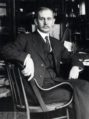