File:Streeter1957 fig04-23.jpg: Difference between revisions
From Embryology
mNo edit summary |
|||
| (2 intermediate revisions by the same user not shown) | |||
| Line 3: | Line 3: | ||
Several sections in each embryo were combined to show the form and dimensions of the optic nerve. x50. | Several sections in each embryo were combined to show the form and dimensions of the optic nerve. x50. | ||
Scale bar 0.5 mm | Carnegie stage {{CS23}} Carnegie Embryo {{CE5725}} Scale bar 0.5 mm | ||
{{Streeter1957 fig4 gallery}} | |||
<br> | |||
{{Carnegie_stage_table_1}} | |||
<br> | |||
{{Carnegie stage 23 links}} | |||
<br> | |||
{{Carnegie_stages}} | |||
<br> | |||
{{Streeter1957 figures}} | {{Streeter1957 figures}} | ||
[[Category:Carnegie Embryo 5725]] | [[Category:Carnegie Embryo 5725]] | ||
[[Category:Carnegie Stage 23]][[Category:Week 8]][[Category:Vision]] | [[Category:Carnegie Stage 23]][[Category:Week 8]][[Category:Vision]] | ||
Latest revision as of 22:59, 17 April 2018
Fig. 4. Drawing of sections through the eye and optic nerve in Stage 23
Several sections in each embryo were combined to show the form and dimensions of the optic nerve. x50.
Carnegie stage 23 Carnegie Embryo 5725 Scale bar 0.5 mm
- Eye and Optic Nerve (week 7-8)


| Week: | 1 | 2 | 3 | 4 | 5 | 6 | 7 | 8 |
| Carnegie stage: | 1 2 3 4 | 5 6 | 7 8 9 | 10 11 12 13 | 14 15 | 16 17 | 18 19 | 20 21 22 23 |
- Carnegie Stages: 1 | 2 | 3 | 4 | 5 | 6 | 7 | 8 | 9 | 10 | 11 | 12 | 13 | 14 | 15 | 16 | 17 | 18 | 19 | 20 | 21 | 22 | 23 | About Stages | Timeline
| Historic Disclaimer - information about historic embryology pages |
|---|
 |
- Links: 1 Graph Embryos 11-23 | 4 Eye and optic nerve 19-23 | Plate 1 - Cornea | Plate 2 - Hypophysis
Reference
Streeter GL. Developmental Horizons In Human Embryos Description Or Age Groups XIX, XX, XXI, XXII, And XXIII, Being The Fifth Issue Of A Survey Of The Carnegie Collection. (1957) Carnegie Instn. Wash. Publ. 611, Contrib. Embryol., 36: 167-196.
Cite this page: Hill, M.A. (2024, April 26) Embryology Streeter1957 fig04-23.jpg. Retrieved from https://embryology.med.unsw.edu.au/embryology/index.php/File:Streeter1957_fig04-23.jpg
- © Dr Mark Hill 2024, UNSW Embryology ISBN: 978 0 7334 2609 4 - UNSW CRICOS Provider Code No. 00098G
File history
Click on a date/time to view the file as it appeared at that time.
| Date/Time | Thumbnail | Dimensions | User | Comment | |
|---|---|---|---|---|---|
| current | 09:56, 1 September 2016 | 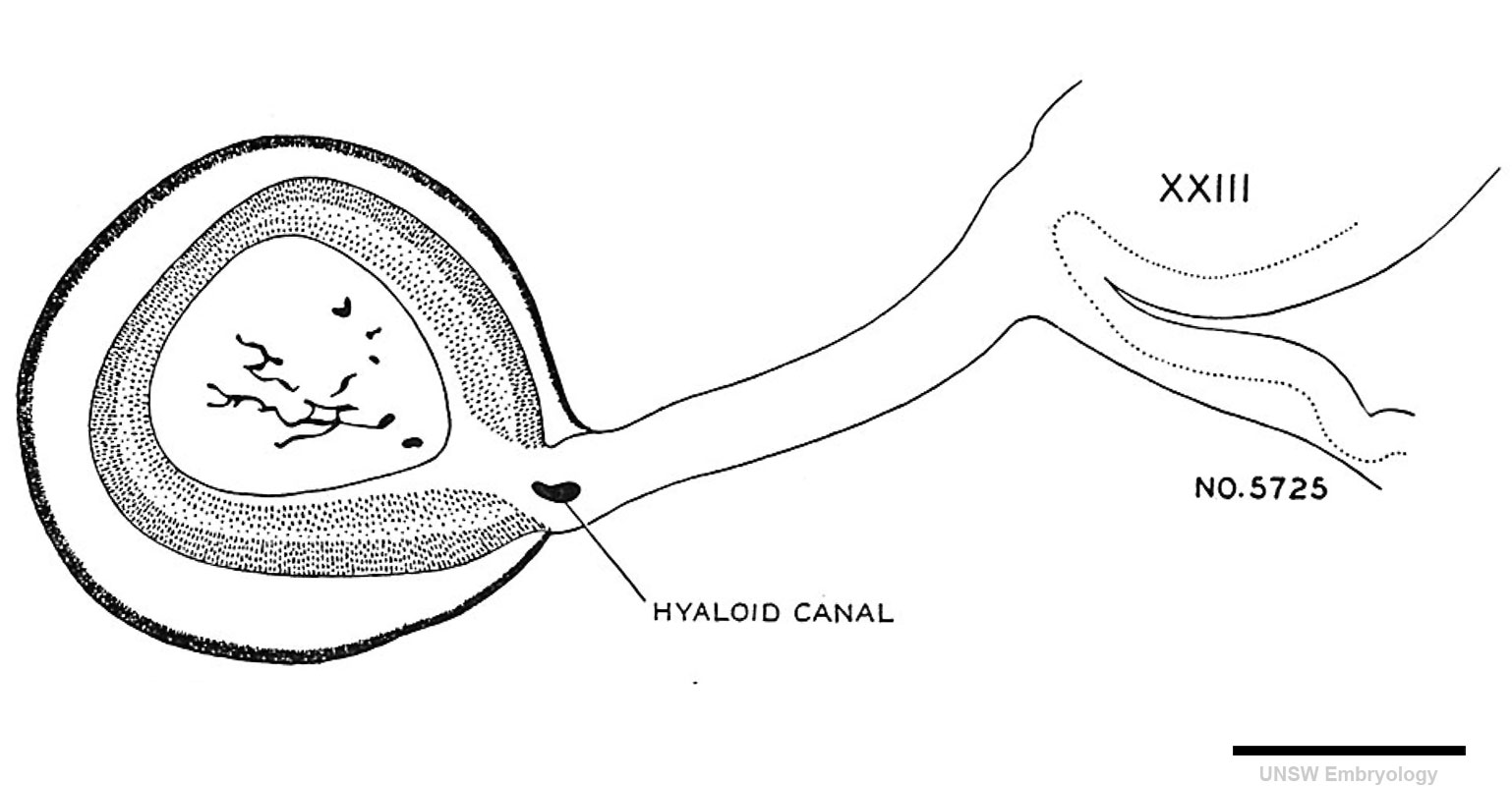 | 1,538 × 800 (115 KB) | Z8600021 (talk | contribs) |
You cannot overwrite this file.
File usage
The following 9 pages use this file: