File:Placental membranes.jpg: Difference between revisions
From Embryology
mNo edit summary |
mNo edit summary |
||
| Line 5: | Line 5: | ||
* '''amniotic sac''' - formed by the amniotic membrane (ectoderm and extra-embryonic mesoderm) completely surrounding the surrounding the embryo. | * '''amniotic sac''' - formed by the amniotic membrane (ectoderm and extra-embryonic mesoderm) completely surrounding the surrounding the embryo. | ||
* '''yolk sac''' - the yolk membrane (endoderm and extra-embryonic mesoderm) attached to the embryo at the umbilicus and continuous with the midgut. | * '''yolk sac''' - the yolk membrane (endoderm and extra-embryonic mesoderm) attached to the embryo at the umbilicus and continuous with the midgut. | ||
* '''chorionic cavity''' - represented by the | * '''chorionic cavity''' - has been removed but is represented by the black space outside the amniotic and yolk sac. | ||
Revision as of 07:43, 15 May 2014
Embryo with Placental Membranes
By the external appearance of the embryo appears to be about Week 7 (GA week 9) either Carnegie stage 18 or Carnegie stage 19
- amniotic sac - formed by the amniotic membrane (ectoderm and extra-embryonic mesoderm) completely surrounding the surrounding the embryo.
- yolk sac - the yolk membrane (endoderm and extra-embryonic mesoderm) attached to the embryo at the umbilicus and continuous with the midgut.
- chorionic cavity - has been removed but is represented by the black space outside the amniotic and yolk sac.
Image Source: UNSW Embryology, no reproduction without permission.
- Carnegie Stages: 1 | 2 | 3 | 4 | 5 | 6 | 7 | 8 | 9 | 10 | 11 | 12 | 13 | 14 | 15 | 16 | 17 | 18 | 19 | 20 | 21 | 22 | 23 | About Stages | Timeline
Cite this page: Hill, M.A. (2024, April 26) Embryology Placental membranes.jpg. Retrieved from https://embryology.med.unsw.edu.au/embryology/index.php/File:Placental_membranes.jpg
- © Dr Mark Hill 2024, UNSW Embryology ISBN: 978 0 7334 2609 4 - UNSW CRICOS Provider Code No. 00098G
File history
Click on a date/time to view the file as it appeared at that time.
| Date/Time | Thumbnail | Dimensions | User | Comment | |
|---|---|---|---|---|---|
| current | 23:36, 16 August 2009 | 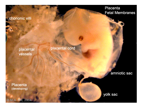 | 600 × 450 (99 KB) | S8600021 (talk | contribs) | Image Source: UNSW Embryology, no reproduction without permission. PlMembraneW600.jpg http://embryology.med.unsw.edu.au/Notes/images/placenta/plMembraneW600.jpg |
You cannot overwrite this file.
File usage
The following 16 pages use this file:
- 2009 Lecture 8
- 2010 BGD Lecture - Development of the Embryo/Fetus 1
- 2010 Group Project 3
- 2010 Lecture 8
- ANAT2341 Lab 4 - Implantation and Villi Development
- ASA Meeting 2013 - Placenta
- BGDA Lecture - Development of the Embryo/Fetus 1
- BGDA Practical 3 - Extraembryonic Spaces
- BGDA Practical Placenta - Villi Development
- Foundations Practical - Week 1 to 8
- Human System Development
- Lecture - Early Vascular Development
- P
- Placenta - Membranes
- Placenta Development
- Yolk Sac Development