File:Low 01.jpg: Difference between revisions
From Embryology
(==Fig. 1. - Right half of a transverse section of the embryo through the region of the eleventh mesodermic somite== Ec., ectoderm; XI. M.S., mesodermic somite; St., segmental tubule; Coe., coelom; d.Ao., dorsal aorta; Sp.c., medullary canal; E) |
mNo edit summary |
||
| (One intermediate revision by one other user not shown) | |||
| Line 1: | Line 1: | ||
==Fig. 1. - Right half of a transverse section of the embryo through the region of the eleventh mesodermic somite== | ==Fig. 1. - Right half of a transverse section of the embryo through the region of the eleventh mesodermic somite== | ||
Ec., ectoderm; XI. | * Ec., ectoderm; XI. | ||
* M.S., mesodermic somite | |||
* St., segmental tubule | |||
* Coe., coelom | |||
* d.Ao., dorsal aorta | |||
* Sp.c., medullary canal | |||
* En.,entoderm | |||
* Ch.,notochord. | |||
<br> | |||
{{Carnegie stage 11 links}} | |||
<br> | |||
{{Carnegie_stage_table_1}} | |||
<br> | |||
{{Low 1908}} | |||
Latest revision as of 02:44, 29 May 2017
Fig. 1. - Right half of a transverse section of the embryo through the region of the eleventh mesodermic somite
- Ec., ectoderm; XI.
- M.S., mesodermic somite
- St., segmental tubule
- Coe., coelom
- d.Ao., dorsal aorta
- Sp.c., medullary canal
- En.,entoderm
- Ch.,notochord.
| Week: | 1 | 2 | 3 | 4 | 5 | 6 | 7 | 8 |
| Carnegie stage: | 1 2 3 4 | 5 6 | 7 8 9 | 10 11 12 13 | 14 15 | 16 17 | 18 19 | 20 21 22 23 |
A 13-14 somite stage embryo would be similar to a Carnegie stage 11 (23 - 26 days) Somite Number 13 - 20.
- 13-14 Somite Paper: Plate 1 | Plate 2 | Plate 3 | Fig 1 | Fig 2 | Fig 3 | Fig 4 | Fig 5 | Fig 6 | Fig 7 | Fig 8 | Fig 9 | Fig 10 | Fig 11 | Fig 12 | Fig 13 | Fig 14 | Fig 15
| Historic Disclaimer - information about historic embryology pages |
|---|
 |
Reference
Low A. Description of a human embryo of 13-14 mesodermic somites. (1908) J Anat Physiol. 42(3): 237-51. PMID 17232769 | PMC1289161
Cite this page: Hill, M.A. (2024, April 27) Embryology Low 01.jpg. Retrieved from https://embryology.med.unsw.edu.au/embryology/index.php/File:Low_01.jpg
- © Dr Mark Hill 2024, UNSW Embryology ISBN: 978 0 7334 2609 4 - UNSW CRICOS Provider Code No. 00098G
File history
Click on a date/time to view the file as it appeared at that time.
| Date/Time | Thumbnail | Dimensions | User | Comment | |
|---|---|---|---|---|---|
| current | 23:38, 20 February 2012 | 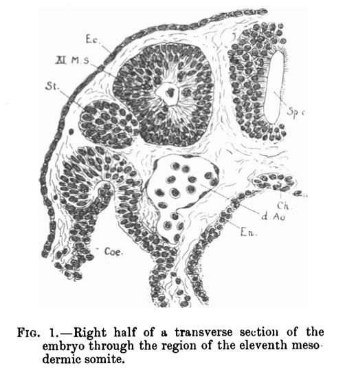 | 502 × 535 (60 KB) | S8600021 (talk | contribs) | ==Fig. 1. - Right half of a transverse section of the embryo through the region of the eleventh mesodermic somite== Ec., ectoderm; XI. M.S., mesodermic somite; St., segmental tubule; Coe., coelom; d.Ao., dorsal aorta; Sp.c., medullary canal; E |
You cannot overwrite this file.
File usage
The following page uses this file: