File:Keith1921 fig045.jpg

Original file (808 × 821 pixels, file size: 84 KB, MIME type: image/jpeg)
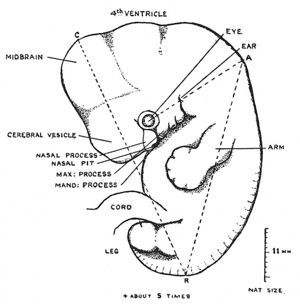
Fig. 45. Outline of an embryo, although only 11 mm long
Yet showing changes characteristic of an early stage of the 7th week. (After Broman.)
- The line C-R indicates the manner in which the crown-rump diameter is measured
- The line A-R shows the neck-rump length, A being found by drawing a line backward through the rudiments of the eye and ear.
During the 7th week the embryo becomes a foetus — the transformation being well shown in Fig. 43. In its crown-rump length the embryo expands from 11 to 17 mm., but the characteristic changes are seen in the face, head and limbs. An early stage of the 7th week is shown in Fig. 45 ; the basal parts of the face are being laid down. Under the eye are seen the nasal processes carrying the open nasal cavities backwards into the region of the mouth, while growing forwards beneath the eye are to be observed the maxillary processes which will provide the bases of the upper jaw. Still further back in the pharynx ( Fig. 45) are seen two comparatively small processes — the mandibular (1st arch) and hyoid (2nd arch). Behind the hyoid arch there is a depression marking the cervical sinus. By the end of the 7th week Fig. 43) the nasal, maxillary and mandibular processes have united to form a relatively small face ; at the upper end of the postmandibular cleft has appeared the rudiment of an ear. The changes in the head itself are also apparent ; at the end of the 7th week the cylindrical cranial form is being replaced by one more distinctly globular ; the forehead in particular has become enlarged. These changes are due to the rapid expansion of the cerebral vesicles during the 7th week. The changes in the limbs are also very evident ; they are now folded on the belly-wall, palm towards palm and sole towards sole ; the digits are demarcated. The tail is disappearing. The head is no longer bent forwards with the forehead touching the root of the umbilical cord, but is lifted up, for the embryonic flexure of the cervical region is being undone and a narrowing of the post-cranial region to form a neck becomes apparent. The heart is now completely divided into right and left chambers and the growth of the neck is lifting the pharyngeal region away from the heart. With these changes in the facial region, in the head, neck, limbs and heart the embryo of the 6th week becomes the foetus of the 7th. One other very important event also characterises this stage of transformation : the cellular blastema of the skeleton begins to change into cartilage and into bone. It also becomes possible to distinguish the ovary from the testicle.
| Week: | 1 | 2 | 3 | 4 | 5 | 6 | 7 | 8 |
| Carnegie stage: | 1 2 3 4 | 5 6 | 7 8 9 | 10 11 12 13 | 14 15 | 16 17 | 18 19 | 20 21 22 23 |
- Chapter 4 Figures: 42 | 43 | 44 | 45 | 46 | All Figures
| Historic Disclaimer - information about historic embryology pages |
|---|
 |
Reference
Keith A. Human Embryology and Morphology. (1921) New York, Longmans, Green & Co. London: Edward Arnold.
Human Embryology and Morphology: 1 Early Ovum and Embryo | 2 Connection between Foetus and Uterus | 3 Primitive Streak Notochord and Somites | 4 Age Changes | 5 Spinal Column and Back | 6 Body Segmentation | 7 Spinal Cord | 8 Mid- and Hind-Brains | 9 Fore-Brain | 10 Fore-Brain Cerebral Vesicles | 11 Cranium | 12 Face | 13 Teeth and Mastication | 14 Nasal and Olfactory | 15 Sense OF Sight | 16 Hearing | 17 Pharynx and Neck | 18 Tongue, Thyroid and Pharynx | 19 Organs of Digestion | 20 Circulatory System | 21 Circulatory System (continued) | 22 Respiratory System | 23 Urogenital System | 24 Urogenital System (Continued) | 25 Body Wall and Pelvic Floor | 26 Limb Buds | 27 Limbs | 28 Skin and Appendages | Figures
Cite this page: Hill, M.A. (2024, April 26) Embryology Keith1921 fig045.jpg. Retrieved from https://embryology.med.unsw.edu.au/embryology/index.php/File:Keith1921_fig045.jpg
- © Dr Mark Hill 2024, UNSW Embryology ISBN: 978 0 7334 2609 4 - UNSW CRICOS Provider Code No. 00098G
File history
Click on a date/time to view the file as it appeared at that time.
| Date/Time | Thumbnail | Dimensions | User | Comment | |
|---|---|---|---|---|---|
| current | 12:40, 23 December 2014 | | 808 × 821 (84 KB) | Z8600021 (talk | contribs) | |
| 12:37, 23 December 2014 | 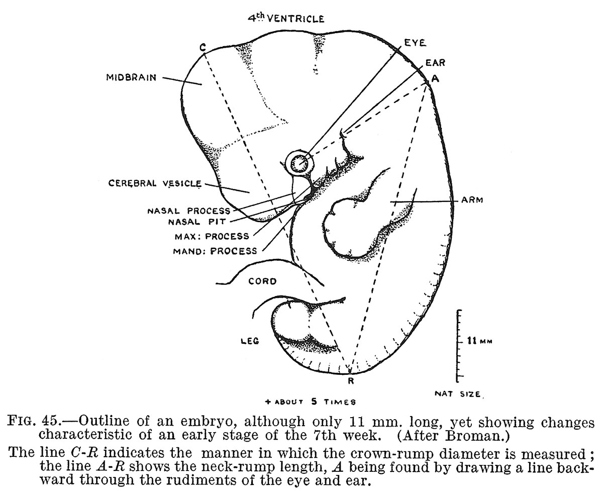 | 1,200 × 987 (153 KB) | Z8600021 (talk | contribs) |
You cannot overwrite this file.
File usage
The following 3 pages use this file: