File:Gilbert1957 plate04.jpg

Original file (2,165 × 2,232 pixels, file size: 694 KB, MIME type: image/jpeg)
Plate 4. Four drawings of a model of the eye, eye-muscle primordia, and associated nerves,
Embryo No. 6258, horizon xvii.
Fig. 14. The eye-muscle primordia of embryo no. 6258 are superimposed on the brain of another embryo, no. 6520, of approximately the same age and size. The relation of the principal branches of cranial nerve V to the four peripheral condensations into which the four rectus muscles grow is illustrated. Lateral aspect. X 15.
Fig. 15. A portion of the brain, the eye, principal nerves, peripheral condensations, and the eye—muscle primordia. Dorso-lateral aspect. No. 6258, X 30.
Fig. 16. Dorsocranial aspect of the same model illustrated in figure 15. X30.
Fig. 17. Ventrocaudal aspect of the same model illustrated in figure 15. X30.
Attention is called to the four peripheral condensations, about the outer margin of the optic vesicle, into which the primordia of the four rectus muscles have grown. Cranial nerves III, IV, and VI have reached their respective eye-muscle primordia: the primordium of the inferior oblique has appeared as a conspicuous condensation at the distal end of the inferior rectus; a prominent bend (at the point where the trochlea will subsequently develop) has appeared near the distal end of the superior oblique primordium, and the proximal end of the superior oblique has begun to shift medially.
| Week: | 1 | 2 | 3 | 4 | 5 | 6 | 7 | 8 |
| Carnegie stage: | 1 2 3 4 | 5 6 | 7 8 9 | 10 11 12 13 | 14 15 | 16 17 | 18 19 | 20 21 22 23 |
| Abbreviations Used in Figures | |
| ao., aorta
ao. :1. I, aortic arch I ao. (I. II, aortic arch II (to. (I. III, aortic arch III a. p. c., anterior peripheral condensation c. g., ciliary ganglion fix, forebrain fg., foregut fr. 9., frontal nerve g. V, Gasserian ganglion hb., hindbrain h. c., head cavity i. c. a., internal carotid artery inn, intermediate mass i. o., inferior oblique i. p. c., inferior peripheral condensation I. r., inferior rectus I. p., lens placode I. p. s., levator palpebrae superioris I. r., lateral rectus I. an, lens vesicle |
m. a., mandibular arch
nmmI. V, mandibular branch of cranial nerve V max. V, maxillary branch of cranial nerve V mb., midbrain m. m. m., maxillomandibular mesoderm m. r., medial rectus N., notochord o. III, oculomotor nerve 11. IV, trochlear nerve 11. VI, abducens nerve m. N., nasociliary nerve op. S., optic stalk op. V., optic vesicle op. V, ophthalmic branch of cranial nerve V p. b., prechorclal bridge p. 6., premandibular condensation p. pl., prechordal plate p. p. c., posterior peripheral condensation s. o., superior oblique s. p. c, superior peripheral condensation s. r., superior rectus t., trochlea v. c. a., vena cerebralis anterior v. c. m., vena capitis medialis v. o. i., vena orhitalis inferior |
Reference
Gilbert PW. The origin and development of the human extrinsic ocular muscles. (1957) Carnegie Instn. Wash. Publ. 611, Contrib. Embryol., Carnegie Inst. Wash. 36: 59-78.
Cite this page: Hill, M.A. (2024, April 27) Embryology Gilbert1957 plate04.jpg. Retrieved from https://embryology.med.unsw.edu.au/embryology/index.php/File:Gilbert1957_plate04.jpg
- © Dr Mark Hill 2024, UNSW Embryology ISBN: 978 0 7334 2609 4 - UNSW CRICOS Provider Code No. 00098G
File history
Click on a date/time to view the file as it appeared at that time.
| Date/Time | Thumbnail | Dimensions | User | Comment | |
|---|---|---|---|---|---|
| current | 23:30, 1 June 2016 | | 2,165 × 2,232 (694 KB) | Z8600021 (talk | contribs) | |
| 23:29, 1 June 2016 | 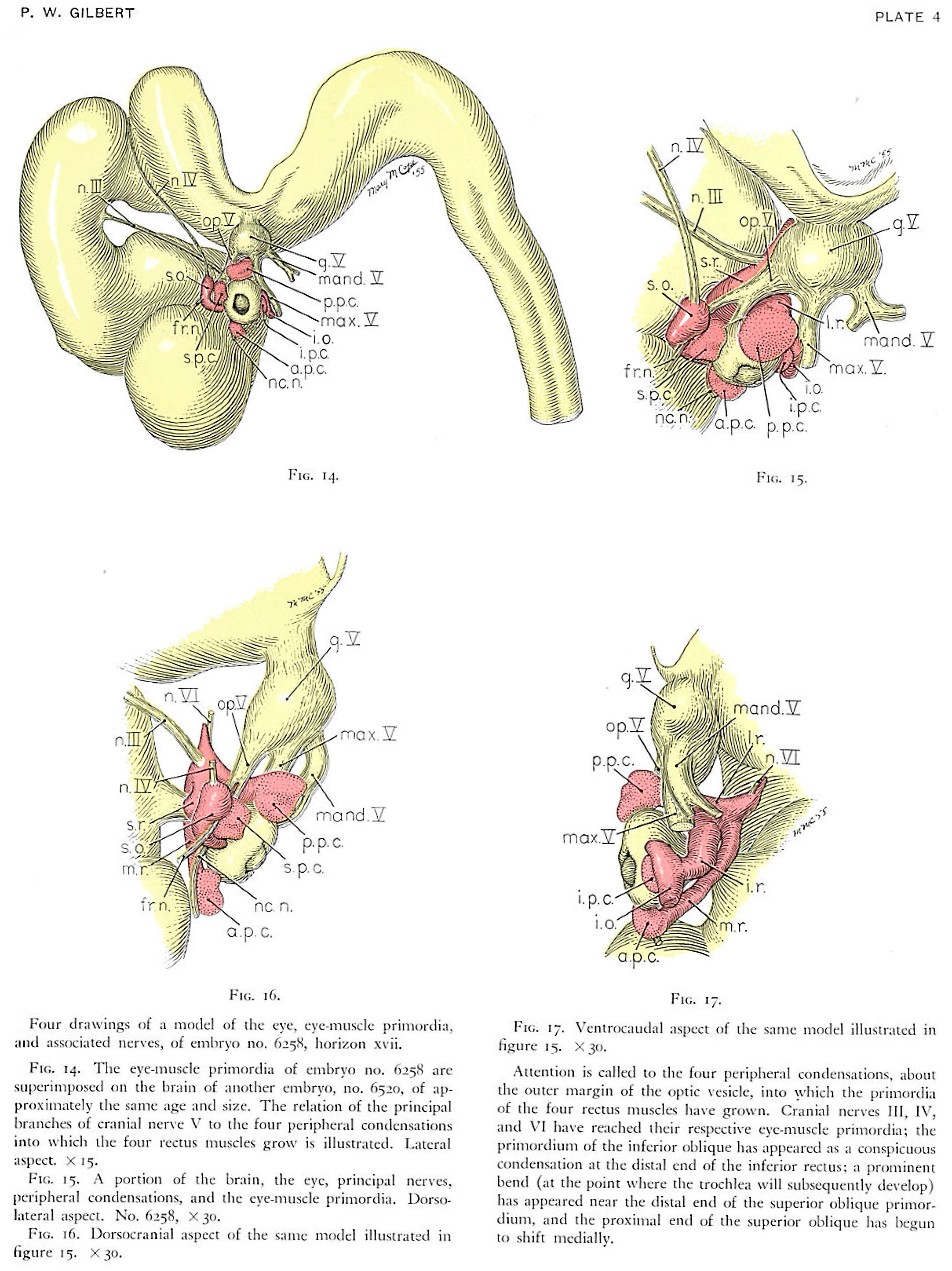 | 2,165 × 2,890 (970 KB) | Z8600021 (talk | contribs) | ==Plate 4. == ((Gilbert1957 figures}} ===Reference=== {{Ref-Gilbert1957}} {{Footer}} |
You cannot overwrite this file.
File usage
The following page uses this file: