File:Bartelmez1923 fig04.jpg
Original file (1,416 × 1,155 pixels, file size: 170 KB, MIME type: image/jpeg)
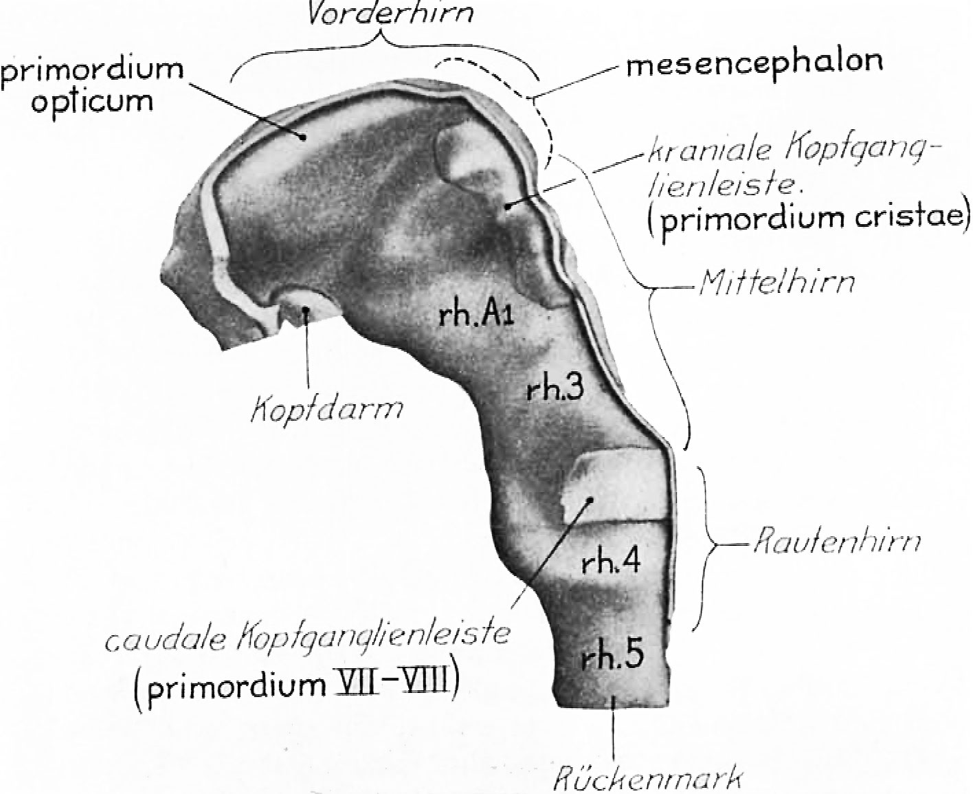
Fig. 4 A copy of figure 9 of Veit and Esch
Veit and Esch (’22, p.354). Their interpretation is indicated by the light-faced type, ours by the heavy-faced. The same divisions of the brain are present as in ‘DuGa’ (fig. 3). The embryo has eight somites completely separated off and the ninth differentiated. Modeled at 200 diameters and reduced one half in reproduction.
It is obvious from the excellent description and figures of Veit and Esch (’22) that their eight—somite embryo is but little younger than ‘DuGa.’ Their figure 9 is here reproduced as figure 4 with both the original labels and, in heavier type, the interpretation we place upon the enlargements of the neural folds. The otic segment rh.4 has the characteristic form; there is the great lateral fossa occupied in part by the acoustico-facial primordium (caudale Kopfganglienleiste) and the marked dip in the floor of the neural groove. figure 11 of Veit and Esch also shows this rhombomere clearly. The midbrain is readily identified through the cranial fiexure. Between these two we find the same divisions as in ‘DuGa.’ The figures of the nervous system show only one postotic neuromere, but the general reconstructions (Taf. X and XI) make it clear that there is at least room for another similar segment between it and the first pair of somites. The Veit embryo is, then, very like ‘DuGa’ so far as the subdivisions of the nervous system are concerned, and indeed in most other details of this system. The most striking difference between the two is the greater size of the midbrain in ‘DuGa.’ Associated, no doubt, with the rapid growth of the mesencephalon is the more obtuse cranial flexure in this specimen. figure 4 shows the neural crest (kraniale Kopfganglienleiste) extending to the rostral end of the midbrain. The forebrain folds are thickened laterally, so that a longitudinal ridge appears in the lateral View (of. fig. 4, primordium opticum). We are inclined to interpret this as the optic primordium, not only because of the general relations, but because Professor Veit’s photomicrographs show that its appearance in section is very like that of the eight-somite embryo H87, when one takes into consideration the difference in the plane of section in the two cases. The basis for this is a series of excellent photomicrographs comprising every third section, made at a magnification of 75 diameters, which Professor Veit was so good as to send us.
Online Editor - Veit and Esch embryo, 8 somite embryo corresponds to Carnegie stage 10 in Week 4. Veit O. and Esch P. Untersuchung eines in situ fixierten, operativ gewonnenen menschlichen Eies der vierten Woche. (Examination of a fixed in situ, surgically obtained human egg the fourth week) (1922) Z Anat. Entw., 63: 343-414.
| Stage 10 Links: Week 4 | Gastrulation | Lecture | Practical | Image Gallery | Carnegie Embryos | Embryos | Category:Carnegie Stage 10 | Next Stage 11 |
| Historic Papers: 1910 | 1917 | 1926 | 1939 | 1943 | 1957 | 1985 |
| Week: | 1 | 2 | 3 | 4 | 5 | 6 | 7 | 8 |
| Carnegie stage: | 1 2 3 4 | 5 6 | 7 8 9 | 10 11 12 13 | 14 15 | 16 17 | 18 19 | 20 21 22 23 |
| Historic Disclaimer - information about historic embryology pages |
|---|
 |
Reference
Bartelmez GW. The subdivisions of the neural folds in man. (1923) J. Comp. Neural., 35: 231-247.
Cite this page: Hill, M.A. (2024, April 27) Embryology Bartelmez1923 fig04.jpg. Retrieved from https://embryology.med.unsw.edu.au/embryology/index.php/File:Bartelmez1923_fig04.jpg
- © Dr Mark Hill 2024, UNSW Embryology ISBN: 978 0 7334 2609 4 - UNSW CRICOS Provider Code No. 00098G
File history
Click on a date/time to view the file as it appeared at that time.
| Date/Time | Thumbnail | Dimensions | User | Comment | |
|---|---|---|---|---|---|
| current | 22:53, 7 June 2016 | | 1,416 × 1,155 (170 KB) | Z8600021 (talk | contribs) | Fig. 4 A copy of figure 9 of Veit and Esch (’22, p.354). Their interpretation is indicated by the light-faced type, ours by the heavy-faced. The same divisions of the brain are present as in ‘DuGa’ (fig. 3). The embryo has eight somites completel... |
You cannot overwrite this file.
File usage
The following 2 pages use this file: