File:Stage13 sem3.jpg
Original file (818 × 1,000 pixels, file size: 81 KB, MIME type: image/jpeg)
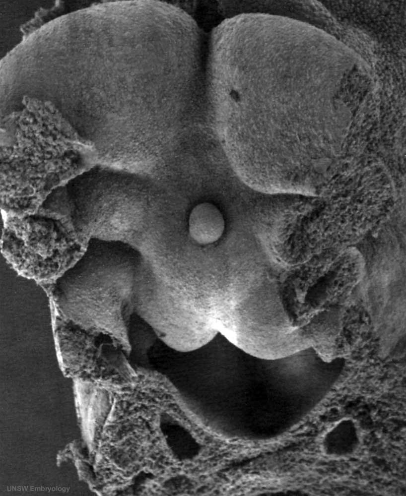
Human Embryo Carnegie stage 13
Carnegie stage 13, 28 day, 30 somite pairs
Scanning EM pharynx internal view of head region showing internal detail of pharyngeal arches from full embryo SEM.
- yolk sac and amnion removed
- pharyngeal arches
- pharynx
- thyroid
- 4th pharyngeal arch arteries
Related Images: Carnegie stage 13 | Embryo Head region SEM | Full Embryo SEM | Bright Field image version
| Week: | 1 | 2 | 3 | 4 | 5 | 6 | 7 | 8 |
| Carnegie stage: | 1 2 3 4 | 5 6 | 7 8 9 | 10 11 12 13 | 14 15 | 16 17 | 18 19 | 20 21 22 23 |
- Carnegie Stages: 1 | 2 | 3 | 4 | 5 | 6 | 7 | 8 | 9 | 10 | 11 | 12 | 13 | 14 | 15 | 16 | 17 | 18 | 19 | 20 | 21 | 22 | 23 | About Stages | Timeline
Image Source: Scanning electron micrographs of the Carnegie stages of the early human embryos are reproduced with the permission of Prof Kathy Sulik, from embryos collected by Dr. Vekemans and Tania Attié-Bitach. Images are for educational purposes only and cannot be reproduced electronically or in writing without permission.
File history
Yi efo/eka'e gwa ebo wo le nyangagi wuncin ye kamina wunga tinya nan
| Gwalagizhi | Nyangagi | Dimensions | User | Comment | |
|---|---|---|---|---|---|
| current | 10:48, 4 September 2009 | | 818 × 1,000 (81 KB) | S8600021 (talk | contribs) | Stage13day28somite30-pharynx-cut-sem3.jpg |
You cannot overwrite this file.
File usage
The following 5 pages use this file: