Category:Neural
From Embryology
This Embryology category shows pages and media related to Neural System Development. This includes related topics and undergraduate classes as well as pages and sub-categories describing specific components formed from the original ectoderm neural tube.
Subcategories
This category has the following 12 subcategories, out of 12 total.
Pages in category 'Neural'
The following 200 pages are in this category, out of 934 total.
(previous page) (next page)N
- Neural Exam - 18 Behaviour 4
- Neural Exam - 18 Cranial Nerves
- Neural Exam - 18 month Deep Tendon Reflex
- Neural Exam - 18 month Motor 1
- Neural Exam - 18 month Motor 2
- Neural Exam - 18 month Motor 3
- Neural Exam - 18 month Motor 4
- Neural Exam - 18 month Motor 5
- Neural Exam - 18 month Motor 6
- Neural Exam - 18 month Motor 7
- Neural Exam - 18 month Motor 8
- Neural Exam - 3 month - hand movements
- Neural Exam - 3 month - head and trunk control
- Neural Exam - 3 month - prone position
- Neural Exam - 3 month - supine position
- Neural Exam - 3 month behaviour
- Neural Exam - 3 month cranial nerves
- Neural Exam - 3 month positions - ventral suspension
- Neural Exam - 3 month positions - vertical suspension
- Neural Exam - 3 month reflexes - asymmetric tonic neck
- Neural Exam - 3 month reflexes - deep tendon reflexes
- Neural Exam - 3 month reflexes - Galant
- Neural Exam - 3 month reflexes - grasp
- Neural Exam - 3 month reflexes - Moro
- Neural Exam - 3 month reflexes - plantar reflex
- Neural Exam - 3 month reflexes - root
- Neural Exam - 3 month tone - lower extremity
- Neural Exam - 3 month tone - upper extremity
- Neural Exam - 30 month Behaviour 1
- Neural Exam - 30 month Behaviour 2
- Neural Exam - 30 month Behaviour 3
- Neural Exam - 30 month Behaviour 4
- Neural Exam - 30 month Behaviour 5
- Neural Exam - 30 month Behaviour 6
- Neural Exam - 30 month Deep Tendon Reflex
- Neural Exam - 30 month Motor 1
- Neural Exam - 30 month Motor 2
- Neural Exam - 30 month Motor 3
- Neural Exam - 30 month Motor 4
- Neural Exam - 30 month Motor 5
- Neural Exam - 30 month Motor 6
- Neural Exam - 30 month Motor 7
- Neural Exam - 6 month - hand movements
- Neural Exam - 6 month - head and trunk control
- Neural Exam - 6 month - prone position
- Neural Exam - 6 month - supine position
- Neural Exam - 6 month behaviour
- Neural Exam - 6 month cranial nerves
- Neural Exam - 6 month positions - ventral suspension
- Neural Exam - 6 month positions - vertical suspension
- Neural Exam - 6 month reflexes - deep tendon reflexes
- Neural Exam - 6 month reflexes - plantar reflex
- Neural Exam - 6 month tone - lower extremity
- Neural Exam - 6 month tone - upper extremity
- Neural Exam - Newborn behaviour comparison
- Neural Exam - Newborn cranial nerves
- Neural Exam - Newborn head circumference
- Neural Exam - Newborn head shape and sutures
- Neural Exam - Newborn normal behaviour
- Talk:Neural Exam - Newborn normal behaviour
- Neural Exam - Newborn positions - prone
- Neural Exam - Newborn positions - ventral suspension
- Neural Exam - Newborn positions - vertical suspension
- Neural Exam - Newborn reflexes - deep tendon reflexes
- Neural Exam - Newborn reflexes - Galant
- Neural Exam - Newborn reflexes - grasp
- Neural Exam - Newborn reflexes - Moro
- Neural Exam - Newborn reflexes - plantar reflex
- Neural Exam - Newborn reflexes - stepping
- Neural Exam - Newborn reflexes - suck, root
- Neural Exam - Newborn tone - arm recoil
- Neural Exam - Newborn tone - arm traction
- Neural Exam - Newborn tone - hand position
- Neural Exam - Newborn tone - head control
- Neural Exam - Newborn tone - head lag
- Neural Exam - Newborn tone - heel to ear
- Neural Exam - Newborn tone - leg recoil
- Neural Exam - Newborn tone - leg traction
- Neural Exam - Newborn tone - lower extremity
- Neural Exam - Newborn tone - neck tone
- Neural Exam - Newborn tone - popliteal angle
- Neural Exam - Newborn tone - resting posture
- Neural Exam - Newborn tone - scarf sign
- Neural Exam - Newborn tone - upper extremity
- Neural Exam Movies
- Talk:Neural Exam Movies
- Template:Neural examination
- Template:Neural fetal
- Template:Neural groove
- Template:Neural interkinetic nuclear migration movie
- Template:Neural Links
- Template:Neural Links 2
- Template talk:Neural Links 2
- Template:Neural Links collapsetable2
- Template:Neural plate
- Neural Plate Movie
- Template:Neural postnatal
- Neural System - Abnormalities
- Neural System - Carnegie Stage 22
- Neural System - Fetal
- Neural System - Glial Development
- Neural System - Molecular
- Neural System - Postnatal
- Neural System Development
- Talk:Neural System Development
- Template:Neural Table
- Template:Neural tube
- Neural Tube Closure Movie
- Template:Neural tube defect
- Template:Neural Tube Defects - Potential risk factors table1
- Neural Tube Movie
- Template:Neural tube regions table
- Template:Neural vascular
- Template:NeuroExam 12month
- Template:Neurohypophysis
- Template:Neuron
- Template:Neuropore
- Talk:New
- Template:Normal 12 Month Neural Exam Table
- Template:Normal 30 Month Neural Exam Table
- Template:Normal Abnormal Newborn Neural Exam Table
- Template:Normal Newborn Neural Exam Table
O
P
- Paper - 1879 The Morphology of the Vertebrate Olfactory Organ
- Paper - A case of accidental impregnation of cells in the brain of a human embryo of four months (1912)
- Paper - A comparison of the growth of the body dimensions of anencephalic human fetuses with normal fetal growth as determined by graphic analysis and empirical formulae
- Paper - A contribution to the histogenesis of the sympathetic nervous system (1909)
- Paper - A contribution to the study of the cerebral cortex in man
- Paper - A human foetus exhibiting iniencephaly and other abnormalities (1922)
- Paper - A note concerning the model of the medulla, pons and midbrain of a new-born babe as reproduced by Herr F. Ziegler (1903)
- Paper - A phylogenetic consideration of the optic tectum
- Paper - A quantitative study of the hypophysis of the human anencephalic fetus (1927)
- Paper - A study of the development of certain features of the cerebellum (1920)
- Paper - A three weeks human embryo with especial reference to the brain and nephric system (1905)
- Paper - Abnormal development of the brain in an 8 mm pig embryo (1938)
- Paper - Address Upon The Development Of The Brain
- Paper - An anencephalic embryo of 25 mm CRL
- Paper - An anencephalic human embryo 16.5 mm long
- Paper - An experimental investigation of the motor cortex and pyramidal tract of echidna aculeata (1939)
- Paper - An experimental study of the origin of the meninges (1924)
- Paper - An iconometrographic representation of the growth of the central nervous system in man
- Paper - Anatomy of the floor of the fourth ventricle (1903)
- Paper - Anencephaly and rhachischisis posterior, with the description of a human hemicephalus of 18 mm (1939)
- Paper - Anterior and posterior rhachischisis (1941)
- Paper - Cell columns in the spinal cord of a human foetus of fourteen weeks (1941)
- Paper - Certain developmental relations and fiber connections of the triangular gyrus in primates (1948)
- Paper - Comparative morphology of the ear 3
- Paper - Comparative studies on the growth of the cerebral cortex 1 (1917)
- Paper - Comparative studies on the growth of the cerebral cortex 2 (1917)
- Paper - Comparative studies on the growth of the cerebral cortex 3 (1918)
- Paper - Comparative studies on the growth of the cerebral cortex 4 (1918)
- Paper - Comparative studies on the growth of the cerebral cortex 5 (1918)
- Paper - Comparative studies on the growth of the cerebral cortex 6 (1918)
- Paper - Comparative studies on the growth of the cerebral cortex 7 (1918)
- Paper - Comparative studies on the growth of the cerebral cortex 8 (1918)
- Paper - Comparative studies upon the origin and development of the brachial plexus
- Paper - Complete dysraphism in 14 somite human embryo
- Paper - Contribution to the structure and development of the vertebrate head
- Paper - Contribution to the structure and development of the vertebrate head 1
- Paper - Contribution to the structure and development of the vertebrate head 2
- Paper - Contribution to the structure and development of the vertebrate head 3
- Paper - Correlated changes in nervous tissues in malformations of the central nervous system (1946)
- Paper - Defective development of the septum pellucidum (1932)
- Paper - Description of a model showing the tracts of fibres medullated in a new-born baby’s brain (1911)
- Paper - Description of a young human anencephalic and amyelic embryo
- Paper - Development and homology of the mammalian cerebellar fissures 1
- Paper - Development and homology of the mammalian cerebellar fissures 2
- Paper - Development and variation of the nerves and the musculature of the inferior extremity and of the neighboring regions of the trunk in man
- Paper - Development of the innervation pattern in the upper limb of staged human embryos (1990)
- Paper - Development of the interfore-brain commissures in the human embryo
- Paper - Development of the region of the isthmus rhombencephali (1928)
- Paper - Development of the spinal reflex mechanism in human embryos
- Paper - Development of the tractus solitarius
- Paper - Differentiation of pituicytes in the human foetus
- Paper - Embryological and morphological studies on the mid-brain and cerebellum of vertebrates
- Paper - Embryological and morphological studies on the mid-brain and cerebellum of vertebrates 1
- Paper - Embryological and morphological studies on the mid-brain and cerebellum of vertebrates 2
- Paper - Embryological and morphological studies on the mid-brain and cerebellum of vertebrates 3
- Paper - Embryological stages in the development of spina bifida and myeloschisis
- Paper - Extroversion of the cerebral hemispheres in a human embryo (1934)
- Paper - Factors Involved In The Formation Of The Filum Terminale
- Paper - Further contributions to the study of the evolution of the forebrain 5
- Paper - Further experiments on the development of peripheral nerves (1906)
- Paper - Further observations on the anatomy of the brain in the monotremata
- Paper - Fusion of notochord to neural tube in a human embryo of the sixth week (1946)
- Paper - Head ganglia of an embryo of eight somite pairs
- Paper - Localization and regeneration in the neural plate of amphibian embryos (1910)
- Paper - Malformations of the human body from a new point of view 1+2
- Paper - Meninges histogenesis and structure
- Paper - Morphology of the roof plate of the fore-brain and the lateral choroid plexuses in the human embryo (1916)
- Paper - Morphophysiology of the cerebral cortex
- Paper - Norms for some structural changes in the human cerebellum from birth to old age (1920)
- Paper - Nuclear masses in the lower portion of the human brain-stem (1914)
- Paper - Observations concerning the comparative anatomy of the diencephalon (1912)
- Paper - Observations on the histogenesis of protoplasmic processes and of collaterals, terminating in end bulbs, of the neurones of peripheral sensory ganglia (1913)
- Paper - Observations on the peripheral distribution of the nervus terminalis in mammalia (1913)
Media in category 'Neural'
The following 200 files are in this category, out of 1,070 total.
(previous page) (next page) 03mo 01.jpg 320 × 240; 9 KB
 03mo 02.jpg 320 × 240; 8 KB
 03mo 03.jpg 320 × 240; 10 KB
 03mo 04.jpg 320 × 240; 11 KB
 03mo 05.jpg 320 × 240; 11 KB
 10wkcerebellumB.jpg 347 × 284; 21 KB
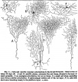
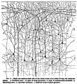
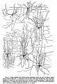
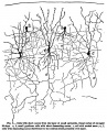
 1899 Cajal 01.jpg 307 × 1,200; 121 KB
 1899 Cajal 02.jpg 1,200 × 646; 177 KB
 1899 Cajal 03.jpg 952 × 1,000; 204 KB
 1899 Cajal 04.jpg 909 × 1,000; 253 KB
 1899 Cajal 05.jpg 683 × 1,000; 164 KB
 1899 Cajal 06.jpg 812 × 1,000; 163 KB
 1899 Cajal 07.jpg 837 × 1,000; 175 KB
 1899 Cajal 08.jpg 329 × 1,000; 101 KB
 2017BGDALecture-Neural.mp4 ; 52.32 MB
 Abnormal81-92-neuron.png 481 × 344; 9 KB
 Adult brain 01.mov ; 1.59 MB
- Adult brain 02.mov ; 434 KB
 Adult brain animation 01.gif 280 × 224; 396 KB
 Adult cochlea cartoon 01.jpg 986 × 800; 123 KB
 Adult cochlea nerve glia cartoon.jpg 1,000 × 725; 85 KB
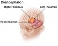 Adult diencephalon.jpg 470 × 376; 20 KB
 Adult human brain movie icon.jpg 717 × 575; 28 KB
 Adult human brain MRI01.jpg 700 × 607; 81 KB
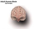
 Adult human brain.jpg 984 × 735; 104 KB
 Adult mouse brain - prosomeric model.jpg 964 × 414; 91 KB
- AEB Histology Prac 171012-3 Ganglion.mp3 ; 1.05 MB
- AEB Histology Prac 171012-5 Nerve.mp3 ; 1.07 MB
 Amin1914 fig01.jpg 1,000 × 652; 120 KB
 Amin1914 fig02.jpg 1,000 × 681; 152 KB
 Amin1914 fig03.jpg 1,000 × 689; 134 KB
 Amin1914 fig04.jpg 1,000 × 727; 139 KB
 Amin1914 fig05.jpg 1,000 × 646; 91 KB
 Anencephaly ultrasound.jpg 900 × 658; 108 KB
 Astrocytes and neonatal hypoxia ischemia.jpg 484 × 705; 309 KB
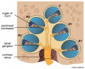
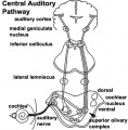 Auditory neural pathway.jpg 450 × 457; 46 KB
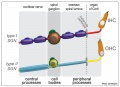
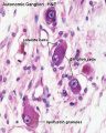 Autonomic ganglion histology 01.jpg 641 × 800; 56 KB
 Baboon- fetal brain.jpg 1,000 × 733; 127 KB
 Bailey358.jpg 854 × 560; 66 KB
 Bailey359.jpg 708 × 572; 46 KB
 Bailey360.jpg 543 × 404; 23 KB
 Bailey361.jpg 815 × 662; 86 KB
 Bailey362.jpg 801 × 354; 49 KB
 Bailey363.jpg 876 × 373; 46 KB
 Bailey364.jpg 809 × 465; 53 KB
 Bailey365.jpg 847 × 595; 94 KB
 Bailey366.jpg 558 × 633; 97 KB
 Bailey367.jpg 1,034 × 440; 101 KB
 Bailey368.jpg 1,074 × 523; 134 KB
 Bailey369.jpg 529 × 446; 33 KB
 Bailey370.jpg 975 × 1,084; 242 KB
 Bailey371.jpg 687 × 997; 97 KB
 Bailey372.jpg 803 × 690; 79 KB
 Bailey373.jpg 894 × 426; 100 KB
 Bailey374.jpg 880 × 490; 75 KB
 Bailey375.jpg 929 × 785; 130 KB
 Bailey376.jpg 922 × 862; 70 KB
 Bailey377.jpg 1,032 × 904; 90 KB
 Bailey378.jpg 1,199 × 700; 69 KB
 Bailey379-382.jpg 671 × 988; 199 KB
 Bailey383.jpg 859 × 455; 118 KB
 Bailey384.jpg 1,532 × 770; 245 KB
 Bailey385.jpg 787 × 683; 165 KB
 Bailey386.jpg 491 × 410; 53 KB
 Bailey387.jpg 507 × 442; 50 KB
 Bailey388.jpg 514 × 438; 68 KB
 Bailey389.jpg 829 × 561; 65 KB
 Bailey390.jpg 583 × 667; 50 KB
 Bailey391.jpg 633 × 485; 56 KB
 Bailey392.jpg 949 × 632; 113 KB
 Bailey393.jpg 680 × 527; 89 KB
 Bailey394.jpg 614 × 563; 70 KB
 Bailey395.jpg 787 × 741; 94 KB
 Bailey396.jpg 829 × 845; 123 KB
 Bailey397.jpg 779 × 995; 162 KB
 Bailey398.jpg 872 × 581; 78 KB
 Bailey399.jpg 832 × 704; 65 KB
 Bailey400.jpg 724 × 749; 66 KB
 Bailey401.jpg 788 × 718; 61 KB
 Bailey402.jpg 640 × 483; 116 KB
 Bailey403.jpg 495 × 617; 118 KB
 Bailey404.jpg 627 × 695; 84 KB
 Bailey405.jpg 745 × 820; 141 KB
 Bailey406.jpg 868 × 763; 116 KB
 Bailey407.jpg 793 × 695; 117 KB
 Bailey408.jpg 862 × 732; 124 KB
 Bailey409.jpg 448 × 408; 32 KB
 Bailey410.jpg 1,412 × 802; 169 KB
 Bailey411.jpg 790 × 809; 104 KB
 Bailey412.jpg 723 × 251; 21 KB
 Bailey413.jpg 674 × 412; 47 KB
 Bailey414.jpg 900 × 717; 123 KB
 Bailey415.jpg 935 × 723; 147 KB
 Bailey416.jpg 815 × 606; 142 KB
 Bailey417.jpg 940 × 494; 116 KB
 Bailey418.jpg 232 × 583; 30 KB
 Bailey419.jpg 805 × 388; 43 KB
 Bailey420.jpg 504 × 348; 34 KB
 Bailey421.jpg 625 × 538; 81 KB
 Bailey422.jpg 495 × 568; 95 KB
 Bailey423.jpg 670 × 581; 56 KB
 Bailey424.jpg 804 × 534; 61 KB
 Bailey425.jpg 957 × 490; 84 KB
 Bailey426.jpg 948 × 758; 80 KB
 Bailey427.jpg 907 × 527; 72 KB
 Bailey428.jpg 756 × 557; 67 KB
 Bailey429.jpg 844 × 484; 81 KB
 Bailey430.jpg 1,072 × 791; 121 KB
 Bailey431.jpg 832 × 535; 75 KB
 Bailey432.jpg 794 × 635; 92 KB
 Bailey433.jpg 723 × 536; 77 KB
 Bailey434.jpg 426 × 436; 44 KB
 Bailey435.jpg 788 × 456; 48 KB
 Bailey436.jpg 552 × 443; 40 KB
 Bailey437.jpg 526 × 397; 50 KB
 Bailey438.jpg 778 × 530; 70 KB
 Bailey439.jpg 932 × 1,016; 125 KB
 Bailey440.jpg 820 × 434; 54 KB
 Bailey441.jpg 786 × 520; 70 KB
 Bailey442.jpg 525 × 504; 38 KB
 Bailey443.jpg 666 × 539; 49 KB
 Bailey444.jpg 777 × 590; 127 KB
 Bailey445.jpg 900 × 655; 89 KB
 Bailey446.jpg 709 × 437; 40 KB
 Bailey447.jpg 795 × 423; 45 KB
 Bailey448.jpg 722 × 416; 55 KB
 Bailey449.jpg 777 × 374; 45 KB
 Bailey450.jpg 680 × 419; 45 KB
 Bailey451-452.jpg 753 × 862; 151 KB
 Bailey453.jpg 461 × 740; 69 KB
 Bailey454.jpg 732 × 527; 119 KB
 Bailey455.jpg 744 × 547; 124 KB
 Bailey456.jpg 591 × 168; 20 KB
 Bailey457.jpg 659 × 335; 40 KB
 Bailey458-459.jpg 741 × 386; 42 KB
 Bailey460.jpg 718 × 423; 55 KB
 Bailey461.jpg 751 × 394; 54 KB
 Bailey462.jpg 463 × 411; 45 KB
 Bailey463.jpg 679 × 345; 52 KB
 Bailey464.jpg 869 × 592; 67 KB
 Bailey465.jpg 806 × 931; 142 KB
 Bailey466.jpg 859 × 683; 139 KB
 Bailey467.jpg 946 × 515; 159 KB
 Bailey468.jpg 774 × 519; 60 KB
 Bailey469.jpg 484 × 401; 50 KB
 Bailey470.jpg 537 × 644; 88 KB
 Bailey475.jpg 1,302 × 852; 213 KB
 Bailey476.jpg 688 × 648; 110 KB
 Bailey477.jpg 593 × 509; 57 KB
 Bailey478.jpg 809 × 620; 81 KB
 Bailey479.jpg 742 × 363; 56 KB
 Bailey480.jpg 799 × 553; 61 KB
 Bailey481.jpg 842 × 752; 109 KB
 Bailey482.jpg 709 × 419; 57 KB
 Baileytable08.jpg 968 × 570; 76 KB
 Baileytable09.jpg 606 × 150; 18 KB
 Bardeen1906-fig02.jpg 1,598 × 1,183; 228 KB
 Bardeen1906-fig03.jpg 1,598 × 1,166; 231 KB
 Bardeen1906-plate01.jpg 1,565 × 2,322; 238 KB
 Bardeen1906-plate02.jpg 1,719 × 2,302; 512 KB
 Bardeen1906-plate06.jpg 1,568 × 2,299; 379 KB
 Bardeen1906-plate31.jpg 1,571 × 2,330; 257 KB
 Bardeen1906-plate32.jpg 1,588 × 2,341; 292 KB
 Bardeen1906-plate41.jpg 1,555 × 2,323; 261 KB
 Bardeen1906-plate42.jpg 1,570 × 2,331; 240 KB
 Bardeen1906-plate51.jpg 1,570 × 2,330; 392 KB
 Bardeen1906-plate52.jpg 1,596 × 2,350; 404 KB
 Bartelmez1922-fig01.jpg 900 × 770; 131 KB
 Bartelmez1922-fig02.jpg 1,203 × 1,700; 317 KB
 Bartelmez1922-fig03.jpg 885 × 1,000; 169 KB
 Bartelmez1922-fig04.jpg 1,300 × 750; 108 KB
 Bartelmez1922-fig05.jpg 800 × 750; 164 KB
 Bartelmez1922-fig06.jpg 749 × 1,000; 179 KB
 Bartelmez1922-fig07.jpg 1,200 × 620; 116 KB
 Bartelmez1922-fig08.jpg 1,000 × 1,255; 251 KB
 Bartelmez1922-fig09.jpg 1,200 × 1,558; 287 KB
 Bartelmez1922-fig10.jpg 1,000 × 1,136; 209 KB
 Bartelmez1923 fig01.jpg 1,312 × 1,919; 245 KB
 Bartelmez1923 fig02.jpg 1,295 × 2,189; 218 KB
 Bartelmez1923 fig03.jpg 1,105 × 1,350; 202 KB
 Bartelmez1923 fig04.jpg 1,416 × 1,155; 170 KB
 Bartelmez1923 fig05.jpg 1,102 × 815; 195 KB
 Bartelmez1923 fig06.jpg 1,280 × 809; 136 KB
 Bat - neural development 01.jpg 733 × 498; 33 KB
 BAW icon 2012.jpg 200 × 280; 17 KB
 Blood-brain barrier cartoon.jpg 765 × 1,000; 75 KB
 Blood-brain barrier EM01.jpg 1,656 × 810; 250 KB
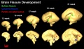
 Brain fissure development 01.jpg 1,157 × 502; 47 KB
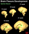
 Brain fissure development 02.jpg 1,000 × 583; 52 KB
 Brain fissure development 03.jpg 600 × 691; 33 KB
 Brain growth and birth size.jpg 800 × 492; 70 KB
 Brain histology 01.jpg 480 × 600; 125 KB
 Brain histology 02.jpg 480 × 600; 51 KB
 Brain stem subdivisions 01.jpg 1,530 × 520; 168 KB
 Brain tract development 01.jpg 720 × 1,023; 76 KB
 Brain tract development 02.jpg 1,000 × 424; 29 KB
 Brain tract development 06.jpg 1,000 × 605; 35 KB


