Category:Neural
From Embryology
This Embryology category shows pages and media related to Neural System Development. This includes related topics and undergraduate classes as well as pages and sub-categories describing specific components formed from the original ectoderm neural tube.
Subcategories
This category has the following 12 subcategories, out of 12 total.
Pages in category 'Neural'
The following 200 pages are in this category, out of 934 total.
(previous page) (next page)N
- Neural Exam - 6 month positions - vertical suspension
- Neural Exam - 6 month reflexes - deep tendon reflexes
- Neural Exam - 6 month reflexes - plantar reflex
- Neural Exam - 6 month tone - lower extremity
- Neural Exam - 6 month tone - upper extremity
- Neural Exam - Newborn behaviour comparison
- Neural Exam - Newborn cranial nerves
- Neural Exam - Newborn head circumference
- Neural Exam - Newborn head shape and sutures
- Neural Exam - Newborn normal behaviour
- Talk:Neural Exam - Newborn normal behaviour
- Neural Exam - Newborn positions - prone
- Neural Exam - Newborn positions - ventral suspension
- Neural Exam - Newborn positions - vertical suspension
- Neural Exam - Newborn reflexes - deep tendon reflexes
- Neural Exam - Newborn reflexes - Galant
- Neural Exam - Newborn reflexes - grasp
- Neural Exam - Newborn reflexes - Moro
- Neural Exam - Newborn reflexes - plantar reflex
- Neural Exam - Newborn reflexes - stepping
- Neural Exam - Newborn reflexes - suck, root
- Neural Exam - Newborn tone - arm recoil
- Neural Exam - Newborn tone - arm traction
- Neural Exam - Newborn tone - hand position
- Neural Exam - Newborn tone - head control
- Neural Exam - Newborn tone - head lag
- Neural Exam - Newborn tone - heel to ear
- Neural Exam - Newborn tone - leg recoil
- Neural Exam - Newborn tone - leg traction
- Neural Exam - Newborn tone - lower extremity
- Neural Exam - Newborn tone - neck tone
- Neural Exam - Newborn tone - popliteal angle
- Neural Exam - Newborn tone - resting posture
- Neural Exam - Newborn tone - scarf sign
- Neural Exam - Newborn tone - upper extremity
- Neural Exam Movies
- Talk:Neural Exam Movies
- Template:Neural examination
- Template:Neural fetal
- Template:Neural groove
- Template:Neural interkinetic nuclear migration movie
- Template:Neural Links
- Template:Neural Links 2
- Template talk:Neural Links 2
- Template:Neural Links collapsetable2
- Template:Neural plate
- Neural Plate Movie
- Template:Neural postnatal
- Neural System - Abnormalities
- Neural System - Carnegie Stage 22
- Neural System - Fetal
- Neural System - Glial Development
- Neural System - Molecular
- Neural System - Postnatal
- Neural System Development
- Talk:Neural System Development
- Template:Neural Table
- Template:Neural tube
- Neural Tube Closure Movie
- Template:Neural tube defect
- Template:Neural Tube Defects - Potential risk factors table1
- Neural Tube Movie
- Template:Neural tube regions table
- Template:Neural vascular
- Template:NeuroExam 12month
- Template:Neurohypophysis
- Template:Neuron
- Template:Neuropore
- Talk:New
- Template:Normal 12 Month Neural Exam Table
- Template:Normal 30 Month Neural Exam Table
- Template:Normal Abnormal Newborn Neural Exam Table
- Template:Normal Newborn Neural Exam Table
O
P
- Paper - 1879 The Morphology of the Vertebrate Olfactory Organ
- Paper - A case of accidental impregnation of cells in the brain of a human embryo of four months (1912)
- Paper - A comparison of the growth of the body dimensions of anencephalic human fetuses with normal fetal growth as determined by graphic analysis and empirical formulae
- Paper - A contribution to the histogenesis of the sympathetic nervous system (1909)
- Paper - A contribution to the study of the cerebral cortex in man
- Paper - A human foetus exhibiting iniencephaly and other abnormalities (1922)
- Paper - A note concerning the model of the medulla, pons and midbrain of a new-born babe as reproduced by Herr F. Ziegler (1903)
- Paper - A phylogenetic consideration of the optic tectum
- Paper - A quantitative study of the hypophysis of the human anencephalic fetus (1927)
- Paper - A study of the development of certain features of the cerebellum (1920)
- Paper - A three weeks human embryo with especial reference to the brain and nephric system (1905)
- Paper - Abnormal development of the brain in an 8 mm pig embryo (1938)
- Paper - Address Upon The Development Of The Brain
- Paper - An anencephalic embryo of 25 mm CRL
- Paper - An anencephalic human embryo 16.5 mm long
- Paper - An experimental investigation of the motor cortex and pyramidal tract of echidna aculeata (1939)
- Paper - An experimental study of the origin of the meninges (1924)
- Paper - An iconometrographic representation of the growth of the central nervous system in man
- Paper - Anatomy of the floor of the fourth ventricle (1903)
- Paper - Anencephaly and rhachischisis posterior, with the description of a human hemicephalus of 18 mm (1939)
- Paper - Anterior and posterior rhachischisis (1941)
- Paper - Cell columns in the spinal cord of a human foetus of fourteen weeks (1941)
- Paper - Certain developmental relations and fiber connections of the triangular gyrus in primates (1948)
- Paper - Comparative morphology of the ear 3
- Paper - Comparative studies on the growth of the cerebral cortex 1 (1917)
- Paper - Comparative studies on the growth of the cerebral cortex 2 (1917)
- Paper - Comparative studies on the growth of the cerebral cortex 3 (1918)
- Paper - Comparative studies on the growth of the cerebral cortex 4 (1918)
- Paper - Comparative studies on the growth of the cerebral cortex 5 (1918)
- Paper - Comparative studies on the growth of the cerebral cortex 6 (1918)
- Paper - Comparative studies on the growth of the cerebral cortex 7 (1918)
- Paper - Comparative studies on the growth of the cerebral cortex 8 (1918)
- Paper - Comparative studies upon the origin and development of the brachial plexus
- Paper - Complete dysraphism in 14 somite human embryo
- Paper - Contribution to the structure and development of the vertebrate head
- Paper - Contribution to the structure and development of the vertebrate head 1
- Paper - Contribution to the structure and development of the vertebrate head 2
- Paper - Contribution to the structure and development of the vertebrate head 3
- Paper - Correlated changes in nervous tissues in malformations of the central nervous system (1946)
- Paper - Defective development of the septum pellucidum (1932)
- Paper - Description of a model showing the tracts of fibres medullated in a new-born baby’s brain (1911)
- Paper - Description of a young human anencephalic and amyelic embryo
- Paper - Development and homology of the mammalian cerebellar fissures 1
- Paper - Development and homology of the mammalian cerebellar fissures 2
- Paper - Development and variation of the nerves and the musculature of the inferior extremity and of the neighboring regions of the trunk in man
- Paper - Development of the innervation pattern in the upper limb of staged human embryos (1990)
- Paper - Development of the interfore-brain commissures in the human embryo
- Paper - Development of the region of the isthmus rhombencephali (1928)
- Paper - Development of the spinal reflex mechanism in human embryos
- Paper - Development of the tractus solitarius
- Paper - Differentiation of pituicytes in the human foetus
- Paper - Embryological and morphological studies on the mid-brain and cerebellum of vertebrates
- Paper - Embryological and morphological studies on the mid-brain and cerebellum of vertebrates 1
- Paper - Embryological and morphological studies on the mid-brain and cerebellum of vertebrates 2
- Paper - Embryological and morphological studies on the mid-brain and cerebellum of vertebrates 3
- Paper - Embryological stages in the development of spina bifida and myeloschisis
- Paper - Extroversion of the cerebral hemispheres in a human embryo (1934)
- Paper - Factors Involved In The Formation Of The Filum Terminale
- Paper - Further contributions to the study of the evolution of the forebrain 5
- Paper - Further experiments on the development of peripheral nerves (1906)
- Paper - Further observations on the anatomy of the brain in the monotremata
- Paper - Fusion of notochord to neural tube in a human embryo of the sixth week (1946)
- Paper - Head ganglia of an embryo of eight somite pairs
- Paper - Localization and regeneration in the neural plate of amphibian embryos (1910)
- Paper - Malformations of the human body from a new point of view 1+2
- Paper - Meninges histogenesis and structure
- Paper - Morphology of the roof plate of the fore-brain and the lateral choroid plexuses in the human embryo (1916)
- Paper - Morphophysiology of the cerebral cortex
- Paper - Norms for some structural changes in the human cerebellum from birth to old age (1920)
- Paper - Nuclear masses in the lower portion of the human brain-stem (1914)
- Paper - Observations concerning the comparative anatomy of the diencephalon (1912)
- Paper - Observations on the histogenesis of protoplasmic processes and of collaterals, terminating in end bulbs, of the neurones of peripheral sensory ganglia (1913)
- Paper - Observations on the peripheral distribution of the nervus terminalis in mammalia (1913)
- Paper - On the development and nature of the neuroglia
- Paper - On the development of the blood-vessels of the brain in the human embryo (1905)
- Paper - On the development of the hind-brain of the pig 1
- Paper - On the development of the hind-brain of the pig 2
- Paper - On the embryology of the corpus ponto-bulbare and its relation to the development of the pons
- Paper - On the embryology of the corpus ponto-bulbare and its relation to the development of the pons (1909)
- Paper - On the Mechanism of morphological differentiation in the nervous system 1
- Paper - On the mechanism of morphological differentiation in the nervous system 2. The relation between compression and the development of a series of vesicles (1917)
- Paper - On the nature and mode of origin of the foramen of magendie (1937)
- Paper - On the occurrence of sheath cells and the nature of the axone sheaths in the central nervous system
- Paper - On the pineal region in human embryos
- Paper - On the relation of the head chorda to the pharyngeal epithelium in the pig embryo
- Paper - On the transitory or artificial fissures of the human cerebrum
- Paper - Overgrowth of the neural tube in young human embryos
- Paper - Prenatal growth of the human spinal cord
- Paper - Primary neuromeres and head segmentation (1922)
- Paper - Primitive neurons in the embryonic human central nervous system
- Paper - Recurrent branches of the abducens nerve in human embryos
- Paper - Report on an Anencephalic Embryo
- Paper - Sensory nerves in the skin of human fetuses of 8 to 14 weeks of menstrual age correlated with functional capability (1941)
- Paper - Sequential innervation of the intestinal loop in the human embryo
- Paper - Significant features in the early prenatal development of the human brain
- Paper - Significant superficial anastomoses in the arterial blood supply to the human brain (1959)
- Paper - Some observations on myelination in the human central nervous system (1931)
- Paper - Structural organization of the human cerebral cortex prior to the appearance of the cortical plate (1983)
- Paper - Structural plan of the human brain
- Paper - Studies in the growth and differentiation of the telencephalon in man - the fissura hippocampi
- Paper - Studies on the nervus terminalis - Mammals (1918)
- Paper - Teratological studies (1919)
- Paper - The anterior end of the neural tube and the anterior end of the body (1924)
- Paper - The circle of Willis - An examination of 700 specimens (1905)
- Paper - The connexions of the posterior commissure in the human foetus and young infant
- Paper - The corpus ponto-bulbare - a hitherto undeseribed nuclear mass in the human hind brain (1907)
- Paper - The cortex of the brain in the human embryo during the fourth month with special reference to the so-called Papilla of Retzius
- Paper - The Course of the Phrenic Nerve in the Embryo
- Paper - The developing third nerve nucleus in human embryos
- Paper - The development and myelination of the posterior longitudinal bundle in the human (1933)
- Paper - The development and reduction of the tail and of the caudal end of the spinal cord (1920)
- Paper - The development and significance of the cell columns in the ventral horn of the cervical and upper thoracic spinal cord of the rabbit (1941)
- Paper - The development of a medial motor nucleus and an accessory abducens nucleus in the pig (1934)
- Paper - The development of cerebro-spinal fluid pathway in human embryos (1977)
- Paper - The development of nerve endings in the human foetus
- Paper - The development of the auditory nerve in vertebrates (1910)
- Paper - The development of the cerebral cortex
- Paper - The development of the cerebral ventricles in the pig (1913)
- Paper - The Development of the Cranial and Spinal Nerves in the Occipital Region of the Human Embryo
- Paper - The development of the gyri and sulci on the surface of the island of Reil of the human brain (1891)
- Paper - The development of the human brain stage 12
- Paper - The development of the hypoglossal ganglia of pig embryos
Media in category 'Neural'
The following 200 files are in this category, out of 1,070 total.
(previous page) (next page) Jenkins010-012.jpg 1,200 × 896; 193 KB
 Jenkins010.jpg 902 × 643; 157 KB
 Jenkins011.jpg 954 × 406; 51 KB
 Jenkins012.jpg 941 × 735; 105 KB
 Johnson1917 plate02fig01.jpg 800 × 460; 46 KB
 Keibel Mall 2 022.jpg 821 × 950; 113 KB
 Keibel Mall 2 024.jpg 900 × 1,212; 184 KB
 Keibel Mall 2 030.jpg 804 × 1,000; 99 KB
 Keibel Mall 2 032.jpg 1,200 × 750; 139 KB
 Keibel Mall 2 044.jpg 889 × 800; 121 KB
 Keibel Mall 2 082.jpg 868 × 573; 63 KB
 Keibel Mall 2 083.jpg 1,142 × 1,142; 185 KB
 Keibel Mall 2 084.jpg 1,100 × 674; 145 KB
 Keibel Mall 2 085.jpg 1,133 × 911; 175 KB
 Keibel Mall 2 086.jpg 1,200 × 1,308; 312 KB
 Keibel Mall 2 087.jpg 1,040 × 921; 133 KB
 Keibel Mall 2 088.jpg 1,153 × 882; 247 KB
 Keibel Mall 2 089.jpg 1,200 × 1,517; 390 KB
 Keibel Mall 2 099.jpg 1,100 × 1,192; 114 KB
 Keibel Mall 2 259.jpg 715 × 1,000; 95 KB
 Keith1902 fig017.jpg 800 × 445; 78 KB
 Keith1902 fig027.jpg 881 × 800; 93 KB
 Keith1902 fig046.jpg 862 × 800; 116 KB
 Keith1902 fig158.jpg 841 × 700; 101 KB
 Keith1902 fig159.jpg 508 × 800; 53 KB
 Keith1902 fig160.jpg 823 × 600; 77 KB
 Keith1902 fig161.jpg 848 × 700; 108 KB
 Keith1902 fig162.jpg 800 × 439; 65 KB
 Keith1902 fig163.jpg 1,069 × 650; 99 KB
 Keith1902 fig164.jpg 800 × 541; 91 KB
 Keith1902 fig165.jpg 800 × 364; 56 KB
 Keith1902 fig166.jpg 800 × 497; 0 bytes
 Keith1902 fig167.jpg 1,000 × 714; 0 bytes
 Keith1902 fig168.jpg 1,000 × 615; 152 KB
 Keith1902 fig169.jpg 800 × 556; 90 KB
 Keith1902 fig170.jpg 650 × 800; 95 KB
 Keith1902 fig171.jpg 1,000 × 629; 107 KB
 Keith1902 fig172.jpg 1,000 × 567; 119 KB
 Keith1902 fig173.jpg 800 × 608; 96 KB
 Keith1902 fig174.jpg 700 × 446; 46 KB
 Keith1902 fig175.jpg 800 × 570; 89 KB
 Keith1902 fig176.jpg 800 × 576; 82 KB
 Keith1902 fig177.jpg 800 × 734; 75 KB
 Keith1902 fig178.jpg 800 × 527; 93 KB
 Keith1902 fig179.jpg 800 × 708; 86 KB
 Keith1902 fig180.jpg 872 × 800; 141 KB
 Keith1902 fig240.jpg 1,000 × 491; 55 KB
 Keith1902 fig241.jpg 458 × 800; 50 KB
 Keith1921 fig036.jpg 547 × 686; 42 KB
 Keith1921 fig037.jpg 612 × 687; 102 KB
 Keith1921 fig069.jpg 644 × 363; 53 KB
 Keith1921 fig070.jpg 524 × 357; 45 KB
 Keith1921 fig071.jpg 1,056 × 857; 153 KB
 Keith1921 fig072.jpg 621 × 1,118; 93 KB
 Keith1921 fig073.jpg 602 × 1,118; 126 KB
 Keith1921 fig078.jpg 586 × 630; 36 KB
 Keith1921 fig079.jpg 741 × 839; 109 KB
 Keith1921 fig080.jpg 803 × 769; 124 KB
 Keith1921 fig081.jpg 835 × 503; 78 KB
 Keith1921 fig082.jpg 898 × 694; 115 KB
 Keith1921 fig083.jpg 1,200 × 563; 119 KB
 Keith1921 fig084.jpg 1,144 × 758; 147 KB
 Keith1921 fig085.jpg 1,175 × 680; 121 KB
 Keith1921 fig086.jpg 953 × 445; 84 KB
 Keith1921 fig087.jpg 971 × 674; 152 KB
 Keith1921 fig088.jpg 953 × 641; 127 KB
 Keith1921 fig089.jpg 1,113 × 672; 105 KB
 Keith1921 fig090.jpg 1,200 × 720; 157 KB
 Keith1921 fig091.jpg 919 × 812; 216 KB
 Keith1921 fig092.jpg 957 × 924; 161 KB
 Keith1921 fig093.jpg 848 × 788; 134 KB
 Keith1921 fig094.jpg 1,034 × 843; 149 KB
 Keith1921 fig095.jpg 627 × 736; 96 KB
 Keith1921 fig096.jpg 1,200 × 655; 183 KB
 Keith1921 fig097.jpg 960 × 602; 145 KB
 Keith1921 fig098.jpg 879 × 520; 104 KB
 Keith1921 fig099.jpg 446 × 416; 39 KB
 Keith1921 fig100.jpg 608 × 406; 55 KB
 Keith1921 fig101.jpg 1,144 × 652; 128 KB
 Keith1921 fig102.jpg 1,257 × 845; 172 KB
 Keith1921 fig103.jpg 758 × 495; 91 KB
 Keith1921 fig104.jpg 607 × 540; 59 KB
 Keith1921 fig105.jpg 1,200 × 902; 261 KB
 Keith1921 fig106.jpg 842 × 469; 86 KB
 Keith1921 fig107.jpg 1,174 × 598; 174 KB
 Keith1921 fig108.jpg 1,400 × 1,601; 335 KB
 Keith1921 fig109.jpg 1,166 × 790; 179 KB
 Keith1921 fig110.jpg 1,200 × 679; 192 KB
 Keith1921 fig111.jpg 1,057 × 421; 85 KB
 Keith1921 fig112.jpg 1,123 × 439; 99 KB
 Keith1921 fig113.jpg 1,125 × 679; 106 KB
 Keith1921 fig114.jpg 870 × 412; 74 KB
 Keith1921 fig115.jpg 1,200 × 627; 144 KB
 Keith1921 fig116.jpg 1,200 × 667; 113 KB
 Keith1921 fig117.jpg 1,034 × 587; 132 KB
 Keith1921 fig118.jpg 970 × 554; 159 KB
 Keith1921 fig119.jpg 783 × 472; 63 KB
 Keith1921 fig120.jpg 974 × 758; 145 KB
 Keith1921 fig121.jpg 1,178 × 814; 171 KB
 Keith1921 fig122.jpg 1,000 × 1,037; 125 KB
 Keith1921 fig123.jpg 1,126 × 744; 179 KB
 Keith1921 fig124.jpg 906 × 822; 121 KB
 Keith1921 fig125.jpg 1,208 × 706; 122 KB
 Keith1921 fig126.jpg 685 × 594; 87 KB
 Keith1921 fig127.jpg 681 × 594; 105 KB
 Keith1921 fig128.jpg 965 × 551; 128 KB
 Keith1921 fig129.jpg 827 × 683; 96 KB
 Ken Ashwell.jpg 110 × 165; 6 KB
 Kingsbury1922 fig01.jpg 1,000 × 927; 198 KB
 Kingsbury1922 fig05.jpg 1,353 × 1,412; 302 KB
 Kingsbury1922 plate01.jpg 1,876 × 2,227; 579 KB
 Kingsbury1922 plate02.jpg 1,875 × 2,273; 573 KB
 Kingsbury1922 plate03.jpg 1,875 × 2,450; 743 KB
 Kollmann540.jpg 746 × 376; 62 KB
 Kollmann541.jpg 757 × 535; 74 KB
 Kollmann542.jpg 732 × 520; 74 KB
 Kollmann543.jpg 739 × 445; 63 KB
 Kollmann544.jpg 731 × 379; 74 KB
 Kollmann545.jpg 731 × 568; 107 KB
 Kollmann559.jpg 720 × 505; 76 KB
 Kollmann560.jpg 705 × 457; 62 KB
 Kollmann583.jpg 553 × 559; 51 KB
 Kollmann584.jpg 457 × 461; 26 KB
 Kollmann605.jpg 622 × 480; 42 KB
 Kollmann606.jpg 650 × 489; 67 KB
 Kollmann607.jpg 706 × 560; 85 KB
 Kollmann608.jpg 697 × 563; 77 KB
 Kollmann609.jpg 679 × 559; 58 KB
 Kollmann610.jpg 898 × 555; 51 KB
 Kollmann611.jpg 910 × 575; 55 KB
 Kollmann612.jpg 666 × 583; 63 KB
 Kollmann613.jpg 652 × 506; 31 KB
 Kollmann614.jpg 679 × 502; 33 KB
 Kollmann615.jpg 660 × 496; 35 KB
 Kollmann616.jpg 496 × 567; 36 KB
 Kollmann617.jpg 688 × 395; 41 KB
 Kollmann618.jpg 940 × 592; 50 KB
 Kollmann619.jpg 750 × 639; 51 KB
 Kollmann620.jpg 772 × 613; 50 KB
 Kollmann621.jpg 976 × 550; 49 KB
 Kollmann622.jpg 712 × 617; 46 KB
 Kollmann623.jpg 906 × 570; 46 KB
 Kollmann624.jpg 733 × 413; 52 KB
 Kollmann625.jpg 733 × 454; 62 KB
 Kollmann626.jpg 971 × 569; 76 KB
 Kollmann627.jpg 812 × 586; 122 KB
 Kollmann628.jpg 823 × 543; 126 KB
 Kollmann629.jpg 747 × 647; 110 KB
 Kollmann630.jpg 412 × 322; 17 KB
 Kollmann631.jpg 604 × 338; 23 KB
 Kollmann632.jpg 536 × 360; 29 KB
 Kollmann633.jpg 552 × 524; 39 KB
 Kollmann634.jpg 654 × 581; 35 KB
 Kollmann635.jpg 575 × 537; 36 KB
 Kollmann636.jpg 759 × 756; 65 KB
 Kollmann637.jpg 679 × 561; 51 KB
 Kollmann638.jpg 667 × 467; 42 KB
 Kollmann639.jpg 1,068 × 655; 99 KB
 Kollmann640-641.jpg 642 × 758; 61 KB
 Kollmann642.jpg 785 × 1,000; 123 KB
 Kollmann643.jpg 556 × 491; 48 KB
 Kollmann644.jpg 333 × 321; 17 KB
 Kollmann645.jpg 667 × 455; 37 KB
 Kollmann646.jpg 1,000 × 606; 122 KB
 Kollmann647.jpg 897 × 926; 188 KB
 Kollmann648.jpg 810 × 1,000; 158 KB
 Kollmann649.jpg 1,015 × 1,000; 144 KB
 Kollmann650.jpg 631 × 500; 52 KB
 Kollmann651.jpg 489 × 391; 27 KB
 Language perisylvian connections.jpg 600 × 396; 50 KB
 Lewis1920 fig08.jpg 1,000 × 765; 81 KB
 McMurrich1930 fig75.jpg 1,280 × 2,307; 389 KB
 McMurrich1930 fig81.jpg 1,280 × 796; 137 KB
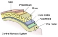 Meninges cartoon.jpg 600 × 385; 38 KB
 Menstrual cycle- brain change measurements.jpg 547 × 893; 88 KB
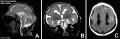 Microlissencephaly MRI-01.jpg 1,280 × 412; 63 KB
 Minot1897 391.jpg 720 × 750; 83 KB
 Minot1897 392.jpg 976 × 900; 145 KB
 Minot1897 393.jpg 981 × 419; 80 KB
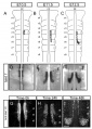
 Mouse - forebrain Robo3 expression.jpg 675 × 1,280; 289 KB
 Mouse - rhombomere boundaries.jpg 669 × 1,000; 147 KB
 Mouse cerebellar foliation defects.jpg 574 × 600; 115 KB
 Mouse cerebellum.jpg 600 × 591; 200 KB
 Mouse cortex and hippocampus development 01.jpg 761 × 622; 112 KB
 Mouse embryo cortex radial expansion.jpg 1,200 × 624; 249 KB
 Mouse neural tube 01 movie icon.jpg 486 × 512; 59 KB
 Mouse neural tube 01.mp4 ; 770 KB
 Mouse neural tube 01.ogg ; 316 KB
 Mouse neural tube bending model.jpg 1,000 × 471; 79 KB
 Mouse posterior neuropore Axd mutant.jpg 475 × 1,074; 95 KB
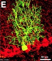 Mouse Purkinje neuron-01.jpg 498 × 578; 152 KB
 Mouse REN expression 01.jpg 904 × 1,000; 293 KB
 Mouse telencephalon radial glia model.jpg 1,000 × 816; 80 KB
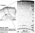 Mouse- adult cortex.jpg 464 × 436; 34 KB
 Mouse- cerebellum axons.jpg 600 × 695; 129 KB
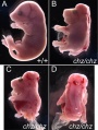 Mouse- chuzhoi mutant.jpg 754 × 1,000; 114 KB
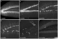
 Mouse- facial branchiomotor neuron migration.jpg 600 × 841; 104 KB
 Mouse- hippocampus dentate granule cells.jpg 1,000 × 671; 130 KB
 Mouse- spinal cord axons.jpg 600 × 693; 127 KB


