Cat Development: Difference between revisions
mNo edit summary |
mNo edit summary |
||
| Line 20: | Line 20: | ||
* '''Development of external genitalia in fetal and neonatal domestic cats'''<ref><pubmed>19262023</pubmed></ref> "The female urogenital folds budded from each side of the genital tubercle and, gradually extended to the tip of the genital tubercle by the 6.8 cm stage in crown-rump length. Then, the well-developed urogenital folds ensheathed completely the genital tubercle to form the prepuce of clitoris and the labia, flanking the external opening of vagina as the folds of skin which were equivalent to the labia minora in humans. The genital swellings known to become the labia majora in humans were clearly recognized in the caudolateral region of the genital tubercle during the fetal stage. These swellings became flat and obscure after birth. Thus, in cats the genital swellings did not join to the formation of the labia in the same way as in humans. The sex difference in the external genitalia was first observed at the 3.2-3.3 cm stages. In the male, the anogenital raphe appeared and the caudal portion of the genital swellings moved and fused each other at the caudal region of the genital tubercle. In the female, both features were not easy to observe." | * '''Development of external genitalia in fetal and neonatal domestic cats'''<ref><pubmed>19262023</pubmed></ref> "The female urogenital folds budded from each side of the genital tubercle and, gradually extended to the tip of the genital tubercle by the 6.8 cm stage in crown-rump length. Then, the well-developed urogenital folds ensheathed completely the genital tubercle to form the prepuce of clitoris and the labia, flanking the external opening of vagina as the folds of skin which were equivalent to the labia minora in humans. The genital swellings known to become the labia majora in humans were clearly recognized in the caudolateral region of the genital tubercle during the fetal stage. These swellings became flat and obscure after birth. Thus, in cats the genital swellings did not join to the formation of the labia in the same way as in humans. The sex difference in the external genitalia was first observed at the 3.2-3.3 cm stages. In the male, the anogenital raphe appeared and the caudal portion of the genital swellings moved and fused each other at the caudal region of the genital tubercle. In the female, both features were not easy to observe." | ||
|} | |} | ||
{| class="wikitable collapsible collapsed" | {| class="wikitable mw-collapsible mw-collapsed" | ||
! More recent papers | ! More recent papers | ||
|- | |- | ||
Revision as of 15:22, 18 June 2014
| Embryology - 30 Apr 2024 |
|---|
| Google Translate - select your language from the list shown below (this will open a new external page) |
|
العربية | català | 中文 | 中國傳統的 | français | Deutsche | עִברִית | हिंदी | bahasa Indonesia | italiano | 日本語 | 한국어 | မြန်မာ | Pilipino | Polskie | português | ਪੰਜਾਬੀ ਦੇ | Română | русский | Español | Swahili | Svensk | ไทย | Türkçe | اردو | ייִדיש | Tiếng Việt These external translations are automated and may not be accurate. (More? About Translations) |
Introduction


Cats (Felis catus) are seasonally polyestrous animals that have multiple estrous cycles only during certain periods of the year.
- Links: Estrous Cycle | Toxoplasmosis | Category:Cat
| Animal Development: axolotl | bat | cat | chicken | cow | dog | dolphin | echidna | fly | frog | goat | grasshopper | guinea pig | hamster | horse | kangaroo | koala | lizard | medaka | mouse | opossum | pig | platypus | rabbit | rat | salamander | sea squirt | sea urchin | sheep | worm | zebrafish | life cycles | development timetable | development models | K12 |
Some Recent Findings
|
| More recent papers |
|---|
 This table allows an automated computer search of the external PubMed database using the listed "Search term" text link.
More? References | Discussion Page | Journal Searches | 2019 References | 2020 References Search term: Cat Embryology <pubmed limit=5>Cat Embryology</pubmed> |
Developmental Timeline

Twenty-two stages have been described for the prenatal development of the domestic cat.[4]
The following data on early development is based upon the time after copulation[5]
oviduct embryo development
- 64 hours - 1 to 4 cells (17 of 20; 85.0%)
- 76 hours - 5 to 8 cells (18 of 28; 64.3% )
- 100 hours - 9 to 16 cells (14 of 24; 58.3%)
- 124 hours - morulae (15 of 21; 71.4% )
uterine embryo development
- 148 hours - compact morulae or early blastocysts
- days 12-14 - implantation occurs
Oocyte and Spermatozoa
The following scanning electron micrographs are from a recent paper on fresh and frozen cat oocytes.[6] Scale bar is 10 microns.


Genetics
Lineage: Eukaryota; Opisthokonta; Metazoa; Eumetazoa; Bilateria; Coelomata; Deuterostomia; Chordata; Craniata; Vertebrata; Gnathostomata; Teleostomi; Euteleostomi; Sarcopterygii; Tetrapoda; Amniota; Mammalia; Theria; Eutheria; Laurasiatheria; Carnivora; Feliformia; Felidae; Felinae; Felis; Felis catus
- Nucleus - not much genomic sequencing information available for the cat.
- Mitochondria - entire mitochondrial genome 17,009 bp has been sequenced.
- Links: Mitochondrial Genome
Placenta
- zonary placenta without cotyledons
- relatively small marginal hematoma
- materno-fetal barrier is endothelial-chorial
- superficially invasive into the endometrium but not into the myometrium
- placental labryrinth has characteristic giant cells
Placental cord
- two pairs of vessels in the cord
- two arteries and two veins
- allantoic duct
- cord average length 2 to 3 cm and 0.3 to 0.5 cm in diameter
- inserts at the margin of the zonary organ
- no spirals, no vitelline duct, and no additional vessels or structures
Additional Images
Historic Images
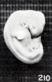
Fig. 210. Normal well-preserved cat fetus
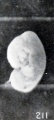
Fig. 211. Normal poorly preserved cat fetus of approximately the same length
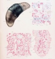
Plate 4. Cat Fetus and Placenta
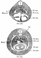
Fig. 297. Transverse section through the thoracic region of a cat embryo of 25 mm
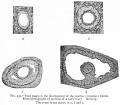
Fig. 331. Sections of a cat's ovary.
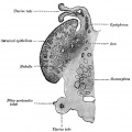
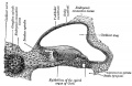
Fig. 903. Cat cochlear duct and ganglia


References
- ↑ <pubmed>21798582</pubmed>
- ↑ <pubmed>19262023</pubmed>
- ↑ <pubmed>20003339</pubmed>| Reprod Biol Endocrinol.
- ↑ <pubmed>11841356</pubmed>
- ↑ <pubmed>7803616</pubmed>| PDF
- ↑ <pubmed>17908298</pubmed>| Acta Vet Scand.
Articles
<pubmed>19151510</pubmed> <pubmed>19262023</pubmed> <pubmed>18405438</pubmed> <pubmed>12606460</pubmed> <pubmed>11841356</pubmed>
Search Pubmed: cat development | feline development
| Animal Development: axolotl | bat | cat | chicken | cow | dog | dolphin | echidna | fly | frog | goat | grasshopper | guinea pig | hamster | horse | kangaroo | koala | lizard | medaka | mouse | opossum | pig | platypus | rabbit | rat | salamander | sea squirt | sea urchin | sheep | worm | zebrafish | life cycles | development timetable | development models | K12 |
Glossary Links
- Glossary: A | B | C | D | E | F | G | H | I | J | K | L | M | N | O | P | Q | R | S | T | U | V | W | X | Y | Z | Numbers | Symbols | Term Link
Cite this page: Hill, M.A. (2024, April 30) Embryology Cat Development. Retrieved from https://embryology.med.unsw.edu.au/embryology/index.php/Cat_Development
- © Dr Mark Hill 2024, UNSW Embryology ISBN: 978 0 7334 2609 4 - UNSW CRICOS Provider Code No. 00098G