BGDB Gastrointestinal - Late Embryo: Difference between revisions
mNo edit summary |
mNo edit summary |
||
| Line 3: | Line 3: | ||
==Week 8== | ==Week 8== | ||
{| | {| | ||
| We have now reached late embryonic development. Start by looking briefly the process of how the definitive GIT tube is formed and then at the overview of the Carnegie stage | | We have now reached late embryonic development. Start by looking briefly the process of how the definitive GIT tube is formed and then at the overview of the Carnegie stage {{CS22}} embryo GIT from one end to the other. | ||
Then work through the listed specific serial sections of the embryo identifying the GIT features. Alternatively step through the serial sections yourself identifying the tract, its associated mesentries, organs and spaces. Note you should also be comparing the GIT appearance with the earlier embryonic (13/14) Carnegie stage. | Then work through the listed specific serial sections of the embryo identifying the GIT features. Alternatively step through the serial sections yourself identifying the tract, its associated mesentries, organs and spaces. Note you should also be comparing the GIT appearance with the earlier embryonic (13/14) Carnegie stage. | ||
| Line 236: | Line 236: | ||
'''Human (week 8, Stage 22) pancreas''' | '''Human (week 8, Stage 22) pancreas''' | ||
* Functions- exocrine (amylase, alpha-fetoprotein) and endocrine (pancreatic islets) | * Functions - exocrine (amylase, alpha-fetoprotein) and endocrine (pancreatic islets) | ||
* Pancreatic buds- endoderm, covered in splanchnic mesoderm | * Pancreatic buds - endoderm, covered in splanchnic mesoderm | ||
* Pancreatic bud formation - duodenal level endoderm, splanchnic mesoderm forms dorsal and ventral mesentery, dorsal bud (larger, first), ventral bud (smaller, later) | * Pancreatic bud formation - duodenal level endoderm, splanchnic mesoderm forms dorsal and ventral mesentery, dorsal bud (larger, first), ventral bud (smaller, later) | ||
* Duodenum growth/rotation -brings ventral and dorsal buds together, fusion of buds | * Duodenum growth/rotation - brings ventral and dorsal buds together, fusion of buds | ||
* Pancreatic duct - ventral bud duct and distal part of dorsal bud, exocrine function | * Pancreatic duct - ventral bud duct and distal part of dorsal bud, exocrine function | ||
* Islet cells- cords of endodermal cells form ducts, which cells bud off to form islets | * Islet cells- cords of endodermal cells form ducts, which cells bud off to form islets | ||
| Line 246: | Line 246: | ||
==Teeth== | ==Teeth== | ||
Epitheilal/mesenchymal (ectoderm first pharyngeal arch and neural crest ectomesenchymal cells) interactions in development and has a major contribution from the neural crest. This has been described as 5 stages of development from late embryonic through early fetal period forming the deciduous teeth. Humans have 2 sets of teeth the deciduous and adult teeth that replace them. (More? | Epitheilal/mesenchymal (ectoderm first pharyngeal arch and neural crest ectomesenchymal cells) interactions in development and has a major contribution from the neural crest. This has been described as 5 stages of development from late embryonic through early fetal period forming the deciduous teeth. Humans have 2 sets of teeth, the deciduous and then the adult teeth that replace them. (More? {{tooth}}) | ||
{{Tooth stages table01}} | {{Tooth stages table01}} | ||
| Line 252: | Line 252: | ||
{{GIT terms}} | {{GIT terms}} | ||
==Additional Information== | ==Additional Information== | ||
{{Med Prac additional Information}} | {{Med Prac additional Information}} | ||
Revision as of 10:51, 28 April 2018
Week 8
| We have now reached late embryonic development. Start by looking briefly the process of how the definitive GIT tube is formed and then at the overview of the Carnegie stage 22 embryo GIT from one end to the other.
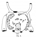
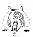
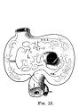
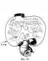
Then work through the listed specific serial sections of the embryo identifying the GIT features. Alternatively step through the serial sections yourself identifying the tract, its associated mesentries, organs and spaces. Note you should also be comparing the GIT appearance with the earlier embryonic (13/14) Carnegie stage. GIT tube has a different appearance at different levels; stomach, duodenum, midgut and hindgut midgut herniated at the umbilicus, lying outside the ventral body wall, connected by mesentry large liver lying directly under the diaphragm and occupying the entire ventral body cavity with organs "embedded" within it the developing pancreas lying in the loop between stomach and duodenum |
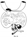
MRI of human midgut herniation at the end of embryonic development i week 8. |
Observe:

|

| |
| Human Embryo (Carnegie stage 22, week 8) | A 3D reconstruction of the gastrointestinal tract. | The developing esophagus. |
| Section | Name | Description |
|---|---|---|

|
E6L | Liver. Ductus venosus.
Cardio-oesophageal junction (cf. E5). Inferior vena cava. |

|
E7L | Stomach body, with mucosa, submucosa and muscularis externa.
Lesser sac. Lesser omentum. Pyloroduodenal junction. Folded duodenal mucosa. Inferior vena cava. Portal vein. Hepatic ducts. Gallbladder. |

|
F1L | Stomach body. Spleen. Pyloric canal. Duodenum.
Pancreas. Small intestine loop (jejunum) cut tangentially, ventral to liver. Portal vein. |

|
F2L | Stomach, spleen. Superior mesenteric artery.
Superior mesenteric vein crossing cranial to body of pancreas. Tail of pancreas. Duodenum. Small intestinal loop herniating from abdominal cavity into the coelom of the umbilical cord (remnant of extra-embryonic coelom). |

|
F4L | Greater curvature of stomach (tangential section). Lesser sac. Greater omentum. Duodenal/jejunal junction.
Note colon (small lumen, darkly-staining wall) and its mesocolon. Note the sections of small and large intestine within the umbilical cord coelom and their mesenteries. Note the thickened jelly to one side of the umbilical cord, containing umbilical vein and R umbilical artery. |

|
F5L | Lesser sac. Greater omentum. Duodenum. Jejunum (cut twice with mesentery in between). Colon and mesocolon. |

|
F6L | Greater omentum and lesser sac.
Jejunum with mesentery. Colon with mesocolon. Three layers of abdominal muscles. Both umbilical arteries now inside abdominal cavity with urachus between them. |

|
F7L | In abdominal cavity - colon with mesocolon, jejunum. Greater omentum and lesser sac.
Umbilical cord - containing umbilical arteries and small dark allantois. Umbilical cord coelom containing mainly, small intestinal loops with their mesentery. |

|
G1L
|
Umbilical cord and coelom containing small intestine loops.
Colon and mesocolon. Jejunum (G1 only). Bladder with umbilical arteries either side. Knees. |

|
G3L | Rectum.
Bladder. Umbilical arteries arising from common iliac arteries. |

|
G4L | Rectum. |

|
G5L | Recto-anal junction with rectovesical pouch of peritoneal cavity. |

|
G6L | Anal canal with triangular lumen. |


Lumen Development
| <html5media height="480" width="255">File:Gastrointestinal tract growth 02.mp4</html5media> |
This is a simplified animation showing how the gastrointestinal tract wall changes during the late embryonic period.
Week 8 - By the end of this week the GIT endoderm tube is a tube once more. Week 9 - (early fetal) the endoderm of this now hollow tube differentiates into the mucosal epithelium (endoderm). |
| Page | Play |
- Splanchnic mesoderm will form the submucosa connective tissue and smooth muscle (circular and longitudinal) layers (mesoderm).
- Neural crest cells migrate into this tissue and will form the nerve plexus innervation (ectoderm).
Organs

Note that while the spleen is not a gastrointestinal tract organ (part of the Immune System), it is often described with this system as it develops within the dorsal mesentery.
Liver
| Feature | ||
|---|---|---|
| hepatic diverticulum development (ductal plate) | ||
| cell differentiation
septum transversum forming liver stroma hepatic diverticulum forming hepatic trabeculae | ||
| epithelial cord proliferation enmeshing stromal capillaries | ||
| hepatic gland and its vascular channels enlarge
hematopoietic function appeared | ||
| obturation due to epithelial proliferation
bile ducts became reorganized (continuity between liver cells and gut) | ||
| biliary ductules developed in periportal connective tissue
produces ductal plates that receive biliary capillaries | ||
| Human data[1], see also liver development in the rat embryonic period (Carnegie stages 15-23).[2] (More? Detailed Timeline | Timeline human development) | ||

|
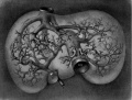
E3 Overview of liver region for selected high power views shown below. Note the position and size of the developing liver spanning the entire abdomen and within the liver the large central ductus venosus. |

|
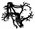
E4 Central veins of liver. Radiating appearance of hepatic sinusoids. unlabeled version |

|
E5 Central vein with endothelial lining, containing nucleated erythrocytes, fetal red blood cells. The fetal liver has an important haemopoietic role. unlabeled version |

|
The Adult Liver Lobule |

- Embryonic Liver and Vasculature
4.3 mm CRL
4.5 mm CRL
6.6 mm CRL
7 mm CRL
9 mm CRL
11 mm CRL
20 mm CRL


Pancreas
Exocrine Function - Pancreatic amylase digests starch to maltose. Postnatally, a blood test to detect amylase can be used to diagnose and monitor acute or chronic pancreatitis (pancreas inflammation).
Pancreatic Duct


The initial formation of the pancreas as two separate lobes each with their own duct that fuses leads a range of anatomical variations in the adult exocrine pancreatic duct. Pancreatic duct five variation classification: common, ansa pancreatica, branch fusion, looped, and separated. Accessory pancreatic duct (APD, of Santorini) in the embryo is the main drainage duct of the dorsal pancreatic bud emptying into the minor duodenal papilla. In the adult it has been further classified as either long-type (joins main pancreatic duct at pancreas neck portion) and short-type (joins main pancreatic duct near first inferior branch).
- Main Pancreatic Duct (MPD or Wirsung's duct) forms within the dorsal pancreatic bud and is present in the body and tail of the pancreas. Discovered by Johann Georg Wirsung (1589 - 1643) a German physician who worked as a prosector in Padua.
- Accessory Pancreatic Duct (APD or Santorini’s duct) is present mainly in the head of the pancreas. Originally dissected and delineated by Giovanni Domenico Santorini (1681 - 1737) an Italian anatomist.
- Endoscopic Retrograde Cholangiopancreatography (ERCP) is a medical procedure which allows an injected dye to display the duct system on an x ray (pancreatograms).

Human (week 8, Stage 22) pancreas
- Functions - exocrine (amylase, alpha-fetoprotein) and endocrine (pancreatic islets)
- Pancreatic buds - endoderm, covered in splanchnic mesoderm
- Pancreatic bud formation - duodenal level endoderm, splanchnic mesoderm forms dorsal and ventral mesentery, dorsal bud (larger, first), ventral bud (smaller, later)
- Duodenum growth/rotation - brings ventral and dorsal buds together, fusion of buds
- Pancreatic duct - ventral bud duct and distal part of dorsal bud, exocrine function
- Islet cells- cords of endodermal cells form ducts, which cells bud off to form islets
Teeth
Epitheilal/mesenchymal (ectoderm first pharyngeal arch and neural crest ectomesenchymal cells) interactions in development and has a major contribution from the neural crest. This has been described as 5 stages of development from late embryonic through early fetal period forming the deciduous teeth. Humans have 2 sets of teeth, the deciduous and then the adult teeth that replace them. (More? tooth)
| Stage | Human (weeks) |
Mouse (days) | |
| lamina | 
|
Week 6 | E 11 |
| placode | 
|
Week 7 | E 11.5 |
| bud | 
|
Week 8 | E 12.5 |
| cap | 
|
Week 11 | E 14.5 |
| bell | 
|
Week 14 | E 15.5 |
| Tooth Stages | |||
|---|---|---|---|
| Stage | Human (weeks) |
Mouse (days) | |
| lamina |
|
Week 6 | E11 |
| placode |
|
Week 7 | E11.5 |
| bud |
|
Week 8 | E12.5 |
| cap |
|
Week 11 | E14.5 |
| bell |
|
Week 14 | E15.5 |
| Gastrointestinal Tract Terms | ||
|---|---|---|
| ||
|
Additional Information
| Additional Information - Content shown under this heading is not part of the material covered in this class. It is provided for those students who would like to know about some concepts or current research in topics related to the current class page. |
Embryonic Liver Timeline
The table below is a detailed timeline overview of human liver development.
| Carnegie Stage | Age (days) | CRL (mm) | Biliary system | Vascular | Hepatic parenchyma |
|---|---|---|---|---|---|
| 14 | 33 | 7 |
|
|
|
| 18 | 46 | 15 |
|
|
|
| 21 | 53 | 22.5 | Bile duct morphology as earlier stage. Common bile duct empties at the level of the proximal duodenum. |
|
Hepatic parenchyma a large rounded mass. |
| 23 | 58 | 27 | Bile duct morphology as earlier stage. |
|
|
| Data from a recent human study[3] Links: liver | Carnegie stage 14 | 18 | 21 | 23 | simple embryonic timeline | Timeline human development | |||||
Cholangiocytes
Epithelial cells that line the intra- and extrahepatic ducts of the biliary tree. These cells modify the hepatocyte-derived bile, and are regulated by hormones, peptides, nucleotides, neurotransmitters, and other molecules.

- Three-dimensional reconstructions of intrahepatic bile duct tubulogenesis in human liver[4]
- initial transition of primitive hepatocytes into cholangiocytes shaping the ductal plate
- process of maturation and remodeling where the intrahepatic biliary tree develops through an asymmetrical form of cholangiocyte tubulogenesis.
Teeth Genetics
- Review - PAX9 gene mutations and tooth agenesis[5] "Paired box 9 (PAX9) is one of the best-known transcription factors involved in the development of human dentition. Mutations in PAX9 gene could, therefore, seriously influence the number, position and morphology of the teeth in an affected individual. To date, over 50 mutations in the gene have been reported as associated with various types of dental agenesis (congenitally missing teeth) and other inherited dental defects or variations. The most common consequence of PAX9 gene mutation is the autosomal-dominant isolated (non-syndromic) oligodontia or hypodontia. In the present review, we are summarizing all known PAX9 mutations as well as their nature and precise loci in the DNA sequence of the gene." PMID 28155232
- ↑ Godlewski G, Gaubert-Cristol R, Rouy S & Prudhomme M. (1997). Liver development in the rat and in man during the embryonic period (Carnegie stages 11-23). Microsc. Res. Tech. , 39, 314-27. PMID: 9407542 <314::AID-JEMT2>3.0.CO;2-H DOI.
- ↑ Godlewski G, Gaubert-Cristol R, Rouy S & Prudhomme M. (1997). Liver development in the rat during the embryonic period (Carnegie stages 15-23). Acta Anat (Basel) , 160, 172-8. PMID: 9718390
- ↑ Lhuaire M, Tonnelet R, Renard Y, Piardi T, Sommacale D, Duparc F, Braun M & Labrousse M. (2015). Developmental anatomy of the liver from computerized three-dimensional reconstructions of four human embryos (from Carnegie stage 14 to 23). Ann. Anat. , 200, 105-13. PMID: 25866917 DOI.
- ↑ <pubmed>21943389</pubmed>
- ↑ <pubmed>28155232</pubmed>
BGDB: Lecture - Gastrointestinal System | Practical - Gastrointestinal System | Lecture - Face and Ear | Practical - Face and Ear | Lecture - Endocrine | Lecture - Sexual Differentiation | Practical - Sexual Differentiation | Tutorial
Glossary Links
- Glossary: A | B | C | D | E | F | G | H | I | J | K | L | M | N | O | P | Q | R | S | T | U | V | W | X | Y | Z | Numbers | Symbols | Term Link
Cite this page: Hill, M.A. (2024, April 26) Embryology BGDB Gastrointestinal - Late Embryo. Retrieved from https://embryology.med.unsw.edu.au/embryology/index.php/BGDB_Gastrointestinal_-_Late_Embryo
- © Dr Mark Hill 2024, UNSW Embryology ISBN: 978 0 7334 2609 4 - UNSW CRICOS Provider Code No. 00098G