AACP Meeting 2013 - Face Embryology: Difference between revisions
mNo edit summary |
mNo edit summary |
||
| Line 303: | Line 303: | ||
==Abnormalities== | ==Abnormalities== | ||
===Pharyngeal Abnormalities=== | |||
There are typically four different terms for the different types of pharyngeal abnormalities, all of these except clefting are relatively rare. | |||
* '''Sinuses''' - a pharyngeal groove defect, when a portion of the groove persists and opens to the skin surface, located laterally on the neck. | |||
* '''Fistula''' - a pharyngeal membrane defect, a tract extends from pharynx (tonsillar fossa) beween the carotid arteries (internal and external) to open on side of neck. | |||
* '''Cysts''' -a cervical sinus defect, remants of the cervical sinus remains as a fluid-filled cyst lined by an epithelium. | |||
* '''Vestiges''' - a cartilaginous or bony developmental remnants that lie under the skin on side of neck. | |||
* '''Clefting''' - the way in which the upper jaw forms from fusion of the smaller upper prominence of the first pharyngeal arch leads to a common congenital defect in this region called "clefting", which may involve either the upper lip, the palate or both structures. | |||
===Cleft Lip and Palate=== | ===Cleft Lip and Palate=== | ||
Revision as of 06:55, 18 May 2013
Face Embryology

2013 Australian Chapter, American Academy of Craniofacial Pain (AACP) Meeting [18 May 2013]

Draft Page - notice removed when complete.
Introduction
This page will be updated and contain the final conference presentation.
| <mediaplayer width='420' height='500' image="http://embryology.med.unsw.edu.au/embryology/images/3/33/Face_001_icon.jpg">file:Face_001.mp4</mediaplayer> |
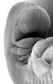
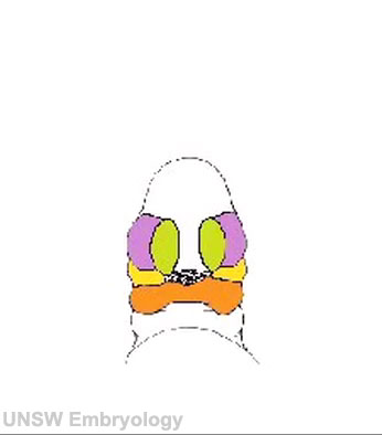
Development of the Face This animation shows a ventral view of development of the human face from approximately week 5 through to neonate. The separate embryonic components that contribute to the face have been colour coded.
The stomodeum is the primordial mouth region and a surface central depression lying between the forebrain bulge and the heart bulge. At the floor of the stomodeum indentation is the buccopharyngeal membrane (oral membrane). Note the complex origin of the maxillary region (upper jaw) requiring the fusion of several embryonic elements, abnormalities of this process lead to cleft lip and cleft palate.
|
Key Concepts
Buccopharyngeal Membrane
These images of the Stage 11 embryo show the breakdown of the buccopharyngeal membrane.

Low power ventral view of the Buccopharyngeal Membrane

Higher power ventrolateral view of the Buccopharyngeal Membrane

Close up view of the degenerating Buccopharyngeal Membrane


The Pharynx


The cavity within the pharyngeal arches forms the pharynx.
- begins at the buccopharyngeal membrane (oral membrane), apposition of ectoderm with endoderm (no mesoderm between)
- expands behind pharyngeal arches
- narrows at glottis and bifurcation of gastrointestinal (oesophagus) and respiratory (trachea) systems
- regions on roof, walls and floor have important contributions to endocrine in oral and neck regions
- also contributes to tongue development
Pharyngeal Arches


Major features to identify for each: arch, pouch, groove and membrane. Contribute to the formation of head and neck and in the human appear at the 4th week. The first arch contributes the majority of upper and lower jaw structures.
- branchial arch (Greek. branchia = gill)
- arch consists of all 3 trilaminar embryo layers
- ectoderm- outside and neural crest
- mesoderm - core of mesenchyme
- endoderm - inside
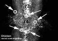
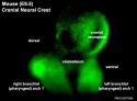
Neural Crest
- Mesenchyme invaded by neural crest generating connective tissue components
- cartilage, bone, ligaments
- arises from midbrain and hindbrain region
|
|
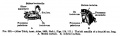
Arch Cartilages


Meckel's cartilage - located within the first pharyngeal arch mandibular prominence, forms a cartilage "template" besides which the mandible develops by the process of intramembranous ossification. It is important to note that this cartilage template does not ossify (endochondral ossification) but provides a transient structure where the mandible will form, and later degenerates.
Week 3
Gestational Age (GA week 5)
These images of the Stage 11 embryo show the breakdown of the buccopharyngeal membrane.
Low power ventral view of the Buccopharyngeal Membrane
Higher power ventrolateral view of the Buccopharyngeal Membrane
Close up view of the degenerating Buccopharyngeal Membrane
Week 4 to 5
Gestational Age (GA week 6 to 7)
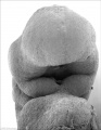
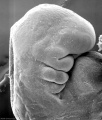
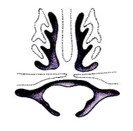
Begins week 4 centered around stomodeum, external depression at oral membrane
5 initial primordia from neural crest mesenchyme (week 4)
- single frontonasal prominence (FNP) - forms forehead, nose dorsum and apex
- nasal placodes develop later bilateral, pushed medially
- paired maxillary prominences - form upper cheek and upper lip
- paired mandibular prominences - lower cheek, chin and lower lip
Stage 11 (25 days)
Stage 12 (26 days)
Stage 13 (28 days)
Stage 14 (32 days)


Cranial Ganglia


Week 6 to 7
Gestational Age (GA week 8 to 9)
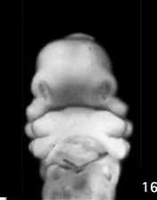
| <mediaplayer width='320' height='420' image="http://embryology.med.unsw.edu.au/embryology/images/9/9c/Stage16-18_face_02.jpg">File:Stage16to18 face 01.mp4</mediaplayer> |  Movie shows a quick animation of the ventral views of the human embryo face, between Carnegie stage 16 to stage 18 (Week 6 to Week 7). Animation based on Kyoto embryos.
|
Week 5 to 8
Gestational Age (GA week 7 to 10)
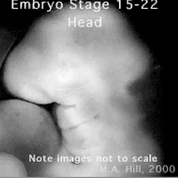
| <mediaplayer width='380' height='400' image="http://embryology.med.unsw.edu.au/embryology/images/9/92/Stage15to22_head_icon.jpg">File:Stage15to22 head 01.mp4</mediaplayer> |
Movie shows a quick animation of the lateral view of the human embryo head, between Carnegie stage 15 to stage 22 (Week 5 to Week 8). Note that these stage images are not to scale.
|
Week 9
Gestational Age (GA week 11)
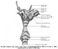
Secondary Palate Development
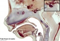
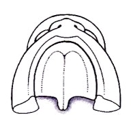
| <mediaplayer width='350' height='350' image="http://embryology.med.unsw.edu.au/embryology/images/4/4f/Palate_001_icon.jpg">File:Palate_001.mp4</mediaplayer> | Animation shows an inferior view of the developmental sequence of secondary palate formation. The lower jaw has been removed and the view shows the roof of the oral cavity and the maxilla (upper jaw) and lip.
|
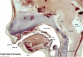
| <mediaplayer width='350' height='350' image="http://embryology.med.unsw.edu.au/embryology/images/a/a3/Palate_002_icon.jpg">File:Palate_002.mp4</mediaplayer> | Animation shows an anterior view of the developmental sequence of secondary palate formation. The frontal region of the head has been removed to show the changes within the oral cavity. Secondary palate formation is the growth of the palatal shelves towards the midline, from top to bottom:
|
Week 10
Gestational Age (GA week 12)

hard palate |

soft palate |
- Fetal Palate Links: Hard and soft palate | Detail - hard and soft palate junction | Detail - hard palate seam | hard palate | hard palate labeled | soft palate | soft palate labeled | Fetal palate movie | MP4 version | GIF version | Palate Development
Image Source: Prof Virginia Diewert
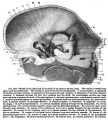
Week 12
Gestational Age (GA week 14)


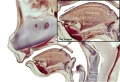
Sagittal unlabeled
Sagittal labeled

Sagittal medial view

Sagittal lateral view
Pituitary unlabeled

Pituitary labeled
Tongue


- 12 Week Images: Sagittal unlabeled | Sagittal labeled | Sagittal medial view | Sagittal lateral view | Pituitary unlabeled | Pituitary labeled | Tongue | Skull Development | Head Development
Week 14
Gestational Age (GA week 16)

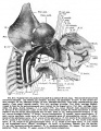
Growth of Head Structures
Maxilla
- First pharyngeal arch - upper maxillary (pair) and lower mandibular prominences
- Late embryonic period - maxillary prominences fuse with frontonasal prominence forming upper jaw (maxilla and upper lip)


- EM Links: Image - stage 16 | Image - stage 17 | Image - stage 18 | Image - stage 19 | Palate Development
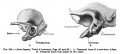
Temporal Bone and Mandible
Image shows growth of both bones from the end of the embryonic period (week 8) through the fetal period of development (to 9 months).

Inner Ear

| Week 5 | Week 8 | ||||
|---|---|---|---|---|---|

|

| ||||
Stage 13 embryo (week 5) showing otocyst that will form the inner ear.
|
Stage 22 embryo (week 8) showing the embryo near the end of the embryonic period.
|
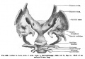
External Ear
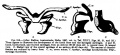
Images of the lateral view of the human embryonic head from week 5 (stage 14) through to week 8 (stage 23) showing development of the auricular hillocks that will form the external ear.
The adult ear is also shown indicating the part of the ear that each hillock contributes.
Images are not to scale.

Fetal Head Growth

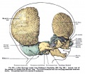
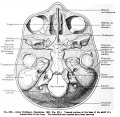
Neonatal Skull
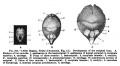
These computed tomography (CT) scans of the normal neonatal skull are shown as as 3D surface-rendered reconstructions.

Abnormalities
Pharyngeal Abnormalities
There are typically four different terms for the different types of pharyngeal abnormalities, all of these except clefting are relatively rare.
- Sinuses - a pharyngeal groove defect, when a portion of the groove persists and opens to the skin surface, located laterally on the neck.
- Fistula - a pharyngeal membrane defect, a tract extends from pharynx (tonsillar fossa) beween the carotid arteries (internal and external) to open on side of neck.
- Cysts -a cervical sinus defect, remants of the cervical sinus remains as a fluid-filled cyst lined by an epithelium.
- Vestiges - a cartilaginous or bony developmental remnants that lie under the skin on side of neck.
- Clefting - the way in which the upper jaw forms from fusion of the smaller upper prominence of the first pharyngeal arch leads to a common congenital defect in this region called "clefting", which may involve either the upper lip, the palate or both structures.
Cleft Lip and Palate

|

|

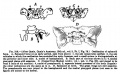
|
| cleft palate | unilateral cleft lip and palate | bilateral cleft lip and palate |
- 300+ different abnormalities, different cleft forms and extent, upper lip and ant. maxilla, hard and soft palate
Victoria
The ten most frequently reported birth defects in Victoria between 2003-2004.
- Hypospadias
- Obstructive Defects of the Renal Pelvis or Obstructive Genitourinary Defects
- Ventricular Septal Defect
- Congenital Dislocated Hip
- Trisomy 21 or Down syndrome
- Hydrocephalus
- Cleft Palate
- Trisomy 18 or Edward Syndrome - multiple abnormalities of the heart, diaphragm, lungs, kidneys, ureters and palate 86% discontinued.
- Renal Agenesis/Dysgenesis - reduction in neonatal death and stillbirth since 1993 may be due to the more severe cases being identified in utero and being represented amongst the increased proportion of terminations (approximately 31%).
- Cleft Lip and Palate - occur with another defect in 33.7% of cases.
| Statistics |
|---|
| USA Selected Abnormalities (CDC National estimates for 21 selected major birth defects 2004–2006) | ||
|---|---|---|
| Birth Defects | Cases per Births (1 in ...) | Estimated Annual Number of Cases |
| anencephaly | 4,859 | 859 |
| spina bifida without anencephaly | 2,858 | 1,460 |
| encephalocele | 12,235 | 341 |
| Anophthalmia/microphthalmia | 5,349 | 780 |
| patent ductus arteriosus/common truncus | 13,876 | 301 |
| transposition of the great vessels | 3,333 | 1,252 |
| Tetralogy of Fallot | 2,518 | 1,657 |
| atrial septal defects/ventricular septal defects | 2,122 | 1,966 |
| hypoplastic left heart | 4,344 | 960 |
| cleft palate without cleft lip | 1,574 | 2,651 |
| cleft lip with and without cleft palate | 940 | 4,437 |
| Esophageal atresia/tracheoesophageal fistula | 4,608 | 905 |
| Rectal and large intestinal atresia/stenosis | 2,138 | 1,952 |
| Reduction deformity, upper limbs | 2,869 | 1,454 |
| Reduction deformity, lower limbs | 5,949 | 701 |
| gastroschisis | 2,229 | 1,871 |
| omphalocele | 5,386 | 775 |
| Diaphragmatic hernia | 3,836 | 1,088 |
| Trisomy 13 | 7,906 | 528 |
| Trisomy 21 (Down syndrome) | 691 | 6,037 |
| Trisomy 18 | 3,762 | 1,109 |
Cleft Palate
- Cleft palate has the International Classification of Diseases code 749.0.
- In Australia the national rate (1982-1992) for this abnormalitity in births was 4.8 - 6/10,000 births, which represented 1,530 infants 5.5% were stillborn and 11.5% liveborn died during neonatal period and slightly more common in twin births than singleton.
Cleft Lip
- The International Classification of Diseases code 749.1 for isolated cleft lip and 749.2 for cleft lip with cleft palate.
- In Australia the national rate (1982-1992) for this abnormalitity was 8.1 - 9.9 /10,000 births. Of 2,465 infants 6.2% were stillborn and 7.8% liveborn died during neonatal period and the rate was similar in singleton and twin births.
- Links: Palate Development
Skull
Cephalic (Greek, kephale = head) are a group of abnormalities that relate to a wide range of skeletal (skull) and neural (brain) associated defects.
| Abnormal Neonatal Skull (CT) | ||
|---|---|---|

Dolichocephaly and Scaphocephaly |

Coronal Synostosis |

Anterior Plagiocephaly |

Turricephaly |
300px
Posterior Plagiocephaly |

Deformational Plagiocepahly |

Trigonocephaly |

Oxycephaly |
- Skull CT Images: Normal overview | Normal vertex and lateral | Normal endocranial and vertex | Normal Vertex - Fontanels | Dolichocephaly and Scaphocephaly | Coronal Synostosis | Anterior Plagiocephaly | Turricephaly | Posterior Plagiocephaly | Deformational Plagiocepahly | Trigonocephaly | Oxycephaly | Computed Tomography
Genetic Syndromes
First Arch Syndrome - There are 2 major types of associated first arch syndromes, Treacher Collins (Mandibulofacial dysostosis) and Pierre Robin (Pierre Robin complex or sequence), both result in extensive facial abnormalites.
Treacher Collins Syndrome

- a rare autosomal dominant craniofacial disorder (1:50,000)
- TCOF1 gene encoding Treacle protein
- caused by frameshift deletions or duplications
- located chromosome 5
- encodes a serine/alanine-rich nucleolar phospho-protein
Features
- hypoplasia of the mandible and zygomatic complex
- down-slanting palpebral fissures
- coloboma of the lower eyelid
- absence of eyelashes medial to the defect
- external and middle ear malformation
- conductive hearing loss
Pierre Robin Syndrome
Also called Pierre Robin sequence.
- Hypoplasia of the mandible, cleft palate, eye and ear defects.
- micrognathia - Initial defect is small mandible resulting in posterior displacement of tongue and a bilateral cleft palate.
- retroglossia
- U-shaped posterior cleft palate


Frontal and lateral views of an infant with Pierre Robin sequence.[2]
DiGeorge Syndrome
- absence of thymus and parathyroid glands, 3rd and 4th pouch do not form
- disturbance of cervical neural crest migration
Mandibular Hypoplasia

One of the most common malformations of the facial skeleton usually associated with a deficient gonial angle, ascending ramus, and mandibular corpus.
- gonial angle - (angle of the jaw, angle of the mandible) the angle formed by the junction of the posterior and lower borders of the human lower jaw.
- ascending ramus - the more or less vertical part of the jaw which carries the joint with the skull.
- mandibular corpus - the horizontal or tooth-bearing portion of the mandible.
Fetal Alcohol Syndrome

(FAS) Due to alcohol in early development (week 3+) leading to both facial and neurological abnormalities. This disorder was clinically described (USA) in humans about 30 years ago (1973), while historically alcohol's teratogenic effects were identified in the early 20th century in a mix with the prohibition cause of the period. Similar effects without the obvious alterations to appearance, but with nervous system effects, are sometimes identified as Fetal Alcohol Effects (FAE). Alcohol is able to cross the placenta from maternal circulation through the placenta into fetal circulation.
FAS Features:
- lowered ears, small face, mild+ retardation
- Microcephaly - leads to small head circumference
- Short Palpebral fissure - opening of eye
- Epicanthal folds - fold of skin at inside of corner of eye
- Flat midface
- Low nasal bridge
- Indistinct Philtrum - vertical grooves between nose and mouth
- Thin upper lip
- Micrognathia - small jaw
Exposure of embryos in vitro to ethanol simulates premature differentiation of prechondrogenic mesenchyme of the facial primordia (1999)
Movies
|
|
|
|
|
Histology

|

|
| Medial view | Lateral view |
Related Pages
Historic
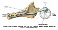
1910 Manual of Human Embryology
Franz Keibel, Franklin P. Mall. (1910) - The The Skull, Hyoid Bone, and Larynx
308
309
310
311
312
313
314
315
316
317
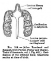
318
319
320
321
322
323
324


1920 Contributions to Embryology Carnegie Institution No.39
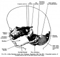
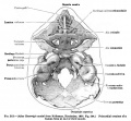
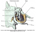
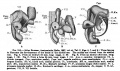
Warren H. Lewis (1920) The Cartilaginous Skull Of A Human Embryo Twenty-One Millimeters In Length
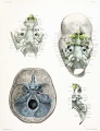
Plate 1
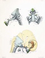
Plate 2
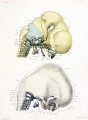
Plate 3
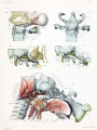
Plate 4
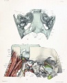
Plate 5


1921 Contributions to Embryology Carnegie Institution No.48
Charles C. Macklin (1921) The skull of a human fetus of 43 millimeters greatest length
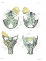
Plate 1
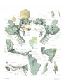
Plate 2
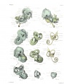
Plate 3
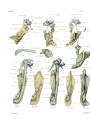
Plate 4


Glossary Links
- Glossary: A | B | C | D | E | F | G | H | I | J | K | L | M | N | O | P | Q | R | S | T | U | V | W | X | Y | Z | Numbers | Symbols | Term Link
Cite this page: Hill, M.A. (2024, April 26) Embryology AACP Meeting 2013 - Face Embryology. Retrieved from https://embryology.med.unsw.edu.au/embryology/index.php/AACP_Meeting_2013_-_Face_Embryology
- © Dr Mark Hill 2024, UNSW Embryology ISBN: 978 0 7334 2609 4 - UNSW CRICOS Provider Code No. 00098G
- ↑ <pubmed>20716376</pubmed>| PMC2931455 | BMC Pregnancy Childbirth.
- ↑ <pubmed>22300418</pubmed>| Ital J Pediatr.