Rabbit Development
| Embryology - 10 Jun 2024 |
|---|
| Google Translate - select your language from the list shown below (this will open a new external page) |
|
العربية | català | 中文 | 中國傳統的 | français | Deutsche | עִברִית | हिंदी | bahasa Indonesia | italiano | 日本語 | 한국어 | မြန်မာ | Pilipino | Polskie | português | ਪੰਜਾਬੀ ਦੇ | Română | русский | Español | Swahili | Svensk | ไทย | Türkçe | اردو | ייִדיש | Tiếng Việt These external translations are automated and may not be accurate. (More? About Translations) |
Introduction

As an embryological tool, the rabbit (Taxon- Oryctolagus cuniculus) along with human was a species which show birth defects with thalidomide (teratogenic effects not detected with prior testing on other species).
These animals are herbivores with a very high breeding rate and number of offspring produced. Rabbit ovulation is induced by mating allowing an accurate staging of embryonic age and pregnancy.
Some Recent Findings
|
| More recent papers |
|---|
 This table allows an automated computer search of the external PubMed database using the listed "Search term" text link.
More? References | Discussion Page | Journal Searches | 2019 References | 2020 References Search term: Rabbit Embryology <pubmed limit=5>Rabbit Embryology</pubmed> |
Taxon
Oryctolagus cuniculus
Taxonomy Id: 9986 Rank: species
Genetic code: Translation table 1 (Standard) Mitochondrial genetic code: Translation table 2 Other names: New Zealand rabbit[includes], rabbits[common name], European rabbit[common name], Japanese white rabbit[common name], domestic rabbit[common name], rabbit[common name], Lepus cuniculus[misnomer]
Lineage( abbreviated ): Eukaryota; Metazoa; Chordata; Craniata; Vertebrata; Mammalia; Eutheria; Lagomorpha; Leporidae; Oryctolagus
Genome
- The Oryctolagus cuniculus haploid genome is estimated to be 3500 Mb.
- The diploid genome is organized in 21 pairs of autosomes and two sex chromosomes.
- Rabbit gene sequences are more similar to human sequences than rodent ones.
- Links: Genome Project Report
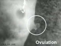
Rabbit Reproductive Cycle
|
|
Developmental Timeline

|
| Early growth of the rabbit morula and blastocyst.[6] |
Early development data from an in vitro development study.[7]
Fertilization - penetration of most ova during the first hour after ovulation.
- 0-16 h - pronuclei
- 16-22 h - 2 cell
- 22-29 h - 4 cell
- 29-32 h - 8 cell
- 32-77h - morula
- 77-98h - blastocyst
- 98h + - hatching blastocyst
- 6 Days - gastrulation starts.
Gonad Development

Timeline of rabbit gonad development.[1]
Day post coïtum (dpc)
- 9 - first germ cells are detected in both sexes.
- 14 - gonad macroscopically evident, the mesonephros and gonads are still connected and interactions between tissues are probable.
- 16 - most germ cells already entered the genital ridges (crests).
- 16 to 25 - regression of the mesonephros.
- 23 - gonadal and mesonephric tissues are separated by connective tissue. Thought to prevent the migration of cells and other substances.
Days post partum (dpp)
- birth - XX gonads first signs of meiosis.
- 50 - XY gonads first signs of meiosis.
- 70 - blood-testis barrier is definitively complete.
(text modified from reference[1])
- Links: Genital System Development
Historic Drawings
The following plates are from Normal Plates of the Development of Vertebrates Vol. 5. 1905 Rabbit (Lepus cuniculus) by Charles S. Minot and Edwing Taylor.

|

|

|
The following drawings were compiled in the textbooks: Bailey, F.R. and Miller, A.M. (1921). Text-Book of Embryology. New York: William Wood and Co; Foster, M., Balfour, F. M., Sedgwick, A., & Heape, W. (1883). The Elements of Embryology. (2nd ed.). London: Macmillan and Co.

|
Rabbit's ovum between 70-90 hours after impregnation, after E. van Beneden. |

|
Historic drawing of a Transverse sections of embryonic disks of rabbit, (a) Kolliker, (b) Rabl.
|

|
Transverse section through primitive groove of rabbit embryo, van Beneden. |

|
Transverse section through primitive groove of rabbit embryo, van Beneden. |

|
Embryo rabbits of about nine days from the dorsal side, Kolliker. |

|
Embryo rabbit of about nine days transverse section through the head, Kolliker. B. is a more highly magnified representation of part of A. |

|
Surface view of area vasculosa of a rabbit embryo of 11 days, van Beneden and Julin. |

|
Advanced embryo of a rabbit (about twelve days), by Mr Weldon. |
| Limb Vasculature (veins) | ||
|---|---|---|

|

|

|
| Rabbit embryo of 14 days (11 mm), modified from Lewis. | Rabbit embryo of 14 days and 18 hours (14.5 mm), modified from Lewis. | Rabbit embryo of 17 days (21 mm), modified from Lewis. |
Rabbit Placentation
Rabbit implantation and placentation is a centric (or fusion) type, where the blastocyst adheres only to the epithelial cells (apical region) by trophectoderm forming projections.[8]
Neural Development
The data below is summarised from an excellent study of early neural development in the rabbit.[9] The same authors have studied neural development in the pig.
- 6 - 8 somite stage - the flat neural plate transforms into a V-shaped neural groove (beginning at rhombo-cervical level)
- 8 and 9 somite stage - multiple closure sites occur simultaneously at three levels
- incipient pros-mesencephalic transition
- incipient mes-rhombencephalic transition
- level of the first pairs of somites
results in four transient neuropores
anterior neuropore
- 9-11 somite stages - anterior and rhombencephalic neuropores close
- mesencephalic neuropore is very briefly present
posterior neuropore
- largest and remains open longest
- 9-10 somite stages - tapered (cranial) portion closes fast within
- wide (caudal) portion closes up to a narrow slit
- further closure slows
- 22 somite stage - full closure occurs
compared with chick and mouse - sequence of multiple site closure resembles that of the mouse embryo, but other important aspects of neurulation resemble those of the chick embryo. In contrast to mouse and chick, no time lag between closure at the three closure sites in the rabbit was seen
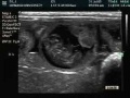
Ultrasound
|
Ultrasound of a 16 day rabbit embryo. |
Rabbit Immune Development
Rabbits generate their antibody repertoire in three stages.[10]
- Neonatal repertoire is generated by B lymphopoiesis in fetal liver and bone marrow (limited by preferential V(H) gene segment usage).
- Between 4 and 8 weeks after birth gut-associated lymphoid tissue (GALT) a complex primary antibody repertoire.
- The primary antibody repertoire is subsequently modified during antigen-dependent immune responses (the secondary repertoire).
Rabbits uniquely develop a primary antibody repertoire through somatic diversification of Ig genes (dependent on intestinal microbial flora).
The esacculus rotundus is located at the ileocaecal junction as an enlargement of the large intestine and contains lymphoid tissue.
Postnatal Rabbit Growth
Postnatal growth data from 2 to 34 weeks of age at biweekly intervals for New Zealand white rabbit.[11]
- 17 male and 12 female rabbits, with the data tabulated separately.
- Skeletal growth was complete at 28 weeks, with the 34 week values mature adult lengths.
Mean body weight
- 2 weeks of age was 6% that at 34 weeks
- 16 weeks was 72% of the weight at 34 weeks
- weight continued to increase in the adult.
Mean body length
- 2 weeks was 40% that at 34 weeks
- 16 weeks was 91% of mature adult
Mean femoral length
- 2 weeks was 38% of adult
- 16 weeks was 95% of adult
Mean tibial length
- 2 weeks was 38% of adult
- 16 weeks was 94% of adult
References
- ↑ 1.0 1.1 1.2 <pubmed>23593221</pubmed> Cite error: Invalid
<ref>tag; name 'PMID23593221' defined multiple times with different content Cite error: Invalid<ref>tag; name 'PMID23593221' defined multiple times with different content - ↑ <pubmed>23181494</pubmed>
- ↑ <pubmed>22669668</pubmed>
- ↑ <pubmed>20047730</pubmed>
- ↑ <pubmed>19732419</pubmed>
- ↑ <pubmed>12683919</pubmed>
- ↑ <pubmed>19245751</pubmed>
- ↑ <pubmed>15579585</pubmed>
- ↑ <pubmed>9543335</pubmed>
- ↑ <pubmed>10933605</pubmed>
- ↑ <pubmed>3712130</pubmed>
Reviews
<pubmed>22580370</pubmed>| Reproduction <pubmed>20150115</pubmed> <pubmed>20150104</pubmed> <pubmed></pubmed>
Articles
<pubmed></pubmed> <pubmed>20874819</pubmed> <pubmed>15271306</pubmed> <pubmed></pubmed>
Search PubMed
- rabbit development - All (26270) Review (1568) Free Full Text (5693)
- rabbit embryo - All (11734) Review (744) Free Full Text (2500)
Search Pubmed: rabbit development | rabbit embryo | Oryctolagus cuniculus development
External Links
External Links Notice - The dynamic nature of the internet may mean that some of these listed links may no longer function. If the link no longer works search the web with the link text or name. Links to any external commercial sites are provided for information purposes only and should never be considered an endorsement. UNSW Embryology is provided as an educational resource with no clinical information or commercial affiliation.
- Food and Agriculture Organization of the United Nations The Rabbit - Husbandry, Health and Production (1997)
| Animal Development: axolotl | bat | cat | chicken | cow | dog | dolphin | echidna | fly | frog | goat | grasshopper | guinea pig | hamster | horse | kangaroo | koala | lizard | medaka | mouse | opossum | pig | platypus | rabbit | rat | salamander | sea squirt | sea urchin | sheep | worm | zebrafish | life cycles | development timetable | development models | K12 | ||
|
Glossary Links
- Glossary: A | B | C | D | E | F | G | H | I | J | K | L | M | N | O | P | Q | R | S | T | U | V | W | X | Y | Z | Numbers | Symbols | Term Link
Cite this page: Hill, M.A. (2024, June 10) Embryology Rabbit Development. Retrieved from https://embryology.med.unsw.edu.au/embryology/index.php/Rabbit_Development
- © Dr Mark Hill 2024, UNSW Embryology ISBN: 978 0 7334 2609 4 - UNSW CRICOS Provider Code No. 00098G