Category:Neural
From Embryology
This Embryology category shows pages and media related to Neural System Development. This includes related topics and undergraduate classes as well as pages and sub-categories describing specific components formed from the original ectoderm neural tube.
Subcategories
This category has the following 12 subcategories, out of 12 total.
Pages in category 'Neural'
The following 200 pages are in this category, out of 934 total.
(previous page) (next page)N
- Neural Exam - 6 month positions - vertical suspension
- Neural Exam - 6 month reflexes - deep tendon reflexes
- Neural Exam - 6 month reflexes - plantar reflex
- Neural Exam - 6 month tone - lower extremity
- Neural Exam - 6 month tone - upper extremity
- Neural Exam - Newborn behaviour comparison
- Neural Exam - Newborn cranial nerves
- Neural Exam - Newborn head circumference
- Neural Exam - Newborn head shape and sutures
- Neural Exam - Newborn normal behaviour
- Talk:Neural Exam - Newborn normal behaviour
- Neural Exam - Newborn positions - prone
- Neural Exam - Newborn positions - ventral suspension
- Neural Exam - Newborn positions - vertical suspension
- Neural Exam - Newborn reflexes - deep tendon reflexes
- Neural Exam - Newborn reflexes - Galant
- Neural Exam - Newborn reflexes - grasp
- Neural Exam - Newborn reflexes - Moro
- Neural Exam - Newborn reflexes - plantar reflex
- Neural Exam - Newborn reflexes - stepping
- Neural Exam - Newborn reflexes - suck, root
- Neural Exam - Newborn tone - arm recoil
- Neural Exam - Newborn tone - arm traction
- Neural Exam - Newborn tone - hand position
- Neural Exam - Newborn tone - head control
- Neural Exam - Newborn tone - head lag
- Neural Exam - Newborn tone - heel to ear
- Neural Exam - Newborn tone - leg recoil
- Neural Exam - Newborn tone - leg traction
- Neural Exam - Newborn tone - lower extremity
- Neural Exam - Newborn tone - neck tone
- Neural Exam - Newborn tone - popliteal angle
- Neural Exam - Newborn tone - resting posture
- Neural Exam - Newborn tone - scarf sign
- Neural Exam - Newborn tone - upper extremity
- Neural Exam Movies
- Talk:Neural Exam Movies
- Template:Neural examination
- Template:Neural fetal
- Template:Neural groove
- Template:Neural interkinetic nuclear migration movie
- Template:Neural Links
- Template:Neural Links 2
- Template talk:Neural Links 2
- Template:Neural Links collapsetable2
- Template:Neural plate
- Neural Plate Movie
- Template:Neural postnatal
- Neural System - Abnormalities
- Neural System - Carnegie Stage 22
- Neural System - Fetal
- Neural System - Glial Development
- Neural System - Molecular
- Neural System - Postnatal
- Neural System Development
- Talk:Neural System Development
- Template:Neural Table
- Template:Neural tube
- Neural Tube Closure Movie
- Template:Neural tube defect
- Template:Neural Tube Defects - Potential risk factors table1
- Neural Tube Movie
- Template:Neural tube regions table
- Template:Neural vascular
- Template:NeuroExam 12month
- Template:Neurohypophysis
- Template:Neuron
- Template:Neuropore
- Talk:New
- Template:Normal 12 Month Neural Exam Table
- Template:Normal 30 Month Neural Exam Table
- Template:Normal Abnormal Newborn Neural Exam Table
- Template:Normal Newborn Neural Exam Table
O
P
- Paper - 1879 The Morphology of the Vertebrate Olfactory Organ
- Paper - A case of accidental impregnation of cells in the brain of a human embryo of four months (1912)
- Paper - A comparison of the growth of the body dimensions of anencephalic human fetuses with normal fetal growth as determined by graphic analysis and empirical formulae
- Paper - A contribution to the histogenesis of the sympathetic nervous system (1909)
- Paper - A contribution to the study of the cerebral cortex in man
- Paper - A human foetus exhibiting iniencephaly and other abnormalities (1922)
- Paper - A note concerning the model of the medulla, pons and midbrain of a new-born babe as reproduced by Herr F. Ziegler (1903)
- Paper - A phylogenetic consideration of the optic tectum
- Paper - A quantitative study of the hypophysis of the human anencephalic fetus (1927)
- Paper - A study of the development of certain features of the cerebellum (1920)
- Paper - A three weeks human embryo with especial reference to the brain and nephric system (1905)
- Paper - Abnormal development of the brain in an 8 mm pig embryo (1938)
- Paper - Address Upon The Development Of The Brain
- Paper - An anencephalic embryo of 25 mm CRL
- Paper - An anencephalic human embryo 16.5 mm long
- Paper - An experimental investigation of the motor cortex and pyramidal tract of echidna aculeata (1939)
- Paper - An experimental study of the origin of the meninges (1924)
- Paper - An iconometrographic representation of the growth of the central nervous system in man
- Paper - Anatomy of the floor of the fourth ventricle (1903)
- Paper - Anencephaly and rhachischisis posterior, with the description of a human hemicephalus of 18 mm (1939)
- Paper - Anterior and posterior rhachischisis (1941)
- Paper - Cell columns in the spinal cord of a human foetus of fourteen weeks (1941)
- Paper - Certain developmental relations and fiber connections of the triangular gyrus in primates (1948)
- Paper - Comparative morphology of the ear 3
- Paper - Comparative studies on the growth of the cerebral cortex 1 (1917)
- Paper - Comparative studies on the growth of the cerebral cortex 2 (1917)
- Paper - Comparative studies on the growth of the cerebral cortex 3 (1918)
- Paper - Comparative studies on the growth of the cerebral cortex 4 (1918)
- Paper - Comparative studies on the growth of the cerebral cortex 5 (1918)
- Paper - Comparative studies on the growth of the cerebral cortex 6 (1918)
- Paper - Comparative studies on the growth of the cerebral cortex 7 (1918)
- Paper - Comparative studies on the growth of the cerebral cortex 8 (1918)
- Paper - Comparative studies upon the origin and development of the brachial plexus
- Paper - Complete dysraphism in 14 somite human embryo
- Paper - Contribution to the structure and development of the vertebrate head
- Paper - Contribution to the structure and development of the vertebrate head 1
- Paper - Contribution to the structure and development of the vertebrate head 2
- Paper - Contribution to the structure and development of the vertebrate head 3
- Paper - Correlated changes in nervous tissues in malformations of the central nervous system (1946)
- Paper - Defective development of the septum pellucidum (1932)
- Paper - Description of a model showing the tracts of fibres medullated in a new-born baby’s brain (1911)
- Paper - Description of a young human anencephalic and amyelic embryo
- Paper - Development and homology of the mammalian cerebellar fissures 1
- Paper - Development and homology of the mammalian cerebellar fissures 2
- Paper - Development and variation of the nerves and the musculature of the inferior extremity and of the neighboring regions of the trunk in man
- Paper - Development of the innervation pattern in the upper limb of staged human embryos (1990)
- Paper - Development of the interfore-brain commissures in the human embryo
- Paper - Development of the region of the isthmus rhombencephali (1928)
- Paper - Development of the spinal reflex mechanism in human embryos
- Paper - Development of the tractus solitarius
- Paper - Differentiation of pituicytes in the human foetus
- Paper - Embryological and morphological studies on the mid-brain and cerebellum of vertebrates
- Paper - Embryological and morphological studies on the mid-brain and cerebellum of vertebrates 1
- Paper - Embryological and morphological studies on the mid-brain and cerebellum of vertebrates 2
- Paper - Embryological and morphological studies on the mid-brain and cerebellum of vertebrates 3
- Paper - Embryological stages in the development of spina bifida and myeloschisis
- Paper - Extroversion of the cerebral hemispheres in a human embryo (1934)
- Paper - Factors Involved In The Formation Of The Filum Terminale
- Paper - Further contributions to the study of the evolution of the forebrain 5
- Paper - Further experiments on the development of peripheral nerves (1906)
- Paper - Further observations on the anatomy of the brain in the monotremata
- Paper - Fusion of notochord to neural tube in a human embryo of the sixth week (1946)
- Paper - Head ganglia of an embryo of eight somite pairs
- Paper - Localization and regeneration in the neural plate of amphibian embryos (1910)
- Paper - Malformations of the human body from a new point of view 1+2
- Paper - Meninges histogenesis and structure
- Paper - Morphology of the roof plate of the fore-brain and the lateral choroid plexuses in the human embryo (1916)
- Paper - Morphophysiology of the cerebral cortex
- Paper - Norms for some structural changes in the human cerebellum from birth to old age (1920)
- Paper - Nuclear masses in the lower portion of the human brain-stem (1914)
- Paper - Observations concerning the comparative anatomy of the diencephalon (1912)
- Paper - Observations on the histogenesis of protoplasmic processes and of collaterals, terminating in end bulbs, of the neurones of peripheral sensory ganglia (1913)
- Paper - Observations on the peripheral distribution of the nervus terminalis in mammalia (1913)
- Paper - On the development and nature of the neuroglia
- Paper - On the development of the blood-vessels of the brain in the human embryo (1905)
- Paper - On the development of the hind-brain of the pig 1
- Paper - On the development of the hind-brain of the pig 2
- Paper - On the embryology of the corpus ponto-bulbare and its relation to the development of the pons
- Paper - On the embryology of the corpus ponto-bulbare and its relation to the development of the pons (1909)
- Paper - On the Mechanism of morphological differentiation in the nervous system 1
- Paper - On the mechanism of morphological differentiation in the nervous system 2. The relation between compression and the development of a series of vesicles (1917)
- Paper - On the nature and mode of origin of the foramen of magendie (1937)
- Paper - On the occurrence of sheath cells and the nature of the axone sheaths in the central nervous system
- Paper - On the pineal region in human embryos
- Paper - On the relation of the head chorda to the pharyngeal epithelium in the pig embryo
- Paper - On the transitory or artificial fissures of the human cerebrum
- Paper - Overgrowth of the neural tube in young human embryos
- Paper - Prenatal growth of the human spinal cord
- Paper - Primary neuromeres and head segmentation (1922)
- Paper - Primitive neurons in the embryonic human central nervous system
- Paper - Recurrent branches of the abducens nerve in human embryos
- Paper - Report on an Anencephalic Embryo
- Paper - Sensory nerves in the skin of human fetuses of 8 to 14 weeks of menstrual age correlated with functional capability (1941)
- Paper - Sequential innervation of the intestinal loop in the human embryo
- Paper - Significant features in the early prenatal development of the human brain
- Paper - Significant superficial anastomoses in the arterial blood supply to the human brain (1959)
- Paper - Some observations on myelination in the human central nervous system (1931)
- Paper - Structural organization of the human cerebral cortex prior to the appearance of the cortical plate (1983)
- Paper - Structural plan of the human brain
- Paper - Studies in the growth and differentiation of the telencephalon in man - the fissura hippocampi
- Paper - Studies on the nervus terminalis - Mammals (1918)
- Paper - Teratological studies (1919)
- Paper - The anterior end of the neural tube and the anterior end of the body (1924)
- Paper - The circle of Willis - An examination of 700 specimens (1905)
- Paper - The connexions of the posterior commissure in the human foetus and young infant
- Paper - The corpus ponto-bulbare - a hitherto undeseribed nuclear mass in the human hind brain (1907)
- Paper - The cortex of the brain in the human embryo during the fourth month with special reference to the so-called Papilla of Retzius
- Paper - The Course of the Phrenic Nerve in the Embryo
- Paper - The developing third nerve nucleus in human embryos
- Paper - The development and myelination of the posterior longitudinal bundle in the human (1933)
- Paper - The development and reduction of the tail and of the caudal end of the spinal cord (1920)
- Paper - The development and significance of the cell columns in the ventral horn of the cervical and upper thoracic spinal cord of the rabbit (1941)
- Paper - The development of a medial motor nucleus and an accessory abducens nucleus in the pig (1934)
- Paper - The development of cerebro-spinal fluid pathway in human embryos (1977)
- Paper - The development of nerve endings in the human foetus
- Paper - The development of the auditory nerve in vertebrates (1910)
- Paper - The development of the cerebral cortex
- Paper - The development of the cerebral ventricles in the pig (1913)
- Paper - The Development of the Cranial and Spinal Nerves in the Occipital Region of the Human Embryo
- Paper - The development of the gyri and sulci on the surface of the island of Reil of the human brain (1891)
- Paper - The development of the human brain stage 12
- Paper - The development of the hypoglossal ganglia of pig embryos
Media in category 'Neural'
The following 200 files are in this category, out of 1,070 total.
(previous page) (next page)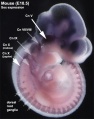 Mouse-E10.5 ganglia Sox10.jpg 540 × 685; 51 KB
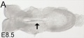 Mouse-E8.5 dorsal view.jpg 788 × 361; 28 KB
 Mouse-E9.0.jpg 800 × 811; 84 KB
 Mouse-olfactory nerve pathway development.jpg 600 × 1,764; 464 KB
 Mouse-optic nerve axons.jpg 600 × 693; 126 KB
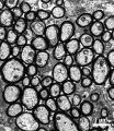
 Mouse-sciatic nerve Schwann cell.jpg 957 × 1,050; 339 KB
 Myelination animation.gif 300 × 200; 77 KB
 Nanagas1925-fig01.jpg 1,319 × 2,000; 251 KB
 Nanagas1925-fig01a.jpg 600 × 765; 40 KB
 Nanagas1925-fig01b.jpg 600 × 765; 41 KB
 Nanagas1925-fig01c.jpg 600 × 765; 34 KB
 Nanagas1925-fig01d.jpg 600 × 765; 45 KB
 Nanagas1925-fig01e.jpg 600 × 765; 52 KB
 Nanagas1925-fig16.jpg 1,300 × 1,901; 451 KB
 Neural - cranial nerves.jpg 800 × 447; 72 KB
 Neural - Human hippocampus marker expression.jpg 600 × 472; 62 KB
 Neural - Sylvian fissure.mov ; 476 KB
 Neural Circuit in the Cerebellum.jpg 293 × 690; 55 KB
 Neural plate movie icon.jpg 100 × 161; 4 KB
 Neural tube - SHH model.jpg 490 × 1,142; 173 KB
 Neural tube defects sex ratio graph 01.png 2,220 × 740; 406 KB
 Neural tube dorsoventral patterning SHH BMP.jpg 1,099 × 891; 148 KB
 Neural tube SHH patterning cartoon.jpg 458 × 594; 92 KB
 Neural- cortex Cajal drawing 01.jpg 800 × 925; 154 KB
 Neural-development.jpg 800 × 535; 66 KB
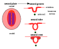 Neuralplate cartoon.png 343 × 284; 5 KB
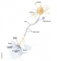 Neuron cartoon.jpg 469 × 500; 11 KB
 Neuroscience 2001.jpg 320 × 400; 23 KB
 Newborn ab 01.flv ; 2.61 MB
 Newborn ab 01.jpg 320 × 240; 11 KB
- Newborn ab 01.mp4 ; 4.4 MB
- Newborn ab 02.flv ; 1.14 MB
 Newborn ab 02.jpg 320 × 240; 12 KB
- Newborn ab 02.mp4 ; 1.8 MB
- Newborn ab 03.flv ; 235 KB
 Newborn ab 03.jpg 320 × 240; 11 KB
- Newborn ab 03.mp4 ; 398 KB
- Newborn ab 04.flv ; 1.41 MB
 Newborn ab 04.jpg 320 × 240; 10 KB
- Newborn ab 04.mp4 ; 1.87 MB
- Newborn ab 05.flv ; 907 KB
 Newborn ab 05.jpg 320 × 240; 12 KB
- Newborn ab 05.mp4 ; 1.23 MB
- Newborn ab 06.flv ; 1.01 MB
 Newborn ab 06.jpg 320 × 240; 13 KB
- Newborn ab 06.mp4 ; 1.52 MB
- Newborn ab 07.flv ; 854 KB
 Newborn ab 07.jpg 320 × 240; 11 KB
- Newborn ab 07.mp4 ; 1.15 MB
- Newborn ab 08.flv ; 894 KB
 Newborn ab 08.jpg 320 × 240; 12 KB
- Newborn ab 08.mp4 ; 1.2 MB
- Newborn ab 09.flv ; 1.76 MB
 Newborn ab 09.jpg 320 × 240; 11 KB
- Newborn ab 09.mp4 ; 2.4 MB
- Newborn ab 10.flv ; 501 KB
 Newborn ab 10.jpg 320 × 240; 11 KB
- Newborn ab 10.mp4 ; 697 KB
- Newborn ab 11.flv ; 702 KB
 Newborn ab 11.jpg 320 × 240; 11 KB
- Newborn ab 11.mp4 ; 1.02 MB
- Newborn ab 12.flv ; 741 KB
 Newborn ab 12.jpg 320 × 240; 11 KB
- Newborn ab 12.mp4 ; 1.06 MB
- Newborn ab 13.flv ; 779 KB
 Newborn ab 13.jpg 320 × 240; 11 KB
- Newborn ab 13.mp4 ; 1,024 KB
- Newborn ab 14.flv ; 416 KB
 Newborn ab 14.jpg 320 × 240; 11 KB
- Newborn ab 14.mp4 ; 601 KB
- Newborn ab 15.flv ; 934 KB
 Newborn ab 15.jpg 320 × 240; 12 KB
- Newborn ab 15.mp4 ; 1.21 MB
- Newborn ab 16.flv ; 922 KB
 Newborn ab 16.jpg 320 × 240; 13 KB
- Newborn ab 16.mp4 ; 1.24 MB
- Newborn ab 17.flv ; 1.57 MB
 Newborn ab 17.jpg 320 × 240; 11 KB
- Newborn ab 17.mp4 ; 2.19 MB
- Newborn ab 18.flv ; 1.36 MB
 Newborn ab 18.jpg 320 × 240; 13 KB
- Newborn ab 18.mp4 ; 1.74 MB
- Newborn ab 19.flv ; 878 KB
 Newborn ab 19.jpg 320 × 240; 12 KB
- Newborn ab 19.mp4 ; 1.13 MB
- Newborn ab 20.flv ; 2.56 MB
 Newborn ab 20.jpg 320 × 240; 11 KB
- Newborn ab 20.mp4 ; 3.43 MB
- Newborn ab 21.flv ; 1.54 MB
 Newborn ab 21.jpg 320 × 240; 11 KB
- Newborn ab 21.mp4 ; 2.27 MB
 Newborn ab 22.jpg 320 × 240; 10 KB
- Newborn ab 22.mp4 ; 1.32 MB
- Newborn ab 23.flv ; 1.18 MB
 Newborn ab 23.jpg 320 × 240; 11 KB
- Newborn ab 23.mp4 ; 1.61 MB
- Newborn ab 24.flv ; 1,008 KB
 Newborn ab 24.jpg 320 × 240; 11 KB
- Newborn ab 24.mp4 ; 1.23 MB
- Newborn ab 25.flv ; 518 KB
 Newborn ab 25.jpg 320 × 240; 13 KB
- Newborn ab 25.mp4 ; 664 KB
- Newborn ab 26.flv ; 1.45 MB
 Newborn ab 26.jpg 320 × 240; 12 KB
- Newborn ab 26.mp4 ; 2.07 MB
- Newborn ab 27.flv ; 1.39 MB
 Newborn ab 27.jpg 320 × 240; 11 KB
- Newborn ab 27.mp4 ; 1.96 MB
- Newborn ab 28.flv ; 920 KB
 Newborn ab 28.jpg 320 × 240; 10 KB
- Newborn ab 28.mp4 ; 1.2 MB
- Newborn n 01.flv ; 2.41 MB
 Newborn n 01.jpg 320 × 240; 11 KB
- Newborn n 01.mp4 ; 3.73 MB
 Newborn n 02.jpg 320 × 240; 13 KB
- Newborn n 02.mp4 ; 3.39 MB
- Newborn n 03.flv ; 410 KB
 Newborn n 03.jpg 320 × 240; 13 KB
- Newborn n 03.mp4 ; 536 KB
- Newborn n 04.flv ; 1.06 MB
 Newborn n 04.jpg 320 × 240; 12 KB
- Newborn n 04.mp4 ; 1.22 MB
- Newborn n 05.flv ; 1.11 MB
 Newborn n 05.jpg 320 × 240; 12 KB
- Newborn n 05.mp4 ; 1.47 MB
- Newborn n 06.flv ; 1.03 MB
 Newborn n 06.jpg 320 × 240; 12 KB
- Newborn n 06.mp4 ; 1.3 MB
- Newborn n 07.flv ; 1.49 MB
 Newborn n 07.jpg 320 × 240; 13 KB
- Newborn n 07.mp4 ; 1.83 MB
- Newborn n 08.flv ; 465 KB
 Newborn n 08.jpg 320 × 240; 14 KB
- Newborn n 08.mp4 ; 645 KB
- Newborn n 09.flv ; 2.4 MB
 Newborn n 09.jpg 320 × 240; 12 KB
- Newborn n 09.mp4 ; 2.81 MB
- Newborn n 10.flv ; 349 KB
 Newborn n 10.jpg 320 × 240; 13 KB
- Newborn n 10.mp4 ; 454 KB
- Newborn n 11.flv ; 542 KB
 Newborn n 11.jpg 320 × 240; 13 KB
- Newborn n 11.mp4 ; 738 KB
- Newborn n 12.flv ; 2.4 MB
 Newborn n 12.jpg 320 × 240; 13 KB
- Newborn n 12.mp4 ; 2.91 MB
- Newborn n 13.flv ; 1.24 MB
 Newborn n 13.jpg 320 × 240; 13 KB
- Newborn n 13.mp4 ; 1.47 MB
- Newborn n 14.flv ; 487 KB
 Newborn n 14.jpg 320 × 240; 14 KB
- Newborn n 14.mp4 ; 553 KB
 Newborn n 15.jpg 320 × 240; 12 KB
- Newborn n 15.mp4 ; 1.02 MB
- Newborn n 16.flv ; 543 KB
 Newborn n 16.jpg 320 × 240; 14 KB
- Newborn n 16.mp4 ; 693 KB
- Newborn n 17.flv ; 1.26 MB
 Newborn n 17.jpg 320 × 240; 13 KB
- Newborn n 17.mp4 ; 1.49 MB
- Newborn n 18.flv ; 732 KB
 Newborn n 18.jpg 320 × 240; 12 KB
- Newborn n 18.mp4 ; 838 KB
- Newborn n 19.flv ; 436 KB
 Newborn n 19.jpg 320 × 240; 11 KB
- Newborn n 19.mp4 ; 496 KB
- Newborn n 20.flv ; 2.14 MB
 Newborn n 20.jpg 320 × 240; 13 KB
- Newborn n 20.mp4 ; 2.45 MB
- Newborn n 21.flv ; 1.14 MB
 Newborn n 21.jpg 320 × 240; 11 KB
- Newborn n 21.mp4 ; 1.53 MB
- Newborn n 22.flv ; 1.52 MB
 Newborn n 22.jpg 320 × 240; 13 KB
- Newborn n 22.mp4 ; 1.93 MB
- Newborn n 23.flv ; 815 KB
 Newborn n 23.jpg 320 × 240; 14 KB
- Newborn n 23.mp4 ; 958 KB
- Newborn n 24.flv ; 744 KB
 Newborn n 24.jpg 320 × 240; 13 KB
- Newborn n 24.mp4 ; 905 KB
- Newborn n 25.flv ; 710 KB
 Newborn n 25.jpg 320 × 240; 13 KB
- Newborn n 25.mp4 ; 813 KB
- Newborn n 26.flv ; 2.12 MB
 Newborn n 26.jpg 320 × 240; 14 KB
- Newborn n 26.mp4 ; 2.73 MB
- Newborn n 27.flv ; 1.44 MB
 Newborn n 27.jpg 320 × 240; 16 KB
- Newborn n 27.mp4 ; 1.88 MB
- Newborn n 28.flv ; 899 KB
 Newborn n 28.jpg 320 × 240; 12 KB
- Newborn n 28.mp4 ; 1.04 MB
 Newborn-cranial-nerves.jpg 320 × 240; 9 KB
 Newborn-normal-behaviour.jpg 320 × 240; 7 KB
 Opioids and neural development timeline.jpg 1,280 × 1,206; 67 KB
 Patten042.jpg 776 × 815; 119 KB
 Patten044.jpg 758 × 521; 93 KB
 Patten045.jpg 800 × 968; 156 KB
- PediNeuroLogic Intro 01.flv ; 848 KB


