Category:Placenta
From Embryology
Pages in category 'Placenta'
The following 197 pages are in this category, out of 197 total.
A
B
- BGDA Practical - Placenta and Fetal Membranes
- Talk:BGDA Practical - Placenta and Fetal Membranes
- Template:BGDA Practical 14 - Abnormalities Interactive
- Template:BGDA Practical 14 - Diagnostic Interactive
- Template:BGDA Practical 14 - Implantation Interactive
- Template:BGDA Practical 14 - Maternal Decidua Interactive
- Template:BGDA Practical 14 - Placental Cord Interactive
- Template:BGDA Practical 14 - Placental Functions Interactive
- Template:BGDA Practical 14 - Villi Interactive
- BGDA Practical Placenta - Abnormalities
- BGDA Practical Placenta - Cord Development
- BGDA Practical Placenta - Diagnostic Techniques
- BGDA Practical Placenta - Implantation and Early Placentation
- BGDA Practical Placenta - Maternal Decidua
- BGDA Practical Placenta - Placental Functions
- BGDA Practical Placenta - Villi Development
- Template:BGDALabPlacenta
- Book - Contributions to Embryology Carnegie Institution No.158
- Book - Contributions to Embryology Carnegie Institution No.51
- Book - Human Embryology and Morphology 8
- Book - Sex and internal secretions (1961) 15
- Book - Text-Book of Embryology 19
- Book - Text-Book of the Embryology of Man and Mammals 13
- Book - Umbilicus (1916)
- Book - Uterine and tubal gestation (1903) 1-12
C
E
H
I
P
- Paper - A contribution to our knowledge of the earliest known stages of placentation and embryonic development in man
- Paper - An anatomical study of the closure of the ductus arteriosus (1942)
- Paper - Anatomy of the placenta (1858)
- Paper - Cytological studies on the internal secretory functions in the human placenta and decidua (1921)
- Paper - development of mammalian ova and placenta formation in mammals
- Paper - development of mammalian ova and placenta formation in mammals 3
- Paper - Development of the human placenta in the first three months of gestation (1960)
- Paper - Early Development and placentation in arvicola (microtus) amphibius (1922)
- Paper - Lectures on the early stages in the development of mammalian ova and on the differentiation of the placenta in different groups of mammals
- Paper - On the placentation of the macaque (Macaca mulatta), from the time of implantation until the formation of the definitive placenta
- Paper - On the structure of the human placenta, and its connexion with the uterus
- Paper - Pathological changes in the placenta associated with erythroblastosis of the fetus (1938)
- Paper - Placenta praevia in a rhesus monkey (1939)
- Paper - Placental circulation
- Paper - Placentation in the rabbit
- Paper - Radioangiographic studies of circulation in the maternal placenta of the rhesus monkey
- Paper - Retrogressive Changes in the Fetal Vessels and the Suspensory Ligament of the Liver
- Paper - The area of the chorionic villi in the full-term placenta (1922)
- Paper - The cytological structure of the human chorionic villus and decidua parietalis
- Paper - The development and structure of the human placenta
- Paper - The form and the functions of the uterine blood vessels in the rhesus monkey
- Paper - The formation of the umbilical cord and the umbilical region of the anterior abdominal wall
- Paper - The histology and cytology of the human and monkey placenta
- Paper - The histology of the umbilical cord of the pig (1919)
- Paper - The interrelations of the mesonephros, kidney, and placenta in different classes of animals (1916)
- Paper - The morphology of human uteroplacental vasculature
- Paper - The origin and occurrence of the single umbilical artery in normal and abnormal human fetuses (1922)
- Template:Persistent right umbilical vein
- Template:Placenta
- Placenta - Abnormalities
- Placenta - Cord
- Placenta - Histology
- Placenta - Maternal Decidua
- Placenta - Membranes
- Placenta - Stage 13
- Placenta - Stage 22
- Placenta - Vascular Beds
- Placenta - Villi Development
- Template:Placenta abnormalities
- Template:Placenta Abnormalities gallery
- Template:Placenta accreta
- Template:Placenta Cord Histology
- Placenta Development
- Template:Placenta EM links
- Template:Placenta histology
- Template:Placenta increta
- Template:Placenta Links
- Template:Placenta percreta
- Template:Placenta previa
- Placenta Quiz
- Template:Placenta terms
- Template:Placenta vascular
- Template:Placenta vascular bed
- Template:Placenta Villi Timeline table
- Template:Placental cord
- Template:Placental membranes
- Template:Placental villi
- Template:Placental villi timeline
- Template:Pre-eclampsia
- Template:Pregnancy hCG Levels table
- Template:Primary villi
R
- Template:Ramsey1972 figures
- Template:Ref-BacsichSmout1938
- Template:Ref-Bartelmez1957
- Template:Ref-Bloxam1840
- Template:Ref-Bremer1916
- Template:Ref-Bremer1918
- Template:Ref-Dalton1858
- Template:Ref-Dawson1922
- Template:Ref-Dodds1922
- Template:Ref-Falkiner1939
- Template:Ref-Fujimura1921
- Template:Ref-Grosser1910
- Template:Ref-HamiltonBoyd1960
- Template:Ref-HarrisRamsey1966
- Template:Ref-HellmanHertig1938
- Template:Ref-Hertig1935
- Template:Ref-Herzog1909
- Template:Ref-Houston1964
- Template:Ref-Hunter1774
- Template:Ref-Ingalls1932
- Template:Ref-Ingalls1932a
- Template:Ref-Ingalls1932b
- Template:Ref-Jordan1919
- Template:Ref-Meyer1918
- Template:Ref-Ninian1939
- Template:Ref-Palmer1939
- Template:Ref-PMID5965440
- Template:Ref-Ramsey1960
- Template:Ref-Ramsey1972
- Template:Ref-Robinson1904a
- Template:Ref-Robinson1904b
- Template:Ref-Robinson1904c
- Template:Ref-Sansom1922
- Template:Ref-Strachan1923
- Template:Ref-Walls1939
- Template:Ref-Wislocki1920
- Template:Ref-Wislocki1921
- Template:Ref-WislockiBennett1943
- Template:Ref-WislockiStreeter1938
- Template:Ref-Wyburn1939
S
T
U
W
Media in category 'Placenta'
The following 164 files are in this category, out of 364 total.
(previous page) (next page) Kollmann108.jpg 800 × 767; 110 KB
 Kollmann109.jpg 698 × 800; 59 KB
 Kollmann110.jpg 754 × 652; 56 KB
 Kollmann111.jpg 800 × 490; 55 KB
 Kollmann112.jpg 800 × 624; 76 KB
 Kollmann113.jpg 800 × 733; 108 KB
 Kollmann114.jpg 800 × 507; 44 KB
 Kollmann553.jpg 724 × 734; 83 KB
 Kollmann555.jpg 605 × 585; 61 KB
 Kollmann562.jpg 670 × 581; 68 KB
 Kyoto16834 stage17-umbilicus.jpg 1,536 × 1,316; 137 KB
 Listeria maternal-fetal barrier.jpg 600 × 522; 49 KB
 Maternal and paternal resource allocation.png 600 × 448; 274 KB
 McMurrich1930 fig84.jpg 1,280 × 1,819; 784 KB
 McMurrich1930 fig85.jpg 1,280 × 1,698; 373 KB
 McMurrich1930 fig86.jpg 1,280 × 970; 166 KB
 McMurrich1930 fig87.jpg 1,280 × 1,612; 267 KB
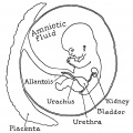
 Minot1889 fig02.jpg 760 × 650; 91 KB
 Minot1889 fig04.jpg 1,000 × 482; 153 KB
 Minot1889 fig26.jpg 952 × 910; 336 KB
 Model male androsterone synthesis.jpg 740 × 518; 92 KB
 Monochorionic twin placenta 01.jpg 600 × 455; 220 KB
 Mouse placenta 01.jpg 429 × 463; 63 KB
 Mouse placenta 02.jpg 464 × 220; 21 KB
 Mouse placenta blood vessel EM01.jpg 1,000 × 739; 132 KB
 Mouse- placenta Hox13 expression.jpg 1,000 × 1,070; 150 KB
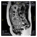
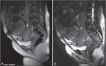
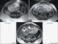 MRI normal placenta different gestational ages.jpg 800 × 609; 75 KB
 MRI Placenta Accreta 01.jpg 600 × 571; 50 KB
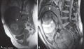 MRI placenta uterus posterior upper segment.jpg 1,200 × 702; 93 KB
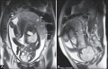 MRI placenta uterus upper segment 37 weeks gestation.jpg 1,200 × 773; 109 KB
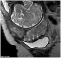
 Multilobed placenta MRI01.jpg 600 × 599; 145 KB
 Ninian1939 plate01.jpg 1,280 × 697; 171 KB
 Peters1899 plate14.jpg 2,000 × 2,527; 343 KB
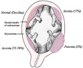 Placenta abnormalities.jpg 450 × 366; 37 KB
 Placenta accreta 01.jpg 1,280 × 960; 440 KB
 Placenta accreta MRI dark intraplacental bands.jpg 2,095 × 1,011; 144 KB
 Placenta accreta ultrasound bladder wall interface.jpg 600 × 537; 48 KB
 Placenta accreta ultrasound retroplacental clear space loss.jpg 600 × 510; 43 KB
 Placenta anchoring villi.jpg 600 × 450; 167 KB
 Placenta and explant model.jpg 800 × 682; 153 KB
 Placenta cartoon.jpg 1,280 × 859; 334 KB
 Placenta drawing 1600.jpg 786 × 1,152; 184 KB
 Placenta gene expression.jpg 925 × 694; 346 KB
 Placenta histology 001.jpg 1,280 × 1,024; 155 KB
 Placenta histology 002.jpg 1,280 × 1,024; 102 KB
 Placenta histology 003.jpg 1,280 × 1,024; 58 KB
 Placenta histology 004.jpg 1,280 × 960; 526 KB
 Placenta histology 005.jpg 1,280 × 960; 433 KB
 Placenta histology 006.jpg 666 × 500; 94 KB
 Placenta histology 007.jpg 1,265 × 437; 207 KB
 Placenta histology 008.jpg 800 × 599; 198 KB
 Placenta Hofbauer cells 01.jpg 934 × 700; 156 KB
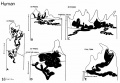
 Placenta humans and guinea-pig cartoon.jpg 1,200 × 889; 562 KB
 Placenta lobule blood flow cartoon.jpg 692 × 800; 134 KB
 Placenta MRI 01.jpg 800 × 516; 60 KB
 Placenta MRI 02.jpg 1,200 × 631; 91 KB
 Placenta MRI01.jpg 1,280 × 1,227; 317 KB
 Placenta oxygen exchange levels.jpg 404 × 401; 18 KB
 Placenta percreta 01.jpg 1,200 × 904; 327 KB
 Placenta percreta 02.jpg 1,000 × 405; 105 KB
 Placenta percreta 03.jpg 600 × 482; 65 KB
 Placenta percreta 04.jpg 700 × 575; 79 KB
 Placenta percreta 05.jpg 900 × 676; 77 KB
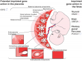 Placenta potential imprinted genes.png 600 × 452; 416 KB
 Placenta previa - anterior.jpg 730 × 547; 107 KB
 Placenta previa 01.jpg 800 × 500; 58 KB
 Placenta previa and increta 01.jpg 960 × 326; 153 KB
 Placenta previa and increta 02.jpg 800 × 544; 48 KB
 Placenta previa ultrasound 01.jpg 739 × 554; 68 KB
 Placenta spiral artery conversion.jpg 592 × 423; 65 KB
 Placenta term anatomy 01.jpg 1,200 × 600; 115 KB
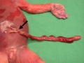
 Placenta velamentous cord 02.jpg 613 × 700; 88 KB
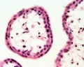 Placenta- first trimester histology x40.jpg 1,000 × 800; 124 KB
 Placenta- umbilical cord torsion.jpg 397 × 298; 29 KB
 Placental arteries week 8 to term.jpg 1,280 × 881; 131 KB
 Placental artery 01.jpg 1,200 × 838; 371 KB
 Placental artery.jpg 600 × 509; 78 KB
 Placental blood flow 01.jpg 1,000 × 556; 63 KB
 Placental chorioangioma 01.jpg 695 × 500; 107 KB
 Placental chorioangioma 02.jpg 695 × 500; 121 KB
 Placental chorioangioma 03.jpg 695 × 500; 111 KB
 Placental chorioangioma ultrasound 01.jpg 1,183 × 837; 101 KB
 Placental chorioangioma ultrasound 02.jpg 1,183 × 837; 92 KB
 Placental cord 01.jpg 600 × 575; 44 KB
 Placental cord blood banks 2009.jpg 800 × 382; 31 KB
 Placental cord cross-section 01.jpg 1,184 × 1,000; 277 KB
 Placental cord cross-section.jpg 525 × 525; 50 KB
 Placental cord epithelium 01.jpg 1,200 × 805; 130 KB
 Placental cord knot.png 300 × 241; 40 KB
 Placental cord ultrasound 01.jpg 585 × 1,309; 192 KB
 Placental cord ultrasound 02.jpg 731 × 479; 101 KB
 Placental cord ultrasound 03.jpg 598 × 646; 61 KB
 Placental cord ultrasound 04.jpg 729 × 572; 65 KB
 Placental cord vessels 01.jpg 650 × 488; 51 KB
 Placental cord vessels 02.jpg 800 × 477; 65 KB
 Placental imaging 01.jpg 695 × 443; 59 KB
 Placental imaging 03.jpg 869 × 830; 227 KB
 Placental imaging 03A.jpg 800 × 738; 181 KB
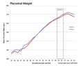 Placental mean weight graph01.jpg 1,500 × 1,200; 114 KB
 Placental membranes.jpg 600 × 450; 99 KB
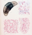
 Placental trophospongium.jpg 567 × 344; 94 KB
 Placental vein.jpg 812 × 392; 59 KB
 Placental villi 1.jpg 1,280 × 1,024; 77 KB
 Placental villi 2.jpg 1,280 × 1,024; 70 KB
 Placental villi 3.jpg 1,280 × 1,024; 99 KB
 Placental villi 4.jpg 1,280 × 1,024; 89 KB
 Placental villi 5.jpg 1,280 × 1,024; 238 KB
 Placental villi 6.jpg 1,000 × 750; 277 KB
 Placental villi.jpg 1,280 × 1,024; 199 KB
 Placental volume - second trimester.jpg 600 × 441; 59 KB
 Placental volume graph.jpg 800 × 628; 54 KB
 Ramsey1960-fig01.jpg 1,000 × 638; 175 KB
 Ramsey1960-fig02.jpg 1,200 × 651; 102 KB
 Ramsey1960-fig03.jpg 1,200 × 641; 85 KB
 Ramsey1960-fig04.jpg 1,000 × 1,957; 208 KB
 Ramsey1972 fig01.jpg 824 × 1,000; 187 KB
 Ramsey1972 fig02.jpg 1,280 × 968; 280 KB
 Ramsey1972 fig03.jpg 828 × 1,000; 221 KB
 Ramsey1972 fig04-16a.jpg 760 × 704; 38 KB
 Ramsey1972 fig04-16b.jpg 760 × 704; 33 KB
 Ramsey1972 fig04-16c.jpg 760 × 704; 47 KB
 Ramsey1972 fig04-16d.jpg 760 × 704; 40 KB
 Ramsey1972 fig04-16e.jpg 760 × 704; 35 KB
 Ramsey1972 fig04.jpg 1,280 × 1,465; 275 KB
 Ramsey1972 fig05a.jpg 900 × 702; 212 KB
 Ramsey1972 fig05b.jpg 900 × 709; 180 KB
 Ramsey1972 fig06.jpg 754 × 1,000; 225 KB
 Ramsey1972 fig07.jpg 1,280 × 767; 143 KB
 Ramsey1972 fig08.jpg 1,280 × 716; 151 KB
 Ramsey1972 fig09.jpg 1,991 × 1,426; 613 KB
 Spiegel1626 table05.jpg 642 × 1,000; 209 KB
 Spiegel1626 table05fig1.jpg 980 × 712; 258 KB
 Spiegel1626 table05fig2.jpg 980 × 712; 263 KB
 Spiegel1626 table07.jpg 707 × 1,000; 251 KB
 Spiral artery trophoblast plug cartoon1.jpg 674 × 616; 159 KB
 Stage 13 image 084.jpg 1,000 × 420; 101 KB
 Stage 13 image 085.jpg 1,000 × 434; 99 KB
 Stage 13 image 086.jpg 1,000 × 499; 97 KB
 Stage 13 image 087.jpg 1,000 × 493; 95 KB
 Stage 13 image 088.jpg 1,000 × 484; 97 KB
 Stage 13 image 089.jpg 1,000 × 498; 104 KB
 Stage 13 image 090.jpg 1,000 × 512; 103 KB
 Stage 13 image 091.jpg 1,000 × 470; 93 KB
 Stage 13 image 092.jpg 1,000 × 481; 86 KB
 Stage 22 image 225.jpg 1,280 × 762; 400 KB
 Stage18 bf10.jpg 1,200 × 897; 151 KB
 Streeter1920 04.jpg 1,000 × 600; 103 KB
 Triplet placenta.jpg 1,260 × 1,080; 220 KB
 Trophoblast cell hCG.jpg 600 × 434; 45 KB
 Trophoblast hCG function.jpg 600 × 1,798; 229 KB
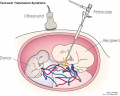
 Twin–twin transfusion syndrome 02.jpg 709 × 398; 31 KB
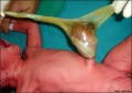
 Twin–twin transfusion syndrome 03.jpg 1,000 × 800; 60 KB
 Ultrasound placenta previa 01.jpg 1,024 × 692; 61 KB
 Umbilical cord hernia 01.jpg 846 × 600; 68 KB
 US Circumvallate placenta 01.jpg 1,024 × 692; 61 KB
 US Circumvallate placenta 02.jpg 1,024 × 692; 72 KB
 Uterine and placental vasculature.jpg 614 × 472; 143 KB
 Uterine vascular anastomoses.jpg 1,200 × 370; 48 KB
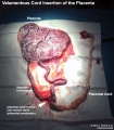
 Velamentous cord insertion 01.jpg 1,024 × 692; 78 KB
 Velamentous cord insertion 02.jpg 1,024 × 692; 104 KB
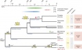 VIT Gene Evolution.jpg 668 × 405; 76 KB
 Waterston02.jpg 661 × 729; 130 KB
 Windle1940 fig43.jpg 1,000 × 1,003; 119 KB
 Wislocki1920 plate 4.jpg 935 × 1,000; 199 KB


