Category:Human
From Embryology
This Embryology category shows pages and media related to human development.
Subcategories
This category has the following 72 subcategories, out of 72 total.
C
- Carnegie Embryo
- Carnegie Embryo 1
- Carnegie Embryo 112
- Carnegie Embryo 1134B
- Carnegie Embryo 116
- Carnegie Embryo 1266
- Carnegie Embryo 1455
- Carnegie Embryo 148
- Carnegie Embryo 172
- Carnegie Embryo 19
- Carnegie Embryo 239
- Carnegie Embryo 2393
- Carnegie Embryo 240
- Carnegie Embryo 248
- Carnegie Embryo 256
- Carnegie Embryo 296
- Carnegie Embryo 3527
- Carnegie Embryo 3956
- Carnegie Embryo 4046
- Carnegie Embryo 4059
- Carnegie Embryo 407
- Carnegie Embryo 4148
- Carnegie Embryo 43
- Carnegie Embryo 4405
- Carnegie Embryo 460
- Carnegie Embryo 463
- Carnegie Embryo 523
- Carnegie Embryo 5541
- Carnegie Embryo 5609
- Carnegie Embryo 5652
- Carnegie Embryo 5682
- Carnegie Embryo 5874
- Carnegie Embryo 6032
- Carnegie Embryo 625
- Carnegie Embryo 6426
- Carnegie Embryo 6581
- Carnegie Embryo 7618
- Carnegie Embryo 7669
- Carnegie Embryo 786
- Carnegie Embryo 808
- Carnegie Embryo 8147
- Carnegie Embryo 8239
- Carnegie Embryo 8370
- Carnegie Embryo 858
- Carnegie Embryo 8630
- Carnegie Embryo 8967
- Carnegie Embryo 9296
- Carnegie Embryo 96
- Carnegie Embryo 963
- Carnegie Embryo 966
- Carnegie Embryo 9697
D
F
H
Pages in category 'Human'
The following 200 pages are in this category, out of 332 total.
(previous page) (next page)A
- Abnormal Development - Cleft Lip and Palate
- Abnormal Development - Cleft Palate
- Abnormal Development - Toxoplasmosis
- Template:Abnormal Newborn Neural Exam Table
- Template:Adipose Timeline table
- Template:Adrenal GA32 Links
- Template:Anderson2016 collapsetable1
- Template:Anderson2016 table1
- Template:AnsonBlack1934 figures
- Aschheim-Zondek Test 1928 Movie
- Template:Australian GIT abnormalities 2002-2003
- Template:Australian Palate abnormalities 2002-2003
B
- Template:B050966
- Template:B100658
- Template:B220849
- Template:Bardeen1906 figures
- Template:Barniville1914 figures
- Template:Bartelmez1922 figures
- Template:BaxterBoyd1939 figures
- BGDA Practical 7 - Week 6
- Birth MRI Movie
- Book - Contributions to Embryology Carnegie Institution No.10
- Book - Contributions to Embryology Carnegie Institution No.112
- Book - Contributions to Embryology Carnegie Institution No.131
- Book - Contributions to Embryology Carnegie Institution No.159
- Book - Contributions to Embryology Carnegie Institution No.21
- Book - Contributions to Embryology Carnegie Institution No.22
- Book - Contributions to Embryology Carnegie Institution No.27
- Book - Contributions to Embryology Carnegie Institution No.29
- Book - Contributions to Embryology Carnegie Institution No.30
- Book - Contributions to Embryology Carnegie Institution No.32
- Book - Contributions to Embryology Carnegie Institution No.33
- Book - Contributions to Embryology Carnegie Institution No.34
- Book - Contributions to Embryology Carnegie Institution No.35
- Book - Contributions to Embryology Carnegie Institution No.38
- Book - Contributions to Embryology Carnegie Institution No.40
- Book - Contributions to Embryology Carnegie Institution No.42
- Book - Contributions to Embryology Carnegie Institution No.43
- Book - Contributions to Embryology Carnegie Institution No.44
- Book - Contributions to Embryology Carnegie Institution No.46
- Book - Contributions to Embryology Carnegie Institution No.47
- Book - Contributions to Embryology Carnegie Institution No.48
- Book - Contributions to Embryology Carnegie Institution No.52
- Book - Contributions to Embryology Carnegie Institution No.55
- Book - Contributions to Embryology Carnegie Institution No.59
- Book - Contributions to Embryology Carnegie Institution No.61
- Book - Contributions to Embryology Carnegie Institution No.65
- Book - Contributions to Embryology Carnegie Institution No.69
- Book - Contributions to Embryology Carnegie Institution No.72
- Book - Contributions to the Development of the Human Brain (1919)
- History:Book - Contributions to the Development of the Human Brain (1919)
- Book - Human embryos of different ages examined in median sections - a contribution to the mechanics of development
- Template:Braune 1877 Plate 2
C
E
F
H
- Template:Hamilton1944 figures
- Harvard Collection
- Template:Hearing EAM timeline
- Hela Apoptosis Movie
- Template:HillH12inks
- Template:HillH13 links
- Template:HillH145 links
- Template:HillH159 links
- Template:HillH202 links
- Template:HillH257 links
- Template:HillH4 links
- Template:HillH5 links
- Template:HillH52
- Template:HillH58 links
- Template:HillH6 links
- Template:HillH8 links
- Template:Hochstadter plates
- Template:Huber1905 table1
- Template:Human 7.5mm Embryo links
- Human Adult Brain Movie
- Human Development Timeline Movie
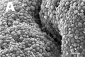
- Human Embryo - Scanning electron microscopy
- Template:Human embryo neck links
- Human Embryo SEM
- Human Fertilization Detail Movie
- Human Fertilization Movie
- Template:Human Fertilization Movie 1 frame table
- Template:Human Fertilization Movie 1 frames
- Template:Human Fertilization Movie 2 frame table
- Template:Human follicles lm and em links
- Template:Human ovary - corpus luteum links
- Template:Human Spermatozoa Statistics collapse table
- Template:Human Spermatozoa Statistics table
- Human Sylvian Fissure Movie
- Template:Human timeline
- Hutchinson-Gilford Progeria Syndrome
J
K
L
M
- Template:Macklin1921 figures
- Template:Mall1912 figures
- Template:Mall1916 figures
- Template:Mall1917 figures
- Menstrual Cycle - Histology
- Model Embryo 1.6mm Movie 1
- Model Embryo 10mm Movie 1
- Model Embryo 3.1mm Movie 1
- Model Embryo 7.5mm Movie 1
- Monosomic Embryo Movie 1
- Template:Morton1949 figures
- Template:Mouse Human lung table
- Movie - Neural Sylvian Fissure
N
P
- Palate Development
- Paper - A human embryo before the appearance of the myotomes (1918)
- Paper - A Human Embryo of Twenty-five Somites
- Paper - A Human Embryo of Twenty-seven Pairs of Somites, Embedded in Decidua
- Paper - A human embryo with head-process and commencing arch enteric canal
- Paper - A Human Embryo with Seven Pairs of Somites Measuring about 2 mm in Length
- Paper - A human embryo with seventeen pairs of somites (1930)
- Paper - A morphological study of testicular descent
- Paper - A Note on the Development of the Septum Transversum and the Liver
- Paper - A presomite human embryo (Shaw) - the implantation
- Paper - A presomite human embryo (Shaw) - the implantation (1942)
- Paper - A presomite human embryo showing a yolk-sac duct
- Paper - A presomite human embryo showing an early stage of the primitive streak
- Paper - A presomite human embryo with a neurenteric canal (embryo R.S.)
- Paper - A study of a 7 mm human embryo with special reference to its peculiar spirally twisted form, and its large aortic cell-clusters
- Paper - A study of the development of certain features of the cerebellum (1920)
- Paper - A very Young Human Embryo found embedded in a "Decidual Cast" of the Uterus
- Paper - A well-preserved human embryo of 10 somites (1929)
- Paper - A Young Human Embryo (Embryo Dobbin) with Head-Process and Prochordal Plate
- Paper - An early human embryo (no. 1285, Manchester Collection) with capsular attachment of the connecting stalk (1935)
- Paper - An Early Human Embryo (No. 1285, Manchester Collection), with Capsular Attachment of the Connecting Stalk
- Paper - An Early Human Embryo, with 0.55 mm long Embryonic Shield
- Paper - An Early Human Ovum (Thomson) in situ
- Paper - An iconometrographic representation of the growth of the central nervous system in man
- Paper - Breech fused twin monster (1934)
- Paper - Changes in fetuses due to formalin preservation
- Special:Badtitle/NS501:Paper - Description of a Human Embryo of 13-14 Mesodermic Somites
- Paper - Description of a Human Embryo of Twenty-three Paired Somites
- History:Paper - Description of a Human Embryo of Twenty-two paired Somites
- Paper - Description of a reconstruction of the head of a thirty-millimetre embryo (1910)
- Paper - Development and variation of the nerves and the musculature of the inferior extremity and of the neighboring regions of the trunk in man
- Paper - Development of the human heart from its earliest appearance to the stage found in embryos of twenty paired somites (1927)
- Paper - Developmental Changes in the Pericardium, the Mesocardia, and the Pleural Sacs in the Human Embryo
Media in category 'Human'
The following 200 files are in this category, out of 2,421 total.
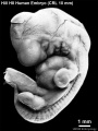
(previous page) (next page)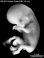 HillH13 Fetus bf01.jpg 1,200 × 1,600; 171 KB
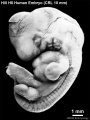
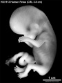 HillH13 Fetus bf02.jpg 1,200 × 1,600; 170 KB
 HillH13 Fetus.gif 450 × 600; 274 KB
 HillH145 Stage 13 bf01.jpg 908 × 1,210; 81 KB
 HillH145 Stage 13 bf02.jpg 908 × 1,210; 83 KB
 HillH145 Stage 13 bf03.jpg 908 × 1,210; 86 KB
 HillH145 Stage 13 bf04.jpg 908 × 1,210; 84 KB
 HillH145 Stage 13 bf05.jpg 1,257 × 1,257; 240 KB
 HillH145 Stage 13 bf06.jpg 1,257 × 1,257; 244 KB
 HillH145 Stage 13 bf07.jpg 1,257 × 1,257; 244 KB
 HillH145 Stage 13 bf08.jpg 1,257 × 1,257; 224 KB
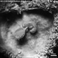
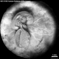
 HillH159 Stage 8 bf01.jpg 1,807 × 1,768; 237 KB
 HillH159 Stage 8 bf02.jpg 1,807 × 1,768; 264 KB
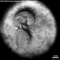
 HillH159 Stage 8 bf03.jpg 1,706 × 1,643; 168 KB
 HillH159 Stage 8 bf04.jpg 1,706 × 1,643; 214 KB
 HillH202 Stage 17 500.gif 500 × 500; 251 KB
 HillH202 Stage 17 600.gif 600 × 600; 346 KB
 HillH202 Stage 17 bf01.jpg 1,200 × 1,166; 173 KB
 HillH202 Stage 17 bf02.jpg 800 × 800; 85 KB
 HillH202 Stage 17 bf03.jpg 800 × 800; 86 KB
 HillH202 Stage 17 bf04.jpg 1,000 × 1,000; 138 KB
 HillH4 Stage 12 bf01.jpg 841 × 1,121; 75 KB
 HillH4 Stage 12 bf02.jpg 841 × 1,121; 75 KB
 HillH5 Stage 16 bf01.jpg 1,125 × 1,500; 142 KB
 HillH5 Stage 16 bf02.jpg 1,125 × 1,500; 142 KB
 HillH5 Stage 16 bf03.jpg 1,125 × 1,500; 148 KB
 HillH5 Stage 16 bf04.jpg 1,125 × 1,500; 149 KB
 HillH5 Stage 16 bf05.jpg 1,125 × 1,500; 116 KB
 HillH5 Stage 16 bf06.jpg 1,125 × 1,500; 111 KB
 HillH5 Stage 16 bf07.jpg 1,125 × 1,500; 151 KB
 HillH5 Stage 16 bf08.jpg 1,125 × 1,500; 149 KB
 HillH5 Stage 16 bf09.jpg 1,125 × 1,500; 109 KB
 HillH5 Stage 16 bf10.jpg 1,125 × 1,500; 110 KB
 HillH5 Stage 16 bf11.jpg 1,125 × 1,500; 147 KB
 HillH5 Stage 16 bf12.jpg 1,125 × 1,500; 154 KB
 HillH5 Stage 16 bf13.gif 450 × 600; 296 KB
 HillH5 Stage 16 bf14.gif 450 × 600; 300 KB
 HillH5 Stage 16 bf15.gif 450 × 600; 184 KB
 HillH5 Stage 16 bf16.gif 450 × 600; 257 KB
 HillH5 Stage 16 bf17.gif 450 × 600; 238 KB
 HillH5 Stage 16 bf18.gif 450 × 600; 314 KB
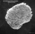
 HillH52 chorionic villi 01.jpg 1,200 × 924; 182 KB
 HillH52 chorionic villi 02.jpg 1,200 × 900; 155 KB
 HillH52 chorionic villi 03.jpg 1,200 × 891; 280 KB
 HillH52 chorionic villi 04.jpg 1,200 × 900; 254 KB
 HillH52 chorionic villi 05.jpg 1,200 × 992; 371 KB
 HillH52 chorionic villi 06.jpg 1,200 × 900; 346 KB
 HillH52 chorionic villi 07.jpg 1,200 × 900; 544 KB
 HillH52 chorionic villi 08.jpg 1,200 × 900; 229 KB
 HillH52 chorionic villi 09.jpg 1,200 × 900; 237 KB
 HillH52 chorionic villi 10.jpg 1,200 × 900; 253 KB
 HillH58 Stage 17 bf01.jpg 1,500 × 1,500; 273 KB
 HillH58 Stage 17 bf02.jpg 1,500 × 1,500; 279 KB
 HillH6 Stage 18 bf01.jpg 1,547 × 2,062; 274 KB
 HillH6 Stage 18 bf02.jpg 1,547 × 2,062; 337 KB
 HillH6 Stage 18 bf03.jpg 1,547 × 2,062; 287 KB
 HillH6 Stage 18 bf04.jpg 1,547 × 2,062; 318 KB
 HillH6 Stage 18 bf05.jpg 1,547 × 2,062; 218 KB
 HillH6 Stage 18 bf06.jpg 1,547 × 2,062; 232 KB
 HillH6 Stage 18 bf51.jpg 1,547 × 2,062; 198 KB
 HillH8 Stage 16 bf01.jpg 1,125 × 1,500; 136 KB
 HillH8 Stage 16 bf02.jpg 1,125 × 1,500; 145 KB
 HillH8 Stage 16 bf03.jpg 1,125 × 1,500; 170 KB
 HillH8 Stage 16 bf04.jpg 1,125 × 1,500; 171 KB
 HillH8 Stage 16 bf05.jpg 1,125 × 1,500; 176 KB
 HillH8 Stage 16 bf06.jpg 1,125 × 1,500; 169 KB
 HillH8 Stage 16 bf07.gif 600 × 800; 490 KB
 HillH8 Stage 16 bf08.gif 450 × 600; 295 KB
 HillH8 Stage 16 bf09.gif 600 × 800; 571 KB
 HillH8 Stage 16 bf10.gif 450 × 600; 338 KB
 HillH8 Stage 16 bf11.gif 600 × 800; 585 KB
 HillH8 Stage 16 bf12.gif 450 × 600; 348 KB
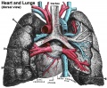 Historic-lungs.jpg 600 × 493; 79 KB
 Hochstadter 1919.jpg 845 × 1,200; 0 bytes
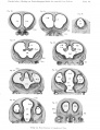
 Hochstadter plate 01.jpg 1,408 × 2,000; 255 KB
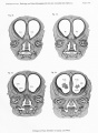
 Hochstadter plate 02.jpg 1,408 × 2,000; 305 KB
 Hochstadter plate 03.jpg 2,000 × 1,408; 258 KB
 Hochstadter plate 04.jpg 1,408 × 2,000; 273 KB
 Hochstadter plate 05.jpg 1,408 × 2,000; 235 KB
 Hochstadter plate 06.jpg 1,408 × 2,000; 280 KB
 Hochstadter plate 07.jpg 1,626 × 2,000; 261 KB
 Hochstadter plate 08.jpg 1,670 × 2,000; 367 KB
 Hochstadter plate 09.jpg 1,692 × 2,000; 307 KB
 Hochstadter plate 10.jpg 1,626 × 2,000; 551 KB
 Hochstadter plate 11.jpg 1,603 × 2,000; 570 KB
 Hochstadter plate 12.jpg 1,541 × 2,000; 543 KB
 Hochstadter plate 13.jpg 1,461 × 2,000; 531 KB
 Hochstadter plate 14.jpg 1,517 × 2,000; 574 KB
 Hochstadter plate 15.jpg 1,462 × 2,000; 450 KB
 Hochstadter plate 16.jpg 1,507 × 2,000; 640 KB
 Hubrecht model-Homo16 4mm.jpg 1,707 × 1,000; 311 KB
 Human - uterine epithelium SEM01.jpg 600 × 400; 37 KB
 Human - uterine epithelium SEM02.jpg 600 × 400; 26 KB
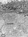
 Human - uterine epithelium TEM01.jpg 600 × 433; 25 KB
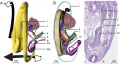
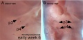
 Human 15 weeks - terminal nerve and vomeronasal organ nerves.jpg 940 × 403; 306 KB
 Human 7.5mm embryo model 01.jpg 747 × 1,000; 149 KB
 Human 7.5mm embryo model 02.jpg 747 × 1,000; 130 KB
 Human 7.5mm embryo model 03.jpg 747 × 1,000; 72 KB
 Human 7.5mm embryo model 04.jpg 747 × 1,000; 56 KB
 Human 7.5mm embryo model 05.jpg 747 × 1,000; 87 KB
 Human 7.5mm embryo model 06.jpg 747 × 1,000; 139 KB
 Human 7.5mm embryo model 07.jpg 747 × 1,000; 118 KB
 Human 7.5mm embryo model 08.jpg 747 × 1,000; 69 KB
 Human 7.5mm embryo model 09.jpg 747 × 1,000; 63 KB
 Human 7.5mm embryo model 10.jpg 747 × 1,000; 106 KB
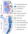 Human and mouse fetal-maternal interface cartoon.jpg 600 × 720; 91 KB
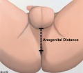 Human anogenital distance.jpg 570 × 499; 23 KB
 Human blastocyst derived stem cells.jpg 1,200 × 825; 198 KB
 Human blastocyst formation-in vitro.jpg 753 × 157; 23 KB
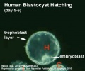 Human blastocyst hatching movie icon.jpg 498 × 414; 29 KB
 Human brain white matter tracts.png 1,200 × 490; 289 KB
 Human cochlea fetal development cartoon.jpg 592 × 1,200; 96 KB
 Human congenital diaphragmatic hernia.jpg 800 × 626; 86 KB
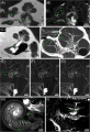
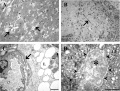 Human corpus luteum - light-and-electron-micrograph.jpg 936 × 711; 208 KB
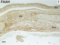
 Human developing lung protein 01.jpg 657 × 1,000; 241 KB
 Human developing lung protein 02.jpg 800 × 302; 82 KB
 Human development 001.mov ; 2 MB
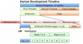
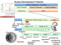 Human development timeline graph 01.jpg 1,000 × 750; 141 KB
 Human development timeline graph 02.jpg 800 × 424; 61 KB
 Human development timeline graph icon.jpg 250 × 188; 16 KB
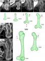 Human embryo femur CS18 to CS23.png 1,200 × 1,624; 1.42 MB
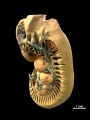
 Human embryo midgut loop 01.jpg 1,423 × 771; 200 KB
 Human embryo neck 01.jpg 534 × 827; 186 KB
 Human embryo neck 02.jpg 529 × 825; 167 KB
 Human embryo neck 03.jpg 558 × 795; 184 KB
 Human embryo parathyroid 01.jpg 1,000 × 495; 63 KB
 Human embryo skin 24 week EGA.jpg 596 × 939; 165 KB
 Human embryo skin 8-9 week EGA desmosomes.jpg 800 × 198; 40 KB
 Human embryo skin 8-9 week EGA.jpg 657 × 872; 188 KB
 Human embryo skin 9-11 week EGA.jpg 623 × 804; 176 KB
 Human Embryo Stage18-19.jpg 600 × 392; 17 KB
 Human embryo thymus and parathyroid 01.jpg 639 × 1,000; 58 KB
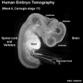 Human embryo tomography Carnegie stage 17.jpg 516 × 516; 35 KB
 Human embryonic shoulder girdle 01.jpg 1,000 × 726; 81 KB
 Human embryonic shoulder girdle 02.jpg 1,025 × 713; 109 KB
 Human embryonic shoulder girdle 04.jpg 1,000 × 755; 71 KB
 Human embryonic stem cell defined conditions 01.jpg 1,200 × 1,132; 372 KB
 Human embryonic stem cell defined conditions 02.jpg 1,091 × 411; 187 KB
 Human embryonic stem cell defined conditions 03.jpg 500 × 376; 73 KB
 Human embryonic tongue 01.jpg 1,200 × 818; 443 KB
 Human embryonic tongue 02.jpg 650 × 850; 220 KB
 Human embryonic tongue 03.jpg 650 × 470; 159 KB
 Human embryonic tongue 04.jpg 650 × 470; 131 KB
 Human embryonic tongue 05.jpg 650 × 470; 146 KB
 Human embryonic tongue 06.jpg 650 × 470; 99 KB
 Human embryonic tongue 07.jpg 650 × 470; 187 KB
 Human embryonic tongue 08.jpg 650 × 470; 185 KB
 Human embryonic tongue 09.jpg 650 × 470; 180 KB
 Human embryonic tongue 10.jpg 650 × 470; 118 KB
 Human embryonic tongue 11.jpg 650 × 470; 155 KB
 Human embryonic-fetal tongue 01.jpg 1,000 × 1,129; 490 KB
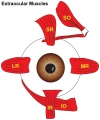 Human extraocular muscles 01.jpg 500 × 600; 47 KB
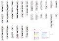
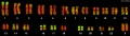 Human female karyotype 01.jpg 2,000 × 562; 118 KB
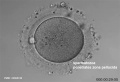
 Human fertilization movie 1 frame 01.jpg 600 × 409; 27 KB
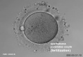
 Human fertilization movie 1 frame 02.jpg 600 × 409; 27 KB
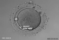
 Human fertilization movie 1 frame 03.jpg 600 × 409; 26 KB
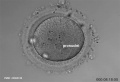
 Human fertilization movie 1 frame 04.jpg 600 × 409; 24 KB
 Human fertilization movie 1 frame 05.jpg 600 × 409; 25 KB
 Human fertilization movie 1 frame 06.jpg 600 × 409; 25 KB
 Human fertilization movie 1 frame 07.jpg 600 × 409; 24 KB
 Human fertilization movie 1 frame 08.jpg 600 × 409; 25 KB
 Human fertilization movie 1 frame 09.jpg 600 × 409; 24 KB
 Human fertilization movie 1 frame 10.jpg 600 × 409; 25 KB
 Human fetal adrenal GA32 large.mp4 ; 2.62 MB
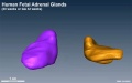 Human fetal adrenal GA32.jpg 800 × 502; 39 KB
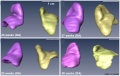 Human fetal adrenal gland 01.jpg 1,266 × 800; 107 KB
 Human fetal cochlea 01.jpg 1,270 × 532; 266 KB
 Human fetal cochlea 02.jpg 1,270 × 532; 271 KB
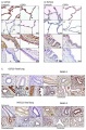
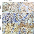 Human fetal gonad retinoid receptor expression.jpg 1,004 × 1,000; 447 KB
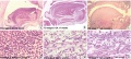
 Human fetal kidney histology 01.jpg 1,280 × 1,024; 481 KB
 Human fetal kidney histology 02.jpg 1,280 × 1,024; 322 KB
 Human fetal kidney histology 03.jpg 1,280 × 1,024; 333 KB
 Human fetal kidney histology 04.jpg 1,280 × 1,024; 307 KB
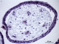
 Human fetal membrane 01.jpg 726 × 545; 69 KB
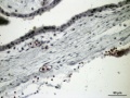
 Human fetal membrane 02.jpg 726 × 545; 87 KB
 Human fetal membrane 03.jpg 726 × 545; 88 KB
 Human fetal neck 01.jpg 549 × 827; 186 KB
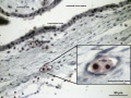
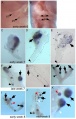
 Human fetal neural aneuploidy.jpg 1,000 × 1,400; 134 KB
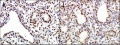
 Human fetal ovary SMAD6 expression.jpg 711 × 535; 167 KB
 Human fetal tongue 01.jpg 1,500 × 672; 350 KB
 Human fetal uterus myometrium.jpg 500 × 554; 86 KB
 Human heart developmental functional networks.jpg 833 × 614; 424 KB
 Human heart SEM1.jpg 1,200 × 330; 47 KB
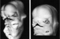
 Human holoprosencephaly cyclopia dissection.jpg 600 × 340; 37 KB
 Human homeobox genes.jpg 1,200 × 840; 222 KB
 Human induced pluripotent stem cells 01.jpg 1,091 × 799; 250 KB
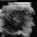
 Human inner ear MicroCT.jpg 2,131 × 3,111; 1,001 KB
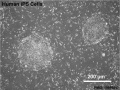 Human iPS cells 01.jpg 598 × 448; 83 KB
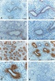
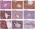 Human liver week 9.jpg 1,200 × 991; 425 KB
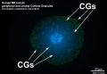
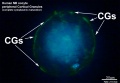
 Human lung pseudoglandular.jpg 672 × 1,000; 121 KB
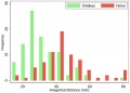 Human male anogenital distance graph.jpg 600 × 429; 28 KB
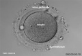
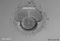
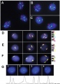
 Human MII oocyte 01.jpg 1,200 × 826; 92 KB
 Human MII oocyte 02.jpg 1,200 × 826; 98 KB
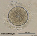
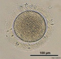 Human oocyte 01.jpg 700 × 675; 98 KB
 Human oocyte 11.jpg 700 × 675; 104 KB
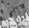 Human oocyte em01.jpg 600 × 589; 65 KB
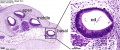
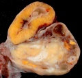
 Human ovary - corpus luteum 01.jpg 1,024 × 979; 162 KB
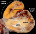
 Human ovary - corpus luteum 02.jpg 837 × 800; 119 KB
 Human ovary - corpus luteum 11.jpg 1,024 × 979; 89 KB


