Category:Histology
From Embryology
Introduction
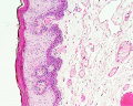
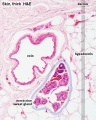
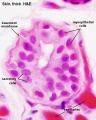
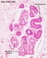
Files and media below relate to histology. Most links are to histological images that relate to tissue structure and development. Many images are sourced from UNSW Histology slide set and UWA Blue Histology online images.
Subcategories
This category has the following 15 subcategories, out of 15 total.
Pages in category 'Histology'
The following 200 pages are in this category, out of 334 total.
(previous page) (next page)A
- Template:Adrenal Histology
- AE Practical - Neural Histology
- Template:Al. coch.
- Alagille Syndrome
- ANAT2241 Blood
- ANAT2241 Bone, Bone Formation and Joints
- ANAT2241 Cardiovascular System
- ANAT2241 Connective Tissue Components
- ANAT2241 Connective Tissue Types
- ANAT2241 Covering and Lining Epithelia
- ANAT2241 Endocrine System
- ANAT2241 Female Reproductive System
- Template:ANAT2241 footer
- ANAT2241 Gastro-Intestinal System
- ANAT2241 Glandular Epithelia
- ANAT2241 Histology - Basic and Systematic
- Talk:ANAT2241 Histology - Basic and Systematic
- ANAT2241 Integumentary System
- ANAT2241 Liver, Gallbladder, and Pancreas
- ANAT2241 Lymphatic Tissue and Immune System
- ANAT2241 Male Reproductive System
- ANAT2241 Muscle Tissue
- ANAT2241 Nervous Tissue
- ANAT2241 Respiratory System
- ANAT2241 Special Senses The Eye
- ANAT2241 The Virtual Microscope
- ANAT2241 Urinary System
- ANAT2241 Virtual Slides - Example Spot Images
- ANAT2511 - Fundamentals of Anatomy
- ANAT2511 Basic Tissues
- ANAT2511 Bones and Joints
- ANAT2511 Circulatory System
- Template:ANAT2511 footer
- ANAT2511 Gastrointestinal Tract
- ANAT2511 Integumentary System
- ANAT2511 Introduction to Histology
- ANAT2511 Muscle Tissue
- ANAT2511 Nervous Tissue
- ANAT2511 Respiratory System
- ANAT2511 Urinary System
- Template:Artifacts
B
- BGD Lecture - Endocrine Histology
- BGD Lecture - Gastrointestinal System Development
- Talk:BGD Lecture - Gastrointestinal System Development
- BGDA Practical - Female Reproductive Tract Histology
- BGDB Gastrointestinal - Abnormalities
- BGDB Practical - Gastrointestinal System Development
- Talk:BGDB Practical - Gastrointestinal System Development
- BGDB Practical - Upper Gastrointestinal Tract Histology
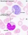
- Blood Cell Histology Movie
- Template:Blood Histology
- Template:Blood Vessel Histology
- Template:Blood vessel histology
- Template:Blue Histology
- Bone Development
- Bone Histology
- Template:Bone Histology
- Template:Bone histology
- Template:Bone Marrow Histology
- Book - A laboratory guide in histology (1917)
- Book - A Laboratory Text-Book of Embryology 8 (1903)
- Book - A text-book of histology arranged upon an embryological basis (1913)
- Book - A text-book of histology arranged upon an embryological basis (1913) 1
- Book - A text-book of histology arranged upon an embryological basis (1913) 1-1
- Book - A text-book of histology arranged upon an embryological basis (1913) 1-2
- Book - A text-book of histology arranged upon an embryological basis (1913) 1-3
- Book - A text-book of histology arranged upon an embryological basis (1913) 2
- Book - A text-book of histology arranged upon an embryological basis (1913) 2-1
- Book - A text-book of histology arranged upon an embryological basis (1913) 2-2
- Book - A textbook of histology, including microscopic technic (1910) General Histology 1
- Book - A textbook of histology, including microscopic technic (1910) General Histology 2
- Book - A textbook of histology, including microscopic technic (1910) microscopic technic
- Book - A textbook of histology, including microscopic technic (1910) Special Histology 1
- Book - A textbook of histology, including microscopic technic (1910) Special Histology 10
- Book - A textbook of histology, including microscopic technic (1910) Special Histology 2
- Book - A textbook of histology, including microscopic technic (1910) Special Histology 3
- Book - A textbook of histology, including microscopic technic (1910) Special Histology 4
- Book - A textbook of histology, including microscopic technic (1910) Special Histology 5
- Book - A textbook of histology, including microscopic technic (1910) Special Histology 6
- Book - A textbook of histology, including microscopic technic (1910) Special Histology 7
- Book - A textbook of histology, including microscopic technic (1910) Special Histology 8
- Book - A textbook of histology, including microscopic technic (1910) Special Histology 9
- Book - Contributions to Embryology Carnegie Institution No.50
- Book - Histology and Embryology 1941
- Book - Liver development
- Book - Stoehr's Histology (1906)
- Book - Stoehr's Histology 1
- Talk:Book - Stoehr's Histology 1
- Book - Stoehr's Histology 1-1
- Book - Stoehr's Histology 1-2
- Book - Stoehr's Histology 1-3
- Book - Stoehr's Histology 2
- Book - Stoehr's Histology Figures
- Book - The microscopic anatomy of the human body, in health and disease
- Template:Bouin
- Buccopharyngeal membrane
- Template:BöhmDavidoffHuber1910 footer
- Template:BöhmDavidoffHuber1910 TOC
C
- Template:CapillaryEM links
- Template:Cardiac muscle EM
- Cardiac Muscle Histology
- Cardiovascular - Arterial Development
- Cardiovascular System - Blood Development
- Cardiovascular System - Heart Histology
- Cardiovascular System - Spleen Development
- Cartilage Histology
- Template:Cartilage Histology
- Template:Cartilage histology
- Cloaca Development
- Colon Histology 2009
- Template:Common Stains collapsetable
- Template:Common Stains table
E
F
G
- Template:Gall Bladder Histology Images
- Gastrointestinal Tract - Abnormalities
- Gastrointestinal Tract - Carnegie Stage 13
- Gastrointestinal Tract - Carnegie Stage 22
- Gastrointestinal Tract - Colon Histology
- Gastrointestinal Tract - Gall Bladder Development
- Gastrointestinal Tract - Gall Bladder Histology
- Gastrointestinal Tract - Gallbladder Development
- Gastrointestinal Tract - Gallbladder Histology
- Gastrointestinal Tract - Histology
- Gastrointestinal Tract - Intestine Development
- Gastrointestinal Tract - Liver Development
- Gastrointestinal Tract - Liver Histology
- Gastrointestinal Tract - Mesentery Development
- Gastrointestinal Tract - Mouth Development
- Gastrointestinal Tract - Oesophagus Development
- Gastrointestinal Tract - Pancreas Development
- Gastrointestinal Tract - Pancreas Histology
- Gastrointestinal Tract - Postnatal
- Gastrointestinal Tract - Stomach Development
- Gastrointestinal Tract Development
- Template:Gastrointestinal Tract Links
- Template:GIT histology links
H
- Template:Hair histology links
- Template:HE
- Template:Heart histology
- Template:HillH52
- Histology
- Template:Histology
- Histology and Embryology 1941 - Bibliography
- Histology and Embryology 1941 - Embryology
- Histology and Embryology 1941 - Embryology 1
- Histology and Embryology 1941 - Embryology 2
- Histology and Embryology 1941 - Histology
- Histology and Embryology 1941 - Histology 1
- Histology and Embryology 1941 - Histology 2
- Histology and Embryology 1941 - Histology 3
- Histology Artifacts
- Histology Fixatives
- Template:Histology Links
- Histology Stains
- Template:Histology Stains
- HM Practical - Blood Vessel Histology
- HM Practical - Cardiac Histology
- Template:Human follicles lm and em links
- Template:Human ovary - corpus luteum links
- Human System Development
I
L
M
P
- Template:Pancreas Histology Images
- Paper - A case of atresia ani in a human embryo of 26 mm
- Paper - A case of atresia of the esophagus combined with traoheoesophageal fistula in a 9 mm human embryo, and its embryological explanation
- Paper - A case of congenital malformations of the intestinal canal (1923)
- Paper - A Contribution to the Embryology of the Liver and Vascular System in Man
- Paper - A contribution to the morphology and development of the mammalian liver
- Paper - A contribution to the morphology and development of the mammalian liver (1908)
- Paper - A histological investigation of the development and structure of the human lung
- Paper - A model demonstrating the changes in position and peritoneal relations of abdominal viscera during development (1912)
Media in category 'Histology'
The following 200 files are in this category, out of 718 total.
(previous page) (next page) Hematopoietic and stromal cell differentiation.jpg 1,000 × 617; 107 KB
 Hill Homo62 fig01.jpg 1,280 × 1,488; 274 KB
 HillH52 chorionic villi 01.jpg 1,200 × 924; 182 KB
 HillH52 chorionic villi 02.jpg 1,200 × 900; 155 KB
 HillH52 chorionic villi 03.jpg 1,200 × 891; 280 KB
 HillH52 chorionic villi 04.jpg 1,200 × 900; 254 KB
 HillH52 chorionic villi 05.jpg 1,200 × 992; 371 KB
 HillH52 chorionic villi 06.jpg 1,200 × 900; 346 KB
 HillH52 chorionic villi 07.jpg 1,200 × 900; 544 KB
 HillH52 chorionic villi 08.jpg 1,200 × 900; 229 KB
 HillH52 chorionic villi 09.jpg 1,200 × 900; 237 KB
 HillH52 chorionic villi 10.jpg 1,200 × 900; 253 KB
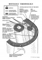 Histology terminology cartoon.jpg 595 × 842; 72 KB
 Histology-fetal liver HEx100.jpg 1,280 × 1,024; 214 KB
 Histology-fetal liver HEx40.jpg 1,000 × 800; 281 KB
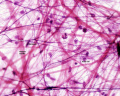
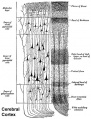 Historic-Cerebral-cortex.jpg 483 × 634; 65 KB
 HMB2011 Gall Bladder Histology 01.mp3 ; 1.05 MB
- HMB2011 Gall Bladder Histology 03.mp3 ; 1.27 MB
- HMB2011 Gall Bladder Histology 04.mp3 ; 1.22 MB
- HMB2011 Liver Histology 01.mp3 ; 1.08 MB
- HMB2011 Liver Histology 02.mp3 ; 870 KB
- HMB2011 Liver Histology 03.mp3 ; 757 KB
- HMB2011 Liver Histology 04.mp3 ; 638 KB
- HMB2011 Liver Histology 05.mp3 ; 1.04 MB
 Holocrine secretion animation.gif 60 × 80; 16 KB
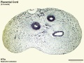 Hubrecht Homo73a cord 1.jpg 1,200 × 900; 427 KB
 Human CS13 otic vesicle 01.jpg 1,028 × 774; 112 KB
 Human CS13-15 otic vesicle 01.jpg 1,574 × 1,779; 364 KB
 Human developing lung protein 01.jpg 657 × 1,000; 241 KB
 Human developing lung protein 02.jpg 800 × 302; 82 KB
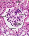
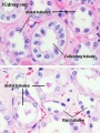
 Human fetal kidney histology 01.jpg 1,280 × 1,024; 481 KB
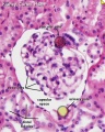
 Human fetal kidney histology 02.jpg 1,280 × 1,024; 322 KB
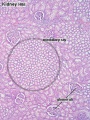
 Human fetal kidney histology 03.jpg 1,280 × 1,024; 333 KB
 Human fetal kidney histology 04.jpg 1,280 × 1,024; 307 KB
 Human fetal membrane 01.jpg 726 × 545; 69 KB
 Human fetal membrane 02.jpg 726 × 545; 87 KB
 Human fetal membrane 03.jpg 726 × 545; 88 KB
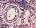
 Human infant ovary follicle 01.jpg 800 × 800; 107 KB
 Human liver week 9.jpg 1,200 × 991; 425 KB
 Human ovary - corpus luteum 01.jpg 1,024 × 979; 162 KB
 Human ovary - corpus luteum 02.jpg 837 × 800; 119 KB
 Human ovary - corpus luteum 11.jpg 1,024 × 979; 89 KB
 Human ovary - corpus luteum 21.jpg 1,024 × 979; 89 KB
 Human ovary follicle basement membrane EM01.jpg 558 × 697; 100 KB
 Human ovary follicles light and electron microscopy 01.jpg 586 × 1,080; 225 KB
 Human placenta cytokeratin 7 expression 01.jpg 800 × 621; 168 KB
 Human placenta SERPINE2 and CK7 expression.jpg 932 × 600; 155 KB
 Human placenta SERPINE2 expression 01.jpg 800 × 601; 134 KB
 Human placenta SERPINE2 expression 02.jpg 1,200 × 419; 229 KB
 Human placenta SERPINE2 expression 03.jpg 800 × 616; 163 KB
 Human uterine glands 01.jpg 726 × 545; 99 KB
 Human- late proliferative uterine endometrium.jpg 509 × 350; 98 KB
 Human- late secretory uterine endometrium.jpg 518 × 350; 93 KB
 Human- menstrual uterine endometrium.jpg 516 × 350; 93 KB
 Human- mid-proliferative uterine endometrium.jpg 513 × 350; 111 KB
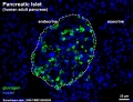 Human- pancreatic adult islet-glucagon.jpg 600 × 460; 67 KB
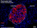 Human- pancreatic adult islet-insulin.jpg 600 × 460; 68 KB
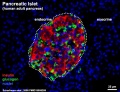 Human- pancreatic adult islet.jpg 600 × 460; 74 KB
 Human- secretory uterine endometrium.jpg 515 × 350; 93 KB
 Human-adrenal gland 01.jpg 596 × 392; 65 KB
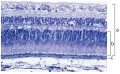 Human-retina-01.jpg 1,000 × 615; 197 KB
 Hyaline cartilage 01.jpg 500 × 626; 71 KB
 Hyaline cartilage 02.jpg 500 × 626; 62 KB
 Hyaline cartilage 03.jpg 500 × 626; 92 KB
 Hyaline cartilage 04.jpg 500 × 626; 101 KB
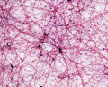
 Hypothalamus histology 001.jpg 500 × 405; 84 KB
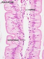
 Ileum histology 01.jpg 480 × 600; 69 KB
 Infant ovary.jpg 943 × 571; 108 KB
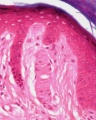
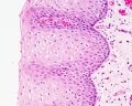
 Integumentary - lip01.jpg 1,280 × 1,024; 253 KB
 Integumentary histology 01.jpg 480 × 600; 70 KB
 Integumentary histology 02.jpg 480 × 600; 58 KB
 Integumentary histology 03.jpg 480 × 600; 85 KB
 Integumentary histology 04.jpg 280 × 700; 65 KB
 Integumentary histology 10.jpg 800 × 1,000; 101 KB
 Integumentary- hair follicle 01.jpg 479 × 599; 66 KB
 Integumentary- hair follicle 02.jpg 479 × 599; 74 KB
 Integumentary- hair follicle 03.jpg 479 × 599; 48 KB
 Integumentary- sebaceous gland histology 01.jpg 400 × 500; 125 KB
 Integumentary- sebaceous gland histology 02.jpg 400 × 500; 150 KB
 Intestinal lymphangiectasia 01.jpg 600 × 452; 138 KB
 Intestine histology 001.jpg 450 × 600; 65 KB
 Intestine histology 002.jpg 800 × 640; 130 KB
 Intestine histology 003.jpg 400 × 533; 64 KB
 Intestine histology 004.jpg 400 × 533; 81 KB
 Intestine histology 005.jpg 400 × 533; 78 KB
 Intestine histology 006.jpg 400 × 533; 77 KB
 Intestine histology 007.jpg 400 × 533; 82 KB
 Intestine villi crypts cartoon.jpg 500 × 334; 29 KB
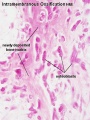 Intramembranous ossification centre.jpg 450 × 600; 69 KB
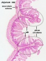
 Jejunum and ileum cartoon.jpg 500 × 704; 45 KB
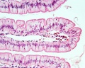
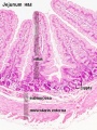
 Jejunum histology 01.jpg 480 × 600; 79 KB
 Lab7 muscle-1.jpg 1,000 × 844; 92 KB
 Lab7 muscle-1c.jpg 400 × 338; 23 KB
 Lab7 muscle-2.jpg 1,000 × 1,007; 164 KB
 Lab7 muscle-2c.jpg 400 × 403; 39 KB
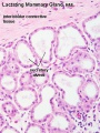
 Lactiferous duct 01.jpg 450 × 600; 58 KB
 Lactobacillus.jpg 320 × 240; 12 KB
 Leydig cells stained for LHCGR1.jpg 404 × 322; 16 KB
 Lingual gland histology 01.jpg 1,280 × 1,024; 325 KB
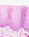
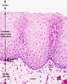
 Lip histology 01.jpg 400 × 533; 63 KB
 Lip histology 02.jpg 1,280 × 1,024; 444 KB
 Liver animated cartoon.gif 300 × 200; 239 KB
 Liver histology 001.jpg 400 × 533; 94 KB
 Liver histology 002.jpg 375 × 500; 54 KB
 Liver histology 003.jpg 375 × 500; 52 KB
 Liver histology 004.jpg 600 × 400; 70 KB
 Liver histology 005.jpg 800 × 664; 166 KB
 Liver histology 006.jpg 1,280 × 1,024; 664 KB
 Liver histology 007.jpg 1,280 × 1,024; 313 KB
 Liver histology 008.jpg 1,280 × 1,024; 214 KB
 Liver histology 009.jpg 1,280 × 1,024; 373 KB
 Liver histology 101.jpg 1,280 × 1,024; 410 KB
 Liver histology 102.jpg 1,280 × 1,024; 475 KB
 Liver histology 103.jpg 1,280 × 1,024; 330 KB
 Liver histology EM01.jpg 1,028 × 708; 141 KB
 Liver histology EM02.jpg 1,028 × 707; 154 KB
 Liver polyploidy 01.jpg 800 × 619; 119 KB
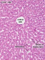
 Liver structure cartoon.jpg 1,000 × 451; 78 KB
 Liver- Kupffer cell and reticular fibre.jpg 600 × 800; 49 KB
 Liver-reticular fibre.jpg 700 × 875; 77 KB
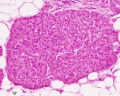
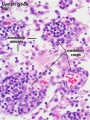
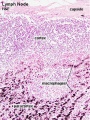
 Lymph node 05.jpg 1,000 × 800; 180 KB
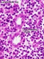
 Lymph node histology 01.jpg 600 × 400; 61 KB
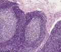
 Lymph node histology 02.jpg 450 × 600; 130 KB
 Lymph node histology 03.jpg 450 × 600; 140 KB
 Lymph node histology 04.jpg 450 × 600; 88 KB
 Lymph node histology 05.jpg 450 × 600; 87 KB
 Lymph node histology 06.jpg 450 × 600; 141 KB
 Lymph node histology01.jpg 800 × 680; 282 KB
 Lymphatic-system-bone-marrow.jpg 800 × 755; 143 KB
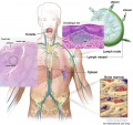
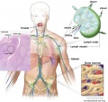
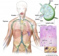 Lymphatic-system-node.jpg 800 × 755; 139 KB
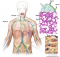
 Lymphatic-system-overview.jpg 800 × 755; 118 KB
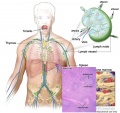 Lymphatic-system-spleen.jpg 800 × 755; 139 KB
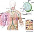 Lymphatic-system-thymus.jpg 800 × 755; 144 KB
 Lymphatic-system-tonsil-MALT.jpg 800 × 755; 144 KB
 Lymphatic-system-tonsil.jpg 800 × 755; 130 KB
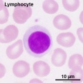
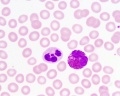
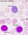 Lymphocyte 01.jpg 400 × 500; 54 KB
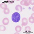
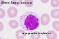 Lymphocyte 02.jpg 500 × 334; 27 KB
 Lymphocyte 03.jpg 600 × 600; 50 KB
 Lymphocyte 04.jpg 600 × 600; 49 KB
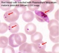 Malaria plasmodium falciparum.jpg 400 × 355; 14 KB
 Male histology 001.jpg 1,012 × 900; 378 KB
 Mammary gland 01.jpg 450 × 600; 75 KB
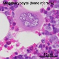 Megakaryocyte 01.jpg 600 × 600; 82 KB
 Meissner corpuscle 01.jpg 793 × 662; 86 KB
 Meissner corpuscle 02.jpg 800 × 1,000; 67 KB
 Merocrine secretion animation.gif 60 × 80; 10 KB
 Mesentery histology 01.jpg 1,280 × 1,024; 139 KB
 Mesentery histology 02.jpg 1,280 × 1,024; 256 KB
 Monkey- ovary primordial follicle.jpg 1,000 × 800; 292 KB
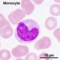
 Monocyte 01.jpg 480 × 600; 39 KB
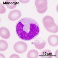
 Monocyte 02.jpg 600 × 600; 52 KB
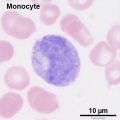
 Monocyte 03.jpg 600 × 600; 54 KB
 Monocyte 04.jpg 600 × 600; 47 KB
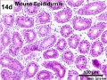
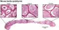
 Mouse epididymis development 01.jpg 1,200 × 909; 486 KB
 Mouse epididymis development 02.jpg 600 × 451; 138 KB
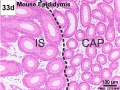 Mouse epididymis development 03.jpg 600 × 451; 119 KB
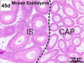 Mouse epididymis development 04.jpg 600 × 451; 120 KB
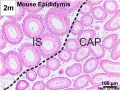 Mouse epididymis development 05.jpg 600 × 451; 112 KB
 Mouse eye and limbal region histology 01.jpg 1,200 × 467; 181 KB
 Mouse eye neural crest cornea 01.jpg 500 × 256; 34 KB
 Mouse eye neural crest cornea 02.jpg 800 × 655; 121 KB
 Mouse lung development 01.jpg 1,000 × 1,254; 791 KB
 Mouse lung development 01a.jpg 800 × 1,003; 495 KB
 Mouse lung development 02.jpg 922 × 922; 239 KB
 Mouse lung development 03.jpg 540 × 1,200; 349 KB
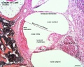
 Mouse organ of corti 01.jpg 1,280 × 1,024; 339 KB
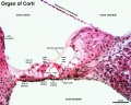
 Mouse organ of corti 02.jpg 1,280 × 1,024; 320 KB
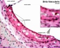
 Mouse organ of corti 03.jpg 1,280 × 1,024; 207 KB
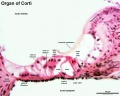
 Mouse organ of corti 04.jpg 1,280 × 1,024; 202 KB
 Mouse organ of corti 05.jpg 1,280 × 1,024; 171 KB
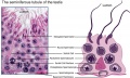
 Mouse seminiferous tubule EM.jpg 1,152 × 862; 352 KB
 Mouse- epididymis histology.jpg 751 × 383; 82 KB
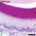
 Mouse- seminiferous tubule histology.jpg 715 × 427; 76 KB
 Mouse- tooth histology.jpg 500 × 503; 42 KB
 Mouse-E17.5 foot.jpg 385 × 500; 29 KB
 Mouse-E17.5 normal and abnormal limb.jpg 661 × 848; 105 KB
 Mouse-E17.5-foot-large.jpg 600 × 779; 53 KB
 Mouse-heart E17.5.jpg 353 × 1,000; 145 KB
 Muscle fiber types.jpg 400 × 250; 49 KB
 Mycoplasma-pneumoniae.jpg 320 × 240; 31 KB
 Myelination animation.gif 300 × 200; 77 KB
 Neisseria-gonorrhoeae.jpg 370 × 278; 22 KB
 Neisseria-meningitidis2.jpg 320 × 240; 14 KB
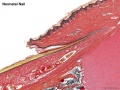 Neonatal nail.jpg 800 × 600; 176 KB
 Nephron histology 01.jpg 400 × 500; 79 KB
 Nephron histology 02.jpg 400 × 500; 77 KB
 Nephron histology 03.jpg 375 × 500; 97 KB
 Nephron histology 04.jpg 375 × 500; 54 KB
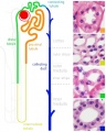 Nephron histology.jpg 400 × 500; 70 KB
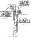 Nephrons-cortical and juxtamedullary.jpg 507 × 600; 39 KB
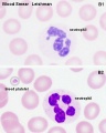 Neutrophil 01.jpg 480 × 600; 42 KB
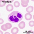 Neutrophil 02.jpg 600 × 600; 54 KB
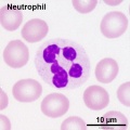 Neutrophil 03.jpg 600 × 600; 53 KB
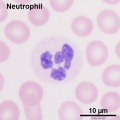 Neutrophil 04.jpg 600 × 600; 48 KB
 Neutrophil and eosinophil 01.jpg 480 × 600; 45 KB
 Neutrophil and eosinophil.jpg 1,280 × 1,024; 125 KB
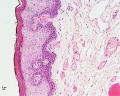
 Oesophagus histology 01.jpg 1,280 × 1,024; 290 KB
 Oesophagus histology 02.jpg 800 × 1,000; 196 KB
 Oesophagus histology 03.jpg 800 × 1,000; 209 KB


