ANAT2341 Lab 10 - Online Assessment 2015: Difference between revisions
No edit summary |
No edit summary |
||
| Line 113: | Line 113: | ||
Link to permalink image: [https://embryology.med.unsw.edu.au/embryology/Slides/Embryo_Stages/Stage22/08-eye/Stage22-08-eye.html?zoom=6&lat=-718.5&lon=3820&layers=B Eyelid] | Link to permalink image: [https://embryology.med.unsw.edu.au/embryology/Slides/Embryo_Stages/Stage22/08-eye/Stage22-08-eye.html?zoom=6&lat=-718.5&lon=3820&layers=B Eyelid] | ||
An eyelid is a thin fold of skin that covers and protects the human eye. Prior to the development of the eyelids, a small sulcus forms both above and below the eye (eyelid groove) at stage 16. And then, these grooves deepen, eyelid folds develop, first below, and then above, the eye. The eyelid folds develop into the eyelids and cover more of the eye as the palpebral fissure takes shape. The upper and the lower eyelids meet at the outer canthus in Stage 19. | |||
'''Embryonic Link''' [[Integumentary System - Eyelid Development]] | '''Embryonic Link''' [[Integumentary System - Eyelid Development]] | ||
Revision as of 20:32, 22 October 2015
| Embryology - 18 Jun 2024 |
|---|
| Google Translate - select your language from the list shown below (this will open a new external page) |
|
العربية | català | 中文 | 中國傳統的 | français | Deutsche | עִברִית | हिंदी | bahasa Indonesia | italiano | 日本語 | 한국어 | မြန်မာ | Pilipino | Polskie | português | ਪੰਜਾਬੀ ਦੇ | Română | русский | Español | Swahili | Svensk | ไทย | Türkçe | اردو | ייִדיש | Tiếng Việt These external translations are automated and may not be accurate. (More? About Translations) |
Individual Assessment
- Place your work on this page under a sub-sub-heading of your ROI.
- Add your own sub-sub-heading below any existing student ROI.
| About this Assessment | ||||||||||||||||||||||||||||||||||||
|---|---|---|---|---|---|---|---|---|---|---|---|---|---|---|---|---|---|---|---|---|---|---|---|---|---|---|---|---|---|---|---|---|---|---|---|---|
| A demonstration of this assessment will be given in the practical class. Below in the collapsible table are examples of links from a virtual slide. There is also a permalink help page.
| ||||||||||||||||||||||||||||||||||||


Student ROIs
This is a sub-sub-heading
Cochlear Duct
link to permalink image:| Cochlear Duct
The cochlear duct is an fluid filled cavity inside the cochlea. It located between the tympanic duct and the vestibular duct, and between the basilr membrane and reissner's memebrane. It derived from otic placode, otic vesicle, and originated from surface ectoderm.
embryology link Sensory - Hearing and Balance Development --Inner Ear
Semicircular Canal
link to permalink image:| Semicircular Canal
The semicircular canals are part of the inner ear.They are lined with cilia and filled with endolymph which is a liquid substance. Every time the head moves, the endolymph moves the cilia and this movements of the cilia are communicated to the brain. As a result, the brain knows how to keep the body balanced, regardless of the posture.
embryology link Sensory - Balance Development -- Inner Ear
Lens of the Eye
Link to permalink image: Anterior portion of the Lens of the embryonic eye
The lens of the eye is derived from surface ectoderm. Said ectoderm forms a lens/optic placode in the head region which then invaginates to form a lens pit and then later a lens vessel. Lens fibres then develop and are surrounded by a lens capsule. The main function of the lens is to focus light onto the retina.
Embryology link Vision - Lens Development --Development Overview
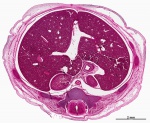
Retina of the Eye
Link to permalink image: Retina of the Eye
The retina is the light sensitive portion of the eye. It contains 10 separate layers, including the photoreceptor layer which is comprised of rods and cones. These rods and cones convert light into signals, which are then communicated to the brain via the optic nerve. Optic cup morphogenesis is responsible for the development of the vertebrate eye, and it is believed that this process significantly contributes to the development of the retina. The image above displays a Carnegie Stage 22 retina. The nerve fibre layer is particularly prominent in this image and is the pale layer closest to the vitreous chamber. The processes of rods, cones and ganglion cells can be observed migrating towards the optic nerve.
Embryology Link Vision - Retina Development
Retinal Pigment Epithelium
Link to permalink image: Retinal Pigment Epithelium
The Retinal Pigment Epithelium (RPE) is a complex differentiation of the retina, is generated from the optic neuroepithelium, and is structurally made up of cuboidal cells and multiple villi on its apical side. Its lateral sides are joined together by gap junctions and adherens and the RPE's basal side is in contact with Bruch's membrane. It lies between the neuronal retina and the choroid. The section shows that in the embryo the pigmented retina is still separated by a space from the neuronal retina. This space will be decreased in the adult and closely appose the two to each other.
Embryology Link Vision - Retina Development#Retinal Pigment Epithelium
Cornea
Permalink: Cornea
The cornea is the front layer of the eye covering the iris, pupil and anterior chamber. The cornea is a transparent layer that accounts for 2/3 of the eyes total optic power by refracting light along with the anterior chamber and lens. The cornea in humans consist of 5 layers as shown in the permalink, the Corneal epithelium, followed by Bowman’s layer, Corneal stroma, Descemet’s membrane and corneal endothelium. The corneal stroma and endothelium are derived from cranial neural crest cells and the corneal epithelium differentiates from ectoderm interacting with the developing lens.
Embryology link: Vision – Cornea Development
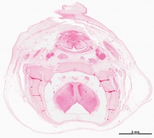
Middle Ear Ossicles
Link to permalink image: Middle Ear Ossicles
The middle ear ossicles named the malleus, incus, and stapes, are involved in transmitting vibrations from the tympanic membrane to the oval window, and ultimately to the inner ear. The are attached to muscles, tensor tympani and stapedius, to assist in reducing sound vibration and oscillations at the oval window. Embryologically, the malleus and incus are derived from the cartilage of the 1st pharyngeal arch, and the stapes is derived from the cartilage of the 2nd pharyngeal arch. In ossicle development, the malleus and incus initially form as a single structure from Meckel's cartilage, that are later separated by joint that forms between them. This process occurs within solid mesenchyme of the pharyngeal arches, therefore the ossicles are not functioning. It is only after birth that elongation of the auditory tube occurs to form the middle ear cavity that the middle ear ossicles are situated in.
Embryology Link Hearing - Middle Ear Development
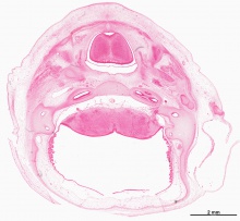
Embryonic Tongue
Link to permalink image: Tongue
The tongue is a muscle and is important for sensing taste. All the pharyngeal arches present in the human embryo contribute to the development of the tongue however, the tongue muscle cells are derived from somites and the muscles of mastication are derived from somitomeres. Each pharyngeal contributes a different portion where arch 1 forms the oral part of the tongue, arch 2 forms the initial transient surface, arch 3 forms the pharyngeal part of the tongue and arch 4 forms the epiglottis and adjacent regions. The superior surface of the tongue comprises of taste buds, various papillae and stratified squamous epithelium. The tongue is innervated by the hypoglossal nerve (CNXII) allowing movement. Tongue Development
Embryonic Link: Tongue Development
Retinal Pigment Epithelium (RPE)
Link to permalink image: Retinal Pigment Epithelium
Retinal pigment epithelium (RPE) cells are generated from the optic neuroepithelium. The choroidal melanocytes, the other pigmented cells, are derived from neural crest cells that have migrated towards the eye. RPE are cuboidal cells with multiple villi on its apical side which are in direct contact with the outer segments of the photoreceptor cells. Its lateral sides are joined together by tight junctions, adherens and gap junctions. The basal side of the retinal pigment epithelium is in contact with the underlying basal membrane which is also known as the Bruch's membrane. The permalink shows that the sensory retina and pigmented epithelium are separated by a space called the optic ventricle. In the adult the optic ventricle will no longer be present and the 2 layers would be closely associated to each other.
Embryology Link Vision - Retina Development#Retinal Pigment Epithelium
Eyelid
Link to permalink image: Eyelid
An eyelid is a thin fold of skin that covers and protects the human eye. Prior to the development of the eyelids, a small sulcus forms both above and below the eye (eyelid groove) at stage 16. And then, these grooves deepen, eyelid folds develop, first below, and then above, the eye. The eyelid folds develop into the eyelids and cover more of the eye as the palpebral fissure takes shape. The upper and the lower eyelids meet at the outer canthus in Stage 19.
Embryonic Link Integumentary System - Eyelid Development
| ANAT2341 Lab 10: Introduction | Early Embryo | Late Embryo | Fetal | Postnatal | Abnormalities | Online Assessment |
- 2015 Course: Week 2 Lecture 1 Lecture 2 Lab 1 | Week 3 Lecture 3 Lecture 4 Lab 2 | Week 4 Lecture 5 Lecture 6 Lab 3 | Week 5 Lecture 7 Lecture 8 Lab 4 | Week 6 Lecture 9 Lecture 10 Lab 5 | Week 7 Lecture 11 Lecture 12 Lab 6 | Week 8 Lecture 13 Lecture 14 Lab 7 | Week 9 Lecture 15 Lecture 16 Lab 8 | Week 10 Lecture 17 Lecture 18 Lab 9 | Week 11 Lecture 19 Lecture 20 Lab 10 | Week 12 Lecture 21 Lecture 22 Lab 11 | Week 13 Lecture 23 Lecture 24 Lab 12 | 2015 Projects: Three Person Embryos | Ovarian Hyper-stimulation Syndrome | Polycystic Ovarian Syndrome | Male Infertility | Oncofertility | Preimplantation Genetic Diagnosis | Students | Student Designed Quiz Questions | Moodle page