Category:Human: Difference between revisions
From Embryology
No edit summary |
mNo edit summary |
||
| Line 1: | Line 1: | ||
This {{Embryology}} category shows pages and media related generally to human development. | |||
[[Category:Human]] | [[Category:Human]] | ||
Revision as of 14:15, 2 July 2015
This Embryology category shows pages and media related generally to human development.
Subcategories
This category has the following 72 subcategories, out of 72 total.
C
- Carnegie Embryo
- Carnegie Embryo 1
- Carnegie Embryo 112
- Carnegie Embryo 1134B
- Carnegie Embryo 116
- Carnegie Embryo 1266
- Carnegie Embryo 1455
- Carnegie Embryo 148
- Carnegie Embryo 172
- Carnegie Embryo 19
- Carnegie Embryo 239
- Carnegie Embryo 2393
- Carnegie Embryo 240
- Carnegie Embryo 248
- Carnegie Embryo 256
- Carnegie Embryo 296
- Carnegie Embryo 3527
- Carnegie Embryo 3956
- Carnegie Embryo 4046
- Carnegie Embryo 4059
- Carnegie Embryo 407
- Carnegie Embryo 4148
- Carnegie Embryo 43
- Carnegie Embryo 4405
- Carnegie Embryo 460
- Carnegie Embryo 463
- Carnegie Embryo 523
- Carnegie Embryo 5541
- Carnegie Embryo 5609
- Carnegie Embryo 5652
- Carnegie Embryo 5682
- Carnegie Embryo 5874
- Carnegie Embryo 6032
- Carnegie Embryo 625
- Carnegie Embryo 6426
- Carnegie Embryo 6581
- Carnegie Embryo 7618
- Carnegie Embryo 7669
- Carnegie Embryo 786
- Carnegie Embryo 808
- Carnegie Embryo 8147
- Carnegie Embryo 8239
- Carnegie Embryo 8370
- Carnegie Embryo 858
- Carnegie Embryo 8630
- Carnegie Embryo 8967
- Carnegie Embryo 9296
- Carnegie Embryo 96
- Carnegie Embryo 963
- Carnegie Embryo 966
- Carnegie Embryo 9697
D
F
H
Pages in category 'Human'
The following 200 pages are in this category, out of 332 total.
(previous page) (next page)A
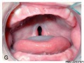
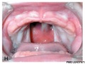
- Abnormal Development - Cleft Lip and Palate
- Abnormal Development - Cleft Palate
- Abnormal Development - Toxoplasmosis
- Template:Abnormal Newborn Neural Exam Table
- Template:Adipose Timeline table
- Template:Adrenal GA32 Links
- Template:Anderson2016 collapsetable1
- Template:Anderson2016 table1
- Template:AnsonBlack1934 figures
- Aschheim-Zondek Test 1928 Movie
- Template:Australian GIT abnormalities 2002-2003
- Template:Australian Palate abnormalities 2002-2003
B
- Template:B050966
- Template:B100658
- Template:B220849
- Template:Bardeen1906 figures
- Template:Barniville1914 figures
- Template:Bartelmez1922 figures
- Template:BaxterBoyd1939 figures
- BGDA Practical 7 - Week 6
- Birth MRI Movie
- Book - Contributions to Embryology Carnegie Institution No.10
- Book - Contributions to Embryology Carnegie Institution No.112
- Book - Contributions to Embryology Carnegie Institution No.131
- Book - Contributions to Embryology Carnegie Institution No.159
- Book - Contributions to Embryology Carnegie Institution No.21
- Book - Contributions to Embryology Carnegie Institution No.22
- Book - Contributions to Embryology Carnegie Institution No.27
- Book - Contributions to Embryology Carnegie Institution No.29
- Book - Contributions to Embryology Carnegie Institution No.30
- Book - Contributions to Embryology Carnegie Institution No.32
- Book - Contributions to Embryology Carnegie Institution No.33
- Book - Contributions to Embryology Carnegie Institution No.34
- Book - Contributions to Embryology Carnegie Institution No.35
- Book - Contributions to Embryology Carnegie Institution No.38
- Book - Contributions to Embryology Carnegie Institution No.40
- Book - Contributions to Embryology Carnegie Institution No.42
- Book - Contributions to Embryology Carnegie Institution No.43
- Book - Contributions to Embryology Carnegie Institution No.44
- Book - Contributions to Embryology Carnegie Institution No.46
- Book - Contributions to Embryology Carnegie Institution No.47
- Book - Contributions to Embryology Carnegie Institution No.48
- Book - Contributions to Embryology Carnegie Institution No.52
- Book - Contributions to Embryology Carnegie Institution No.55
- Book - Contributions to Embryology Carnegie Institution No.59
- Book - Contributions to Embryology Carnegie Institution No.61
- Book - Contributions to Embryology Carnegie Institution No.65
- Book - Contributions to Embryology Carnegie Institution No.69
- Book - Contributions to Embryology Carnegie Institution No.72
- Book - Contributions to the Development of the Human Brain (1919)
- History:Book - Contributions to the Development of the Human Brain (1919)
- Book - Human embryos of different ages examined in median sections - a contribution to the mechanics of development
- Template:Braune 1877 Plate 2
C
E
F
H
- Template:Hamilton1944 figures
- Harvard Collection
- Template:Hearing EAM timeline
- Hela Apoptosis Movie
- Template:HillH12inks
- Template:HillH13 links
- Template:HillH145 links
- Template:HillH159 links
- Template:HillH202 links
- Template:HillH257 links
- Template:HillH4 links
- Template:HillH5 links
- Template:HillH52
- Template:HillH58 links
- Template:HillH6 links
- Template:HillH8 links
- Template:Hochstadter plates
- Template:Huber1905 table1
- Template:Human 7.5mm Embryo links
- Human Adult Brain Movie
- Human Development Timeline Movie
- Human Embryo - Scanning electron microscopy
- Template:Human embryo neck links
- Human Embryo SEM
- Human Fertilization Detail Movie
- Human Fertilization Movie
- Template:Human Fertilization Movie 1 frame table
- Template:Human Fertilization Movie 1 frames
- Template:Human Fertilization Movie 2 frame table
- Template:Human follicles lm and em links
- Template:Human ovary - corpus luteum links
- Template:Human Spermatozoa Statistics collapse table
- Template:Human Spermatozoa Statistics table
- Human Sylvian Fissure Movie
- Template:Human timeline
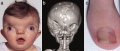
- Hutchinson-Gilford Progeria Syndrome
J
K
L
M
- Template:Macklin1921 figures
- Template:Mall1912 figures
- Template:Mall1916 figures
- Template:Mall1917 figures
- Menstrual Cycle - Histology
- Model Embryo 1.6mm Movie 1
- Model Embryo 10mm Movie 1
- Model Embryo 3.1mm Movie 1
- Model Embryo 7.5mm Movie 1
- Monosomic Embryo Movie 1
- Template:Morton1949 figures
- Template:Mouse Human lung table
- Movie - Neural Sylvian Fissure
N
P
- Palate Development
- Paper - A human embryo before the appearance of the myotomes (1918)
- Paper - A Human Embryo of Twenty-five Somites
- Paper - A Human Embryo of Twenty-seven Pairs of Somites, Embedded in Decidua
- Paper - A human embryo with head-process and commencing arch enteric canal
- Paper - A Human Embryo with Seven Pairs of Somites Measuring about 2 mm in Length
- Paper - A human embryo with seventeen pairs of somites (1930)
- Paper - A morphological study of testicular descent
- Paper - A Note on the Development of the Septum Transversum and the Liver
- Paper - A presomite human embryo (Shaw) - the implantation
- Paper - A presomite human embryo (Shaw) - the implantation (1942)
- Paper - A presomite human embryo showing a yolk-sac duct
- Paper - A presomite human embryo showing an early stage of the primitive streak
- Paper - A presomite human embryo with a neurenteric canal (embryo R.S.)
- Paper - A study of a 7 mm human embryo with special reference to its peculiar spirally twisted form, and its large aortic cell-clusters
- Paper - A study of the development of certain features of the cerebellum (1920)
- Paper - A very Young Human Embryo found embedded in a "Decidual Cast" of the Uterus
- Paper - A well-preserved human embryo of 10 somites (1929)
- Paper - A Young Human Embryo (Embryo Dobbin) with Head-Process and Prochordal Plate
- Paper - An early human embryo (no. 1285, Manchester Collection) with capsular attachment of the connecting stalk (1935)
- Paper - An Early Human Embryo (No. 1285, Manchester Collection), with Capsular Attachment of the Connecting Stalk
- Paper - An Early Human Embryo, with 0.55 mm long Embryonic Shield
- Paper - An Early Human Ovum (Thomson) in situ
- Paper - An iconometrographic representation of the growth of the central nervous system in man
- Paper - Breech fused twin monster (1934)
- Paper - Changes in fetuses due to formalin preservation
- Special:Badtitle/NS501:Paper - Description of a Human Embryo of 13-14 Mesodermic Somites
- Paper - Description of a Human Embryo of Twenty-three Paired Somites
- History:Paper - Description of a Human Embryo of Twenty-two paired Somites
- Paper - Description of a reconstruction of the head of a thirty-millimetre embryo (1910)
- Paper - Development and variation of the nerves and the musculature of the inferior extremity and of the neighboring regions of the trunk in man
- Paper - Development of the human heart from its earliest appearance to the stage found in embryos of twenty paired somites (1927)
- Paper - Developmental Changes in the Pericardium, the Mesocardia, and the Pleural Sacs in the Human Embryo
Media in category 'Human'
The following 200 files are in this category, out of 2,421 total.
(previous page) (next page) Bartelmez1922-fig10.jpg 1,000 × 1,136; 209 KB
 Basic Heart Development Timeline.jpg 1,658 × 556; 73 KB
 BaxterBoyd1939-fig01.jpg 361 × 736; 43 KB
 BaxterBoyd1939-fig02.jpg 459 × 851; 65 KB
 BaxterBoyd1939-fig03.jpg 732 × 1,000; 170 KB
 BaxterBoyd1939-fig04.jpg 642 × 615; 109 KB
 BaxterBoyd1939-fig05.jpg 1,000 × 864; 263 KB
 BaxterBoyd1939-fig06.jpg 489 × 917; 141 KB
 BaxterBoyd1939-fig07.jpg 795 × 917; 242 KB
 BaxterBoyd1939-plate01.jpg 1,680 × 2,400; 786 KB
 BaxterBoyd1939-plate02.jpg 1,681 × 2,400; 860 KB
 BaxterBoyd1939-text-fig01.jpg 1,283 × 1,000; 138 KB
 BaxterBoyd1939-text-fig02.jpg 1,200 × 861; 133 KB
 Bedford01.jpg 734 × 1,000; 82 KB
 Bedford02.jpg 1,069 × 911; 266 KB
 Bicornuate uterus01.jpg 1,452 × 1,691; 209 KB
 Blood test result for glucose and iron.jpg 879 × 345; 54 KB
 Bone histology 014.jpg 1,280 × 1,024; 541 KB
 Bone histology 015.jpg 1,280 × 1,024; 519 KB
 Bone histology 016.jpg 1,280 × 1,024; 379 KB
 Bone-femur.jpg 798 × 1,000; 150 KB
 Boyd collection icon.jpg 400 × 554; 56 KB
 Brain growth and birth size.jpg 800 × 492; 70 KB
 Braune 1877 plate 2 fig1.jpg 960 × 1,000; 181 KB
 Braune 1877 plate 2 fig2.jpg 806 × 1,000; 200 KB
 Braune 1877 plate 2 fig3.jpg 893 × 1,000; 208 KB
 Braune 1877 plate 2 fig4.jpg 809 × 1,000; 213 KB
 Braune 1877 plate 2 fig5.jpg 771 × 1,000; 169 KB
 Braune 1877 plate 2 fig6.jpg 690 × 1,000; 144 KB
 Braune 1877 plate 2 fig7.jpg 893 × 1,000; 241 KB
 Braune 1877 plate 2A.jpg 780 × 1,200; 263 KB
 Braune 1877 plate 2B.jpg 801 × 1,200; 264 KB
 Bremer1914 plate04.jpg 643 × 1,000; 127 KB
 Brown015.jpg 800 × 654; 72 KB
 Brown021.jpg 800 × 783; 94 KB
 Brown024.jpg 558 × 800; 52 KB
 Brown025.jpg 600 × 521; 43 KB
 Brown026.jpg 457 × 423; 25 KB
 Brown028.jpg 700 × 730; 52 KB
 Bryce1908 fig01.jpg 500 × 500; 20 KB
 Bryce1908 fig02.jpg 828 × 1,000; 110 KB
 Bryce1908 fig03.jpg 801 × 500; 40 KB
 Bryce1908 fig04.jpg 600 × 400; 28 KB
 Bryce1908 fig05.jpg 700 × 536; 0 bytes
 Bryce1908 fig06.jpg 600 × 500; 39 KB
 Bryce1908 table01.jpg 837 × 1,000; 141 KB
 Bryce1908 table02.jpg 702 × 1,000; 68 KB
 Bryce1908 table03.jpg 731 × 1,000; 78 KB
 Cardiac Conduction System.jpg 1,201 × 862; 81 KB
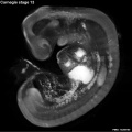
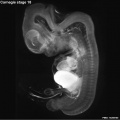
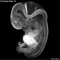
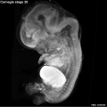
 Carnegie stage 12 OPT.jpg 800 × 801; 46 KB
 Carnegie stage 13 OPT.jpg 800 × 801; 53 KB
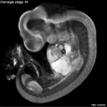 Carnegie stage 14 OPT.jpg 800 × 801; 55 KB
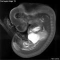 Carnegie stage 15 OPT.jpg 800 × 801; 56 KB
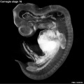 Carnegie stage 16 OPT.jpg 800 × 801; 51 KB
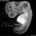 Carnegie stage 17 OPT.jpg 800 × 801; 57 KB
 Carnegie stage 18 OPT.jpg 800 × 801; 43 KB
 Carnegie stage 19 OPT.jpg 800 × 801; 46 KB
 Carnegie stage 20 OPT.jpg 800 × 801; 47 KB
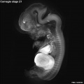 Carnegie stage 21 OPT.jpg 800 × 801; 39 KB
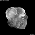 Carnegie stage 22 OPT.jpg 800 × 801; 38 KB
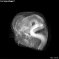 Carnegie stage 23 OPT.jpg 800 × 801; 36 KB
 Caudal duplication syndrome.jpg 700 × 599; 47 KB
 Cerebral blood supply development 01.jpg 1,200 × 460; 67 KB
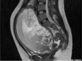
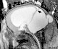
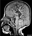 Chiari II malformation MRI01.jpg 723 × 800; 88 KB
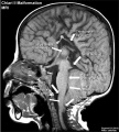 Chiari II malformation MRI02.jpg 723 × 800; 101 KB
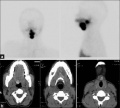
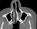 Choanal atresia computed tomography 01.jpg 598 × 477; 35 KB
 Chromosome telomeres.jpg 600 × 471; 33 KB
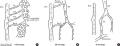
 Classification of Oesophageal atresia.jpg 513 × 726; 55 KB
 Cleft lip 001.jpg 400 × 300; 16 KB
 Cleft lip 002.jpg 400 × 300; 22 KB
 Cleft lip 003.jpg 400 × 300; 19 KB
 Cleft lip 004.jpg 400 × 300; 21 KB
 Cleft lip 005.jpg 400 × 300; 0 bytes
 Cleft lip 006.jpg 400 × 300; 22 KB
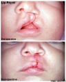 Cleft lip 007.jpg 405 × 504; 38 KB
 Cleft lip 01.jpg 585 × 438; 34 KB
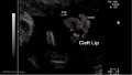 Cleft lip 02.jpg 641 × 362; 22 KB
 Cleft palate 001.jpg 400 × 300; 22 KB
 Cleft palate 002.jpg 400 × 300; 20 KB
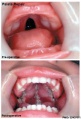 Cleft palate 003.jpg 407 × 600; 46 KB
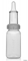 Cleft palate feeder.jpg 276 × 600; 19 KB
 Cockle01.jpg 881 × 1,000; 158 KB
 Cockle02.jpg 1,459 × 929; 266 KB
 Cockle03.jpg 810 × 1,062; 158 KB
 Cockle04.jpg 740 × 1,065; 135 KB
 Common bile duct in duodenal bulb.jpg 367 × 364; 38 KB
 Complete hydatidiform mole 01.jpg 748 × 560; 50 KB
 Complete hydatidiform mole 02.jpg 748 × 560; 60 KB
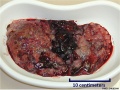 Complete hydatidiform mole 03.jpg 748 × 560; 103 KB
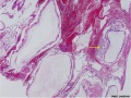 Complete hydatidiform mole 04.jpg 748 × 560; 124 KB
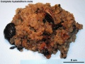 Complete hydatidiform mole 05.jpg 1,280 × 960; 324 KB
 Complete hydatidiform mole 06.jpg 1,280 × 960; 553 KB
 Congdon-table01.jpg 764 × 1,000; 167 KB
 Congdon1922-1-16.jpg 980 × 1,000; 157 KB
 Congdon1922-17.jpg 1,000 × 411; 55 KB
 Congdon1922-18-25.jpg 1,200 × 795; 179 KB
 Congdon1922-18.jpg 494 × 506; 29 KB
 Congdon1922-19.jpg 653 × 471; 31 KB
 Congdon1922-20.jpg 794 × 446; 43 KB
 Congdon1922-21.jpg 578 × 407; 27 KB
 Congdon1922-22.jpg 511 × 489; 27 KB
 Congdon1922-23.jpg 519 × 412; 26 KB
 Congdon1922-24.jpg 790 × 482; 40 KB
 Congdon1922-25.jpg 509 × 358; 25 KB
 Congdon1922-26.jpg 746 × 726; 54 KB
 Congdon1922-27-28.jpg 997 × 612; 68 KB
 Congdon1922-29.jpg 976 × 1,000; 81 KB
 Congdon1922-30.jpg 1,133 × 1,000; 176 KB
 Congdon1922-31.jpg 1,063 × 1,000; 93 KB
 Congdon1922-32.jpg 1,133 × 1,000; 132 KB
 Congdon1922-33.jpg 920 × 1,000; 107 KB
 Congdon1922-34.jpg 920 × 1,000; 122 KB
 Congdon1922-35.jpg 920 × 1,000; 97 KB
 Congdon1922-36.jpg 920 × 1,000; 113 KB
 Congdon1922-37.jpg 1,200 × 838; 163 KB
 Congdon1922-38.jpg 1,187 × 1,000; 165 KB
 Congdon1922-39.jpg 1,200 × 756; 138 KB
 Congdon1922-40.jpg 1,013 × 1,000; 110 KB
 Congdon1922-plate01.jpg 877 × 1,200; 145 KB
 Congdon1922-plate02.jpg 877 × 1,200; 191 KB
 Congdon1922-plate03.jpg 1,200 × 885; 182 KB
 Congenital diaphragmatic hernia 01.jpg 1,000 × 494; 108 KB
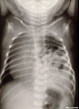 Congenital diaphragmatic hernia 02.jpg 578 × 800; 48 KB
 Congenital diaphragmatic hernia 03.jpg 611 × 800; 82 KB
 Congenital diaphragmatic hernia 04.jpg 637 × 600; 66 KB
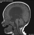 Congenital hydrocephalus MRI01.jpg 595 × 600; 43 KB
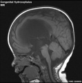 Congenital hydrocephalus MRI02.jpg 595 × 600; 42 KB
 Cord blood induced stem cells 01.jpg 800 × 787; 157 KB
 Cord blood induced stem cells 02.jpg 700 × 966; 189 KB
 Cranial neural crest skeletal fate 01.jpg 800 × 633; 59 KB
 Craniofrontonasal syndrome.jpg 1,280 × 543; 130 KB
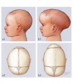 Craniosynostosis .jpg 375 × 430; 85 KB
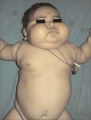 Cushing's syndrome.jpg 450 × 592; 62 KB
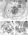
 Cytomegalovirus infected spermatozoa EM01.jpg 990 × 991; 204 KB
 Cytomegalovirus infected spermatozoa.jpg 1,000 × 1,260; 324 KB
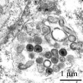
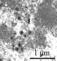 Cytomegalovirus virions EM.jpg 911 × 987; 212 KB
 Dandy Walker malformation MRI 01.jpg 600 × 506; 37 KB
 Dandy1910-plate01.jpg 1,738 × 2,359; 541 KB
 Dandy1910-plate02.jpg 1,754 × 2,400; 951 KB
 Dandy1910-plate03.jpg 1,000 × 1,617; 224 KB
 Dandy1910-plate04.jpg 1,000 × 1,875; 174 KB
 Dandy1910-plate05.jpg 1,000 × 2,034; 238 KB
 Dandy1910-plate06.jpg 1,000 × 2,166; 265 KB
 Davis1927 plate01.jpg 1,357 × 1,500; 501 KB
 Davis1927 plate02.jpg 1,357 × 1,500; 795 KB
 Developing human cerebellum 01.jpg 1,009 × 1,200; 494 KB
 Dichorionic twins ultrasound 01.gif 401 × 282; 2.51 MB
 Differentially expressed RefSeq genes in human trisomy 21.jpg 661 × 847; 171 KB
 Double meningomyelocele.jpg 504 × 800; 49 KB
 Double tetrasomy 18 mosaicism.jpg 600 × 447; 78 KB
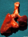
 Duodenal atresia.jpg 600 × 873; 63 KB
 Early human telomere length.jpg 1,800 × 1,034; 70 KB
 Early human telomeres.jpg 1,280 × 1,006; 194 KB
 Ectopia cordis.jpg 800 × 603; 37 KB
 Ectopic molar pregnancy 01.jpg 700 × 535; 60 KB
 Ectopic pregnancy 01.jpg 735 × 596; 45 KB
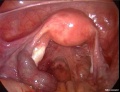
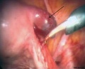
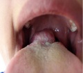 Ectopic thyroid - lingual 01.jpg 600 × 525; 34 KB
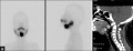 Ectopic thyroid - sublingual and suprahyoid.jpg 1,000 × 383; 42 KB
 Ectopic thyroid - sublingual, suprahyoid and subhyoid.jpg 800 × 721; 59 KB
 Embryo 7.5mm model 01.gif 448 × 600; 903 KB
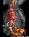
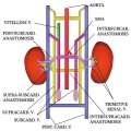 Embryo renal venous cartoon.jpg 600 × 600; 68 KB
 Embryonic neck muscle cartoon.jpg 600 × 570; 45 KB
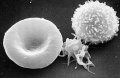 Erythrocyte and lymphocyte SEM01.jpg 800 × 522; 74 KB
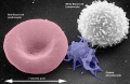 Erythrocyte and lymphocyte SEM02.jpg 800 × 522; 78 KB
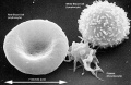 Erythrocyte and lymphocyte SEM03.jpg 800 × 522; 80 KB
 Extravillous trophoblasts week 5.5.jpg 1,280 × 900; 421 KB
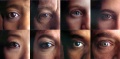 Eye collage 2.jpg 848 × 417; 112 KB
 Fawcett1910 fig01.jpg 891 × 639; 148 KB
 Fawcett1910 fig02.jpg 1,035 × 949; 265 KB
 Fawcett1910 fig03.jpg 905 × 978; 160 KB
 Fawcett1910 fig04.jpg 969 × 1,110; 115 KB
 Fawcett1913 fig01.jpg 793 × 573; 67 KB
 Fawcett1913 fig02.jpg 679 × 579; 42 KB
 Fawcett1913 fig03.jpg 687 × 581; 33 KB
 Fawcett1913 fig04.jpg 928 × 664; 122 KB
 Fawcett1913 fig05.jpg 979 × 671; 98 KB
 Fawcett1913 fig06.jpg 899 × 675; 80 KB
 Fawcett1913 fig07.jpg 1,113 × 763; 97 KB
 Fawcett1913 fig08.jpg 867 × 397; 28 KB
 Female genital and ureter abnormality 01.jpg 766 × 732; 86 KB
 Female genital and ureter abnormality 02.jpg 766 × 733; 78 KB
 Female genital and ureter abnormality 03.jpg 766 × 762; 79 KB
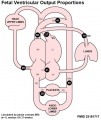
 Fetal blood flow 01.jpg 1,000 × 599; 75 KB
 Fetal blood flow 02.jpg 491 × 599; 38 KB
 Fetal blood flow 03.jpg 500 × 599; 43 KB
 Fetal blood flow 04.jpg 506 × 599; 50 KB
 Fetal blood flow liver and brain.jpg 677 × 790; 69 KB
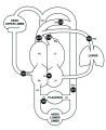
 Fetal cardiac state diagram 01.jpg 1,200 × 321; 29 KB
 Fetal cardiac state diagram 02.jpg 1,200 × 458; 27 KB
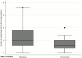 Fetal cells maternal blood graph.jpg 600 × 426; 18 KB
 Fetal ductus venosus pressure wave 01.jpg 706 × 755; 52 KB
 Fetal ductus venosus ultrasound 01.jpg 783 × 1,000; 68 KB
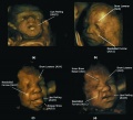 Fetal facial expression 01.jpg 1,200 × 1,094; 122 KB
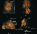 Fetal facial expression 02.jpg 1,914 × 1,762; 205 KB
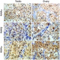 Fetal gonad retinoid receptor expression 01.jpg 1,004 × 1,000; 226 KB
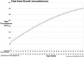 Fetal head growth circumference graph01.jpg 905 × 613; 58 KB
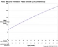 Fetal head growth circumference graph02.jpg 800 × 650; 44 KB
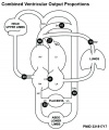
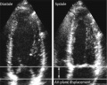 Fetal heart atrioventricular plane displacement 01.jpg 915 × 733; 55 KB
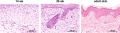 Fetal integumentary histology 01.jpg 800 × 219; 74 KB
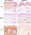 Fetal integumentary histology 02.jpg 600 × 664; 145 KB


