Category:Carnegie Stage 14: Difference between revisions
From Embryology
| Line 21: | Line 21: | ||
* His, embryo B, 7 mm, and embryo A, 7.5 mm. These two embryos are described jointly, in great detail, by His (1880).<ref name="His1880"/> Embryo A is evidently a little more advanced than embryo B and perhaps should be placed in stage 15. | * His, embryo B, 7 mm, and embryo A, 7.5 mm. These two embryos are described jointly, in great detail, by His (1880).<ref name="His1880"/> Embryo A is evidently a little more advanced than embryo B and perhaps should be placed in stage 15. | ||
* Keibel, 6.5-mm embryo “forensis.” Embryo described in the Keibel and Elze Normentafeln.<ref name="KeibelElze1908"/> This embryo was used by Keibel in a series of studies on the development of the urogenital system. | * Keibel, 6.5-mm embryo “forensis.” Embryo described in the Keibel and Elze Normentafeln.<ref name="KeibelElze1908"/> This embryo was used by Keibel in a series of studies on the development of the urogenital system. | ||
* Keibel, 6.8-mm embryo, No. 501. Embryo included in the Keibel and Elze Normentafeln.<ref name="KeibelElze1908"/> It is described in monographic form by Piper (1900). | * Keibel, 6.8-mm embryo, No. 501. Embryo included in the Keibel and Elze Normentafeln.<ref name="KeibelElze1908"/> It is described in monographic form by Piper (1900).<ref>Piper, H. 1900. ''Ein menschlicher Embryo von 6.8 mm. Nakkenlinie.'' Arch. Anat. PhysioL, Anat. Abth. Cited by Streeter (1945).</ref> | ||
* Strabl, 6.75-mm embryo (Walther), Giessen. Described in the Keibel and Elze Normentafeln.<ref name="KeibelElze1908"/>The external form is pictured by Hirschland (1898).<ref>Hirschland, L. 1898. ''Beitrage zur ersten Entwicklung der Mammarorgane beim Menschen''. Anat. Hefte, Abt. I, Heft 34, 35 (Vol. II). Cited by Streeter (1945).</ref> | * Strabl, 6.75-mm embryo (Walther), Giessen. Described in the Keibel and Elze Normentafeln.<ref name="KeibelElze1908"/>The external form is pictured by Hirschland (1898).<ref>Hirschland, L. 1898. ''Beitrage zur ersten Entwicklung der Mammarorgane beim Menschen''. Anat. Hefte, Abt. I, Heft 34, 35 (Vol. II). Cited by Streeter (1945).</ref> | ||
* 6.5-mm embryo, University of Minnesota. Drawings of external views of this and of a 2.9-mm (stage 11) embryo were published by Wells and Kaiser (1959).<ref><pubmed>13843903</pubmed></ref> | * 6.5-mm embryo, University of Minnesota. Drawings of external views of this and of a 2.9-mm (stage 11) embryo were published by Wells and Kaiser (1959).<ref><pubmed>13843903</pubmed></ref> | ||
Revision as of 14:11, 20 July 2015
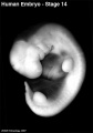
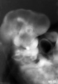
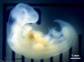
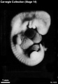
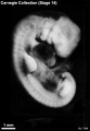
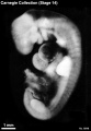
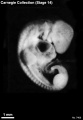
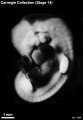
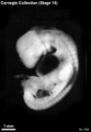
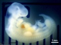
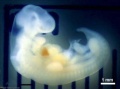
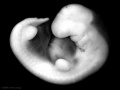
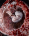
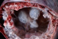
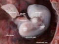
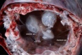
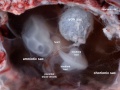
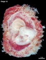
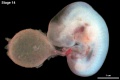
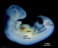
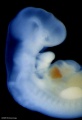
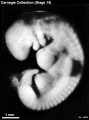
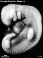
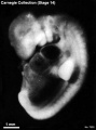
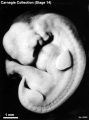
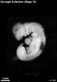
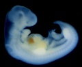
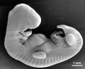
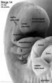
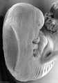
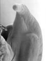
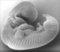
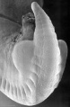
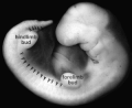
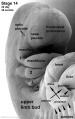
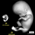
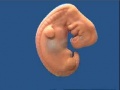
This Embryology category shows pages and media related to Carnegie stage 14 occurring in Week 5, 31 - 35 days, CRL 5 - 7 mm, Gestational Age GA week 7.
- Mesoderm: continued segmentation of paraxial mesoderm (30+ somite pairs), heart prominence
- Head: 1st, 2nd and 3rd pharyngeal arch, forebrain, site of lens placode, site of otic placode, stomodeum
- Body: heart, liver, umbilical, upper and early lower limb bud
- Links: Carnegie Stage 14
| Week: | 1 | 2 | 3 | 4 | 5 | 6 | 7 | 8 |
| Carnegie stage: | 1 2 3 4 | 5 6 | 7 8 9 | 10 11 12 13 | 14 15 | 16 17 | 18 19 | 20 21 22 23 |
- Carnegie Stages: 1 | 2 | 3 | 4 | 5 | 6 | 7 | 8 | 9 | 10 | 11 | 12 | 13 | 14 | 15 | 16 | 17 | 18 | 19 | 20 | 21 | 22 | 23 | About Stages | Timeline
Historic Stage 14 Embryos
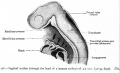
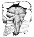
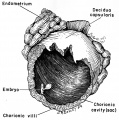
- H. Braus, 6-mm embryo, Heidelberg. Enlarged views of this well-preserved curettage specimen are included in the Hochstetter (1907)[1] Cited by Streeter (1945).[2] portfolio of pictures of the outer form of a series of human embryos. The nasal plate is more advanced than in others of this stage, but the limb buds are like those of the older members of the group.
- E. Gasser, 6.5-mm. Leyding embryo, Marburg Anatomisches Institut. Description of this advanced embryo is in the Keibel and Elze Normentafeln (1908).[3]
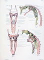
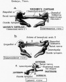
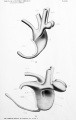
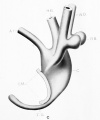
- J. A. Hammar, 5-mm Vestberg embryo, Anatomisches Institut, Uppsala. Embryo described by Hammar in the Keibel and Elze Normentafeln.[3] Typical sections and models of the pharyngeal region and gut are illustrated. This well-preserved embryo has been studied by several investigators, including Hammar on the development of the foregut, salivary glands, and tongue, and Broman on the development of the diaphragm and omental bursa.
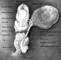
- His, embryo R, 5.5 mm. Illustrated by His (1885).[4] It was concluded by His that this embryo approximated the Fol embryo (stage 13). The lens pit indicates stage 14. This assignment is supported by other features: e.g., the ductus venosus.
- His, embryo B, 7 mm, and embryo A, 7.5 mm. These two embryos are described jointly, in great detail, by His (1880).[4] Embryo A is evidently a little more advanced than embryo B and perhaps should be placed in stage 15.
- Keibel, 6.5-mm embryo “forensis.” Embryo described in the Keibel and Elze Normentafeln.[3] This embryo was used by Keibel in a series of studies on the development of the urogenital system.
- Keibel, 6.8-mm embryo, No. 501. Embryo included in the Keibel and Elze Normentafeln.[3] It is described in monographic form by Piper (1900).[5]
- Strabl, 6.75-mm embryo (Walther), Giessen. Described in the Keibel and Elze Normentafeln.[3]The external form is pictured by Hirschland (1898).[6]
- 6.5-mm embryo, University of Minnesota. Drawings of external views of this and of a 2.9-mm (stage 11) embryo were published by Wells and Kaiser (1959).[7]
- Free Hospital for Women, Brookline, Massachusetts, No. 33, 6 mm, No. 29, 7 mm, and No. 31, 7 mm. The histochemistry of these embryos of stage 14 was studied by McKay et al. (1956).[8]
References
- ↑ Hochstetter, F. 1907. Atlas. Munich.
- ↑ Streeter, G. L. 1945. Developmental horizons in human embryos. Description of age group XIII, embryos about 4 or 5 millimeters long, and age group XTV, period of indentation of the lens vesicle. Carnegie Instn. Wash. Publ. 557, Contrib. Embryol., 31, 27-63.
- ↑ 3.0 3.1 3.2 3.3 3.4 Keibel, F., and Elze, C. 1908. Normentafeln zur Entwicklungsgeschichte der Wirbeltiere.(Normal Plates for Evolution of Vertebrates) 8. Heft Normentafeln zur Entwicklungsgeschichte des Menschen (Vol. 8. Normal Plates of the Development of the Human Embryo) Fisher, Jena., Germany.
- ↑ 4.0 4.1 His, W. 1880-1885. Anatomie menscblicher Embryonen. Vogel, Leipzig.
- ↑ Piper, H. 1900. Ein menschlicher Embryo von 6.8 mm. Nakkenlinie. Arch. Anat. PhysioL, Anat. Abth. Cited by Streeter (1945).
- ↑ Hirschland, L. 1898. Beitrage zur ersten Entwicklung der Mammarorgane beim Menschen. Anat. Hefte, Abt. I, Heft 34, 35 (Vol. II). Cited by Streeter (1945).
- ↑ <pubmed>13843903</pubmed>
- ↑ <pubmed>13403206</pubmed>
Subcategories
This category has the following 48 subcategories, out of 48 total.
C
- Carnegie Embryo 1380
- Carnegie Embryo 1620
- Carnegie Embryo 18
- Carnegie Embryo 187
- Carnegie Embryo 208
- Carnegie Embryo 245
- Carnegie Embryo 3360
- Carnegie Embryo 372
- Carnegie Embryo 380
- Carnegie Embryo 387
- Carnegie Embryo 3960
- Carnegie Embryo 4
- Carnegie Embryo 4154
- Carnegie Embryo 442
- Carnegie Embryo 4629
- Carnegie Embryo 4672
- Carnegie Embryo 4692
- Carnegie Embryo 4805
- Carnegie Embryo 5437
- Carnegie Embryo 552
- Carnegie Embryo 560
- Carnegie Embryo 5654
- Carnegie Embryo 5787
- Carnegie Embryo 6428
- Carnegie Embryo 6500
- Carnegie Embryo 6502
- Carnegie Embryo 6503
- Carnegie Embryo 6739
- Carnegie Embryo 676
- Carnegie Embryo 6830
- Carnegie Embryo 7333
- Carnegie Embryo 7400
- Carnegie Embryo 7522
- Carnegie Embryo 7598
- Carnegie Embryo 7667
- Carnegie Embryo 7829
- Carnegie Embryo 7870
- Carnegie Embryo 80
- Carnegie Embryo 8141
- Carnegie Embryo 8306
- Carnegie Embryo 8308
- Carnegie Embryo 8314
- Carnegie Embryo 8357
- Carnegie Embryo 8552
- Carnegie Embryo 8999
E
W
Pages in category 'Carnegie Stage 14'
The following 82 pages are in this category, out of 82 total.
C
- Template:Carnegie Embryo Stage14
- Carnegie stage 14
- Template:Carnegie stage 14 links
- Carnegie Stage 14 Surface Movie
- Template:CE1380
- Template:CE16
- Template:CE1620
- Template:CE18
- Template:CE187
- Template:CE208
- Template:CE245
- Template:CE2841
- Template:CE3360
- Template:CE372
- Template:CE380
- Template:CE3805
- Template:CE387
- Template:CE3960
- Template:CE4
- Template:CE4154
- Template:CE442
- Template:CE4672
- Template:CE4692
- Template:CE4805
- Template:CE5437
- Template:CE552
- Template:CE560
- Template:CE5654
- Template:CE5787
- Template:CE6428
- Template:CE6500
- Template:CE6502
- Template:CE6503
- Template:CE6739
- Template:CE676
- Template:CE6830
- Template:CE6848
- Template:CE7333
- Template:CE7370
- Template:CE7394
- Template:CE7400
- Template:CE7522
- Template:CE7598
- Template:CE7667
- Template:CE7829
- Template:CE7870
- Template:CE80
- Template:CE8141
- Template:CE8306
- Template:CE8308
- Template:CE8314
- Template:CE8357
- Template:CE8552
- Template:CE873
- Template:CE8999
- Template:CE9695
- Template:CE988
- Template:CS14
P
- Paper - A study of a 7 mm human embryo with special reference to its peculiar spirally twisted form, and its large aortic cell-clusters
- Paper - Developmental horizons in human embryos. Description of age group XIII
- Paper - Histochemical horizons in human embryos - Stage 14
- Paper - The Course of the Phrenic Nerve in the Embryo
- Paper - Two choice human embryos at Streeter's horizons XI and XIV
- Paper - Two choice human embryos at Streeter’s horizons 11 and 14
R
Media in category 'Carnegie Stage 14'
The following 114 files are in this category, out of 114 total.
 600px stage14.jpg 421 × 600; 27 KB
 Anderson2016-fig07a.jpg 800 × 800; 193 KB
 Anderson2016-fig07b.jpg 800 × 800; 304 KB
 Anderson2016-fig09a.jpg 800 × 800; 106 KB
 Anderson2016-fig09b.jpg 800 × 800; 90 KB
 Anderson2016-fig13b.jpg 800 × 800; 191 KB
 B100658-01.jpg 1,388 × 1,040; 677 KB
 B100658-02.jpg 1,068 × 800; 458 KB
 Bailey087.jpg 928 × 803; 132 KB
 Bailey256.jpg 894 × 545; 75 KB
 Bat - neural development 01.jpg 733 × 498; 33 KB
 Carnegie stage 14 OPT.jpg 800 × 801; 55 KB
 Congdon1922-33.jpg 920 × 1,000; 107 KB
 Congdon1922-plate02.jpg 877 × 1,200; 191 KB
 Frazer1910 fig02.jpg 750 × 804; 144 KB
 Gilbert1957 fig01.jpg 1,280 × 1,579; 156 KB
 Heart outflow tract stage 14 01.jpg 2,039 × 996; 274 KB
 Heart outflow tract stage 14 02.jpg 996 × 996; 139 KB
 Heart outflow tract stage 14 03.jpg 989 × 996; 134 KB
 Human CS13-15 otic vesicle 01.jpg 1,574 × 1,779; 364 KB
 Human embryo midgut loop 01.jpg 1,423 × 771; 200 KB
 Human embryonic renal branching 1.jpg 1,280 × 779; 236 KB
 Human embryonic tongue 05.jpg 650 × 470; 146 KB
 Human stage14 heart MRI - ventricular septation.jpg 864 × 652; 39 KB
 Human Stage14 neural01.jpg 1,375 × 2,048; 283 KB
 Human Stage14 neural02.jpg 1,375 × 2,048; 506 KB
 Human Stage14-16 CN5-01.jpg 1,028 × 681; 44 KB
 Keibel Mall 2 576.jpg 1,280 × 767; 154 KB
 Mall1906-fig14.jpg 673 × 699; 50 KB
 Mall1908a fig80a.jpg 785 × 829; 83 KB
 Mall1908a fig80b.jpg 1,000 × 1,040; 244 KB
 Mall1908a fig80c.jpg 1,000 × 709; 127 KB
 Mckay1956 plate01.jpg 1,280 × 1,970; 377 KB
 ME52 001.jpg 1,280 × 874; 148 KB
 ME52 002.jpg 1,000 × 1,449; 213 KB
 Pohlman1911 plate2.jpg 2,050 × 3,230; 331 KB
 Pohlman1911 plate2C.jpg 1,301 × 1,560; 103 KB
 Stage14 bf1.jpg 1,000 × 743; 27 KB
 Stage14 bf10.jpg 416 × 600; 24 KB
 Stage14 bf11.jpg 416 × 600; 24 KB
 Stage14 bf12.jpg 416 × 600; 25 KB
 Stage14 bf13.jpg 416 × 600; 26 KB
 Stage14 bf14.jpg 416 × 600; 22 KB
 Stage14 bf15.jpg 416 × 600; 23 KB
 Stage14 bf16.jpg 416 × 600; 23 KB
 Stage14 bf17.jpg 416 × 600; 23 KB
 Stage14 bf18.jpg 1,800 × 2,250; 549 KB
 Stage14 bf19.jpg 1,200 × 1,500; 287 KB
 Stage14 bf1a.jpg 800 × 594; 20 KB
 Stage14 bf1b.jpg 600 × 446; 13 KB
 Stage14 bf1c.jpg 400 × 297; 8 KB
 Stage14 bf2.jpg 1,000 × 750; 71 KB
 Stage14 bf20.jpg 800 × 1,000; 158 KB
 Stage14 bf21.jpg 2,000 × 1,334; 323 KB
 Stage14 bf22.jpg 1,500 × 1,001; 214 KB
 Stage14 bf23.jpg 1,000 × 667; 122 KB
 Stage14 bf24.jpg 1,231 × 923; 156 KB
 Stage14 bf25.jpg 2,000 × 1,334; 337 KB
 Stage14 bf26.jpg 1,263 × 947; 150 KB
 Stage14 bf27.jpg 1,000 × 1,291; 208 KB
 Stage14 bf28.jpg 1,280 × 848; 115 KB
 Stage14 bf2a.jpg 800 × 600; 45 KB
 Stage14 bf2b.jpg 600 × 450; 26 KB
 Stage14 bf2c.jpg 400 × 300; 13 KB
 Stage14 bf2cl.jpg 400 × 333; 17 KB
 Stage14 bf2l.jpg 1,000 × 834; 66 KB
 Stage14 bf3.jpg 1,368 × 2,000; 159 KB
 Stage14 bf3a.jpg 684 × 1,000; 61 KB
 Stage14 bf3b.jpg 547 × 800; 43 KB
 Stage14 bf4.jpg 2,650 × 2,000; 352 KB
 Stage14 bf4a.jpg 1,325 × 1,000; 137 KB
 Stage14 bf4b.jpg 1,060 × 800; 99 KB
 Stage14 bf5.jpg 458 × 618; 32 KB
 Stage14 bf6.jpg 458 × 618; 35 KB
 Stage14 bf7.jpg 458 × 618; 29 KB
 Stage14 bf8.jpg 458 × 618; 33 KB
 Stage14 bf9.jpg 416 × 600; 18 KB
 Stage14 human.jpg 646 × 530; 32 KB
 Stage14 sem1.jpg 1,000 × 819; 94 KB
 Stage14 sem1c.jpg 400 × 327; 23 KB
 Stage14 sem2.jpg 1,261 × 2,000; 234 KB
 Stage14 sem2a-limb.jpg 504 × 800; 40 KB
 Stage14 sem2a.jpg 631 × 1,000; 83 KB
 Stage14 sem2al.jpg 504 × 800; 68 KB
 Stage14 sem2b-limb.jpg 378 × 600; 26 KB
 Stage14 sem2b.jpg 505 × 800; 58 KB
 Stage14 sem2c-limb.jpg 252 × 400; 14 KB
 Stage14 sem2cl.jpg 252 × 400; 22 KB
 Stage14 sem2l.jpg 630 × 1,000; 96 KB
 Stage14 sem3.jpg 2,114 × 2,997; 443 KB
 Stage14 sem3a.jpg 705 × 1,000; 94 KB
 Stage14 sem3b.jpg 564 × 800; 67 KB
 Stage14 sem3c.jpg 423 × 600; 42 KB
 Stage14 sem4.jpg 1,916 × 2,549; 297 KB
 Stage14 sem4a.jpg 752 × 1,000; 79 KB
 Stage14 sem4b.jpg 602 × 800; 57 KB
 Stage14 sem4c.jpg 452 × 600; 37 KB
 Stage14 sem5.jpg 2,553 × 2,220; 313 KB
 Stage14 sem5a.jpg 1,000 × 870; 85 KB
 Stage14 sem5b.jpg 920 × 800; 75 KB
 Stage14 sem5c.jpg 690 × 600; 48 KB
 Stage14 sem6.jpg 1,569 × 2,394; 311 KB
 Stage14 sem6a.jpg 655 × 1,000; 87 KB
 Stage14 sem6b.jpg 524 × 800; 61 KB
 Stage14 sem6c.jpg 393 × 600; 39 KB
 Stage14 somites limbbuds.png 300 × 245; 24 KB
 Stage14-sem2 limb.jpg 630 × 1,000; 57 KB
 Stage14compare23.jpg 593 × 601; 32 KB
 Stage14modelicon.jpg 321 × 241; 5 KB
 Streeter1922-fig13.jpg 617 × 675; 69 KB
 Streeter1957 fig01.jpg 1,292 × 1,500; 218 KB
 Wells1959 fig01.jpg 1,280 × 1,404; 354 KB
 WellsKaiser1959 fig04.jpg 1,280 × 1,286; 330 KB
 WellsKaiser1959 fig05.jpg 1,280 × 1,260; 312 KB


