Category:Placenta: Difference between revisions
From Embryology
mNo edit summary |
mNo edit summary |
||
| Line 1: | Line 1: | ||
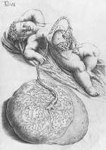
[[File:Spiegel1626_table07.jpg|thumb|150px|alt=Spiegel and Casseri, De formato foetu liber singularis (Dedication dated 1626)|link=Placenta Development|Spiegel and Casseri: De formato foetu liber singularis (Dedication dated 1626).]] | |||
This {{Embryology}} category shows pages and media related to placenta development. | This {{Embryology}} category shows pages and media related to placenta development. | ||
Revision as of 09:49, 26 August 2016
This Embryology category shows pages and media related to placenta development.
Pages in category 'Placenta'
The following 197 pages are in this category, out of 197 total.
A
B
- BGDA Practical - Placenta and Fetal Membranes
- Talk:BGDA Practical - Placenta and Fetal Membranes
- Template:BGDA Practical 14 - Abnormalities Interactive
- Template:BGDA Practical 14 - Diagnostic Interactive
- Template:BGDA Practical 14 - Implantation Interactive
- Template:BGDA Practical 14 - Maternal Decidua Interactive
- Template:BGDA Practical 14 - Placental Cord Interactive
- Template:BGDA Practical 14 - Placental Functions Interactive
- Template:BGDA Practical 14 - Villi Interactive
- BGDA Practical Placenta - Abnormalities
- BGDA Practical Placenta - Cord Development
- BGDA Practical Placenta - Diagnostic Techniques
- BGDA Practical Placenta - Implantation and Early Placentation
- BGDA Practical Placenta - Maternal Decidua
- BGDA Practical Placenta - Placental Functions
- BGDA Practical Placenta - Villi Development
- Template:BGDALabPlacenta
- Book - Contributions to Embryology Carnegie Institution No.158
- Book - Contributions to Embryology Carnegie Institution No.51
- Book - Human Embryology and Morphology 8
- Book - Sex and internal secretions (1961) 15
- Book - Text-Book of Embryology 19
- Book - Text-Book of the Embryology of Man and Mammals 13
- Book - Umbilicus (1916)
- Book - Uterine and tubal gestation (1903) 1-12
C
E
H
I
P
- Paper - A contribution to our knowledge of the earliest known stages of placentation and embryonic development in man
- Paper - An anatomical study of the closure of the ductus arteriosus (1942)
- Paper - Anatomy of the placenta (1858)
- Paper - Cytological studies on the internal secretory functions in the human placenta and decidua (1921)
- Paper - development of mammalian ova and placenta formation in mammals
- Paper - development of mammalian ova and placenta formation in mammals 3
- Paper - Development of the human placenta in the first three months of gestation (1960)
- Paper - Early Development and placentation in arvicola (microtus) amphibius (1922)
- Paper - Lectures on the early stages in the development of mammalian ova and on the differentiation of the placenta in different groups of mammals
- Paper - On the placentation of the macaque (Macaca mulatta), from the time of implantation until the formation of the definitive placenta
- Paper - On the structure of the human placenta, and its connexion with the uterus
- Paper - Pathological changes in the placenta associated with erythroblastosis of the fetus (1938)
- Paper - Placenta praevia in a rhesus monkey (1939)
- Paper - Placental circulation
- Paper - Placentation in the rabbit
- Paper - Radioangiographic studies of circulation in the maternal placenta of the rhesus monkey
- Paper - Retrogressive Changes in the Fetal Vessels and the Suspensory Ligament of the Liver
- Paper - The area of the chorionic villi in the full-term placenta (1922)
- Paper - The cytological structure of the human chorionic villus and decidua parietalis
- Paper - The development and structure of the human placenta
- Paper - The form and the functions of the uterine blood vessels in the rhesus monkey
- Paper - The formation of the umbilical cord and the umbilical region of the anterior abdominal wall
- Paper - The histology and cytology of the human and monkey placenta
- Paper - The histology of the umbilical cord of the pig (1919)
- Paper - The interrelations of the mesonephros, kidney, and placenta in different classes of animals (1916)
- Paper - The morphology of human uteroplacental vasculature
- Paper - The origin and occurrence of the single umbilical artery in normal and abnormal human fetuses (1922)
- Template:Persistent right umbilical vein
- Template:Placenta
- Placenta - Abnormalities
- Placenta - Cord
- Placenta - Histology
- Placenta - Maternal Decidua
- Placenta - Membranes
- Placenta - Stage 13
- Placenta - Stage 22
- Placenta - Vascular Beds
- Placenta - Villi Development
- Template:Placenta abnormalities
- Template:Placenta Abnormalities gallery
- Template:Placenta accreta
- Template:Placenta Cord Histology
- Placenta Development
- Template:Placenta EM links
- Template:Placenta histology
- Template:Placenta increta
- Template:Placenta Links
- Template:Placenta percreta
- Template:Placenta previa
- Placenta Quiz
- Template:Placenta terms
- Template:Placenta vascular
- Template:Placenta vascular bed
- Template:Placenta Villi Timeline table
- Template:Placental cord
- Template:Placental membranes
- Template:Placental villi
- Template:Placental villi timeline
- Template:Pre-eclampsia
- Template:Pregnancy hCG Levels table
- Template:Primary villi
R
- Template:Ramsey1972 figures
- Template:Ref-BacsichSmout1938
- Template:Ref-Bartelmez1957
- Template:Ref-Bloxam1840
- Template:Ref-Bremer1916
- Template:Ref-Bremer1918
- Template:Ref-Dalton1858
- Template:Ref-Dawson1922
- Template:Ref-Dodds1922
- Template:Ref-Falkiner1939
- Template:Ref-Fujimura1921
- Template:Ref-Grosser1910
- Template:Ref-HamiltonBoyd1960
- Template:Ref-HarrisRamsey1966
- Template:Ref-HellmanHertig1938
- Template:Ref-Hertig1935
- Template:Ref-Herzog1909
- Template:Ref-Houston1964
- Template:Ref-Hunter1774
- Template:Ref-Ingalls1932
- Template:Ref-Ingalls1932a
- Template:Ref-Ingalls1932b
- Template:Ref-Jordan1919
- Template:Ref-Meyer1918
- Template:Ref-Ninian1939
- Template:Ref-Palmer1939
- Template:Ref-PMID5965440
- Template:Ref-Ramsey1960
- Template:Ref-Ramsey1972
- Template:Ref-Robinson1904a
- Template:Ref-Robinson1904b
- Template:Ref-Robinson1904c
- Template:Ref-Sansom1922
- Template:Ref-Strachan1923
- Template:Ref-Walls1939
- Template:Ref-Wislocki1920
- Template:Ref-Wislocki1921
- Template:Ref-WislockiBennett1943
- Template:Ref-WislockiStreeter1938
- Template:Ref-Wyburn1939
S
T
U
W
Media in category 'Placenta'
The following 200 files are in this category, out of 364 total.
(previous page) (next page) 2016Lecture-Placenta.mp4 ; 54.16 MB
 Allantois.jpg 600 × 531; 51 KB
 Bailey081.jpg 782 × 755; 118 KB
 Bailey082.jpg 879 × 756; 137 KB
 Bailey206.jpg 973 × 854; 162 KB
 Bailey489.jpg 517 × 617; 82 KB
 Bailey490.jpg 755 × 612; 71 KB
 Bailey491.jpg 728 × 473; 56 KB
 Bailey492.jpg 907 × 695; 123 KB
 Bailey493.jpg 930 × 744; 210 KB
 Bailey494.jpg 875 × 769; 157 KB
 Bailey495.jpg 802 × 730; 142 KB
 Bailey496.jpg 844 × 563; 122 KB
 Bailey497.jpg 649 × 610; 74 KB
 Bailey498.jpg 949 × 751; 180 KB
 Bailey499.jpg 805 × 477; 65 KB
 Bailey500.jpg 1,215 × 677; 159 KB
 Bailey501.jpg 688 × 1,093; 276 KB
 Bailey502.jpg 751 × 547; 74 KB
 Bailey503.jpg 606 × 624; 76 KB
 BakerHookSeveringhaus1944 plate01.jpg 1,534 × 2,295; 228 KB
 BakerHookSeveringhaus1944 plate02.jpg 1,502 × 2,261; 265 KB
 BakerHookSeveringhaus1944 plate03.jpg 1,508 × 2,327; 279 KB
 BakerHookSeveringhaus1944 plate04.jpg 1,563 × 2,278; 347 KB
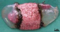
 Bilobed placenta with velamentous cord insertion.jpg 1,339 × 1,000; 274 KB
 Bloxam1840-plate03.jpg 1,200 × 997; 463 KB
 Bloxam1840-plate04.jpg 1,438 × 1,470; 491 KB
 Bloxam1840-plate05.jpg 729 × 1,645; 311 KB
 Boyd collection icon.jpg 400 × 554; 56 KB
 Canine embryo E35-38 image001.jpg 1,200 × 635; 199 KB
 Canine embryo E35-38 image002.jpg 1,000 × 774; 210 KB
 Canine embryo E35-38 image003.jpg 1,000 × 750; 192 KB
 Canine embryo E35-38 image004.jpg 613 × 1,000; 169 KB
 Chronic chorioamnionitis histology.jpg 1,000 × 708; 156 KB
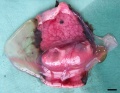
 Circumvallate placenta 01.jpg 1,188 × 1,000; 296 KB
 Cord blood induced stem cells 01.jpg 800 × 787; 157 KB
 Cord blood induced stem cells 02.jpg 700 × 966; 189 KB
 Cullen1916 fig14.jpg 1,280 × 1,293; 434 KB
 Cullen1916 fig15.jpg 1,000 × 1,227; 314 KB
 Cullen1916 fig16.jpg 1,280 × 1,686; 621 KB
 Cullen1916 fig17.jpg 1,280 × 1,134; 365 KB
 Cullen1916 fig19.jpg 1,280 × 1,098; 330 KB
 Cullen1916 fig22.jpg 1,224 × 1,501; 324 KB
 Cullen1916 fig23.jpg 1,000 × 1,266; 346 KB
 Cullen1916 fig33.jpg 1,154 × 2,052; 422 KB
 Cullen1916 fig92.jpg 885 × 637; 109 KB
 DeBruin1910 fig10.jpg 1,422 × 1,100; 208 KB
 Early placental structure.jpg 792 × 367; 90 KB
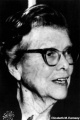 Elizabeth M. Ramsey.jpg 333 × 500; 27 KB
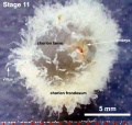 Embryo-membranes stage 11.jpg 600 × 568; 61 KB
 Endotheliochorial placenta EM01.jpg 600 × 602; 134 KB
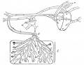
 Extravillous trophoblast cartoon.jpg 1,028 × 829; 249 KB
 Extravillous trophoblasts week 5.5.jpg 1,280 × 900; 421 KB
 Fetal membrane and placenta cartoon.jpg 600 × 429; 125 KB
 Foster117.jpg 797 × 707; 97 KB
 Foster118a.jpg 724 × 1,003; 175 KB
 Foster118b.jpg 809 × 1,077; 232 KB
 Galletti1770 placenta previa.jpg 450 × 450; 36 KB
 Gray0025.gif 293 × 232; 7 KB
 Gray0026.gif 300 × 334; 9 KB
 Gray0027.gif 300 × 298; 10 KB
 Gray0031.jpg 1,371 × 1,309; 270 KB
 Gray0032.gif 500 × 417; 57 KB
 Gray0032.jpg 800 × 667; 159 KB
 Gray0034.jpg 800 × 613; 98 KB
 Gray0035.jpg 450 × 415; 59 KB
 Gray0036.gif 450 × 320; 26 KB
 Gray0036.jpg 500 × 356; 52 KB
 Gray0037.gif 450 × 371; 37 KB
 Gray0037.jpg 500 × 412; 74 KB
 Gray0038.jpg 462 × 600; 45 KB
 Gray0039.gif 500 × 384; 43 KB
 Gray0039.jpg 1,797 × 1,305; 464 KB
 Gray0472.jpg 550 × 653; 57 KB
 Gray0502.jpg 1,000 × 1,329; 215 KB
 Haemomonochorial human placenta EM01.jpg 792 × 775; 79 KB
 Haemomonochorial placenta EM01.jpg 600 × 602; 111 KB
 HamiltonBoyd1960 fig02.jpg 800 × 836; 121 KB
 HamiltonBoyd1960 fig03.jpg 644 × 754; 114 KB
 HamiltonBoyd1960 fig04.jpg 700 × 926; 131 KB
 HamiltonBoyd1960 fig05.jpg 700 × 1,043; 150 KB
 HamiltonBoyd1960 fig06.jpg 800 × 872; 97 KB
 HamiltonBoyd1960 fig07.jpg 800 × 775; 122 KB
 HamiltonBoyd1960 fig08.jpg 1,278 × 1,048; 268 KB
 HamiltonBoyd1960 plate01.jpg 1,000 × 1,021; 350 KB
 HamiltonBoyd1960 plate02.jpg 1,280 × 1,646; 445 KB
 HamiltonBoyd1960 plate03.jpg 1,280 × 1,620; 389 KB
 HamiltonBoyd1960 plate04.jpg 1,280 × 1,710; 515 KB
 HamiltonBoyd1960 plate05.jpg 1,280 × 1,805; 790 KB
 HamiltonBoyd1960 plate06.jpg 1,280 × 1,354; 444 KB
 HamiltonBoyd1960 plate07.jpg 1,280 × 1,536; 608 KB
 HamiltonBoyd1960 plate08.jpg 1,280 × 1,681; 604 KB
 HamiltonBoyd1960 plate09.jpg 1,280 × 1,675; 493 KB
 HamiltonBoyd1960 plate10.jpg 1,280 × 2,316; 499 KB
 HamiltonBoyd1960 plate11.jpg 1,280 × 1,707; 659 KB
 HamiltonBoyd1960 plate12.jpg 1,280 × 1,455; 605 KB
 HamiltonBoyd1960 plate13.jpg 1,280 × 1,923; 462 KB
 Hertig1946b fig17.jpg 800 × 1,155; 161 KB
 Hertig1946b fig17a.jpg 800 × 587; 85 KB
 Hertig1946b fig17b.jpg 800 × 559; 76 KB
 Hertig1946b fig25.jpg 800 × 1,184; 266 KB
 Hertig1946b fig25a.jpg 800 × 528; 126 KB
 Hertig1946b fig25b.jpg 800 × 644; 139 KB
 Hertig1946b fig26.jpg 800 × 1,299; 290 KB
 Hertig1946b fig26a.jpg 800 × 646; 131 KB
 Hertig1946b fig26b.jpg 800 × 638; 198 KB
 Hill Homo62 fig01.jpg 1,280 × 1,488; 274 KB
 HillH5 Stage 16 bf07.jpg 1,125 × 1,500; 151 KB
 HillH52 chorionic villi 01.jpg 1,200 × 924; 182 KB
 HillH52 chorionic villi 02.jpg 1,200 × 900; 155 KB
 HillH52 chorionic villi 03.jpg 1,200 × 891; 280 KB
 HillH52 chorionic villi 04.jpg 1,200 × 900; 254 KB
 HillH52 chorionic villi 05.jpg 1,200 × 992; 371 KB
 HillH52 chorionic villi 06.jpg 1,200 × 900; 346 KB
 HillH52 chorionic villi 07.jpg 1,200 × 900; 544 KB
 HillH52 chorionic villi 08.jpg 1,200 × 900; 229 KB
 HillH52 chorionic villi 09.jpg 1,200 × 900; 237 KB
 HillH52 chorionic villi 10.jpg 1,200 × 900; 253 KB
 Hubrecht Homo73a cord 1.jpg 1,200 × 900; 427 KB
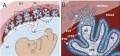
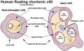
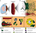
 Human and mouse fetal-maternal interface cartoon.jpg 600 × 720; 91 KB
 Human fetal membrane 01.jpg 726 × 545; 69 KB
 Human fetal membrane 02.jpg 726 × 545; 87 KB
 Human fetal membrane 03.jpg 726 × 545; 88 KB
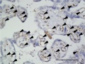
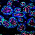
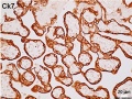
 Human placenta cytokeratin 7 expression 01.jpg 800 × 621; 168 KB
 Human placenta SERPINE2 and CK7 expression.jpg 932 × 600; 155 KB
 Human placenta SERPINE2 expression 01.jpg 800 × 601; 134 KB
 Human placenta SERPINE2 expression 02.jpg 1,200 × 419; 229 KB
 Human placenta SERPINE2 expression 03.jpg 800 × 616; 163 KB
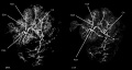 Human placenta vascular 01.jpg 1,200 × 644; 125 KB
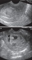
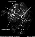 Human placenta vascular CT 01.jpg 938 × 1,000; 126 KB
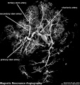 Human placenta vascular MRI 01.jpg 938 × 1,000; 151 KB
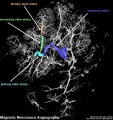 Human placenta vascular MRI 02.jpg 938 × 1,000; 156 KB
 Human placenta vasohibin 1 expression.jpg 1,206 × 908; 168 KB
 Human placenta vasohibin 2 expression.jpg 1,198 × 904; 165 KB
 Human placental villi cartoon 01.jpg 1,084 × 663; 142 KB
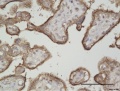
 Human placental villi keratin 19.jpg 800 × 798; 216 KB
 Human placental villi keratin 5.jpg 800 × 798; 148 KB
 Human placental villi keratin 7.jpg 800 × 798; 197 KB
 Human placental villi keratin 8.jpg 800 × 798; 251 KB
 Human term placental villi E-cadherin-vimentin.jpg 1,003 × 1,000; 233 KB
 Human term placental villi keratin 7.jpg 1,000 × 749; 179 KB
 Human trophoblast invasion 01.jpg 985 × 909; 223 KB
 Hydatidiform mole 02.jpg 585 × 1,100; 237 KB
 Hydatidiform mole metastasis 01.jpg 712 × 800; 67 KB
 Hydatidiform mole pulmonary metastasis 01.jpg 1,000 × 750; 105 KB
 Keibel Mall 088-091.jpg 673 × 564; 75 KB
 Keibel Mall 092.jpg 685 × 813; 129 KB
 Keibel Mall 113.jpg 679 × 502; 87 KB
 Keibel Mall 114.jpg 704 × 496; 96 KB
 Keibel Mall 115.jpg 686 × 506; 91 KB
 Keibel Mall 116.jpg 682 × 201; 31 KB
 Keibel Mall 117.jpg 687 × 534; 85 KB
 Keibel Mall 118.jpg 698 × 1,004; 158 KB
 Keibel Mall 119.jpg 684 × 496; 45 KB
 Keibel Mall 120.jpg 688 × 533; 57 KB
 Keibel Mall 121.jpg 683 × 691; 66 KB
 Keibel Mall 122.jpg 678 × 494; 78 KB
 Keibel Mall 123.jpg 709 × 435; 56 KB
 Keibel Mall 124.jpg 712 × 803; 140 KB
 Keibel Mall 125.jpg 743 × 566; 107 KB
 Keibel Mall 126.jpg 706 × 724; 99 KB
 Keibel Mall 127.jpg 730 × 768; 125 KB
 Keibel Mall 128.jpg 751 × 612; 105 KB
 Keibel Mall 129.jpg 702 × 756; 127 KB
 Keibel Mall 130.jpg 715 × 532; 82 KB
 Keibel Mall 131.jpg 703 × 834; 134 KB
 Keibel Mall 132.jpg 698 × 570; 100 KB
 Keibel Mall 133.jpg 690 × 449; 85 KB
 Keibel Mall 134.jpg 687 × 945; 96 KB
 Keibel Mall 135.jpg 686 × 568; 56 KB
 Keibel Mall 136.jpg 688 × 911; 174 KB
 Keibel Mall 137.jpg 684 × 780; 88 KB
 Keibel Mall 138.jpg 667 × 237; 34 KB
 Keibel Mall 139.jpg 690 × 559; 85 KB
 Keibel Mall 140.jpg 691 × 354; 43 KB
 Keith1902 fig074.jpg 500 × 511; 30 KB
 Keith1902 fig075.jpg 1,000 × 708; 118 KB
 Keith1902 fig076.jpg 732 × 800; 108 KB
 Keith1902 fig077.jpg 927 × 600; 96 KB
 Keith1902 fig078.jpg 788 × 600; 81 KB
 Keith1921 fig025.jpg 1,075 × 814; 214 KB
 Keith1921 fig030.jpg 922 × 693; 159 KB
 Keith1921 fig031.jpg 1,000 × 760; 127 KB
 Keith1921 fig032.jpg 1,147 × 1,383; 266 KB
 Keith1921 fig033.jpg 1,170 × 727; 146 KB
 Kellicott 182.jpg 904 × 971; 180 KB
 Kellicott 183.jpg 1,042 × 1,000; 197 KB
 Kellicott 184.jpg 661 × 1,000; 196 KB
 Kellicott 185.jpg 1,000 × 800; 225 KB
 Kollmann096.jpg 800 × 817; 104 KB
 Kollmann097.jpg 800 × 529; 42 KB
 Kollmann098.jpg 789 × 661; 76 KB
 Kollmann099.jpg 800 × 761; 78 KB
 Kollmann100.jpg 800 × 571; 68 KB
 Kollmann102.jpg 800 × 411; 40 KB
 Kollmann103.jpg 800 × 448; 54 KB
 Kollmann104.jpg 800 × 773; 88 KB
 Kollmann105.jpg 800 × 565; 86 KB
 Kollmann106.jpg 800 × 596; 57 KB
 Kollmann107.jpg 800 × 609; 97 KB


