Category:Mouse E13.5: Difference between revisions
mNo edit summary |
mNo edit summary |
||
| (8 intermediate revisions by the same user not shown) | |||
| Line 1: | Line 1: | ||
This {{Embryology}} category shows pages and media related to mouse embryonic day 13.5 ('''E13.5''') of development. This staging by "days" relate to in the female presence of a vaginal plug indicating that the mating occurred, see [[Mouse_Timeline_Detailed#Timed_Pregnancy|timed pregnancy]]. | |||
{{Mouse E days}} | {{Mouse E days}} | ||
'''Search Pubmed:''' [http://www.ncbi.nlm.nih.gov/sites/entrez?db=pubmed&cmd=search&term=Mouse+E13.5 Mouse E13.5] | |||
{{Mouse}} | {{Mouse}} | ||
==Events== | |||
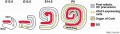
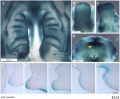
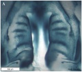
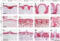
* [[Musculoskeletal_System_Development|Musculoskeletal System]] - Abdominal wall all five the principal muscles observed (rectus abdominis, external oblique, internal oblique, transversus abdominis, and panniculus carnosus). Rectus abdominis not completely segregated from the transversus abdominis and obliques. The panniculus carnosus was formed in the dorsal half of the abdominal wall adjacent to the skin. The myoblasts have migrated approximately three fourths of the distance to the ventral midline. Connective tissue between the muscle layers and dermis comprised the majority of the thickness of the secondary abdominal wall with the layer between the panniculus carnosus and external oblique being the thickest of these layers (470 μm). Directional organization of the myoblasts in the external oblique, internal oblique, and transverses abdominis.{{#pmid:22976993|PMID22976993}} | |||
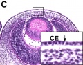
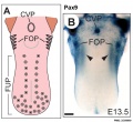
* {{Taste}} - Pax9 expression in tongue circumvallate papilla and foliate papillae [[:File:Mouse tongue Pax9 expression 03.jpg|Image]] | {{Pax}} | |||
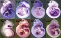
* [[Gastrointestinal Tract - Pancreas Development|Pancreas]] - day 13 to 14 ([[:Category:Mouse E13|E13]], [[:Category:Mouse E13.5|E13.5]], [[:Category:Mouse E14|E14]]) stomach rotation brings the smaller ventral pancreatic bud dorsally to fuse with the larger dorsal pancreatic bud. | |||
===References=== | |||
<references/> | |||
'''Search Pubmed:''' [http://www.ncbi.nlm.nih.gov/sites/entrez?db=pubmed&cmd=search&term=Mouse+E13.5 Mouse E13.5] | '''Search Pubmed:''' [http://www.ncbi.nlm.nih.gov/sites/entrez?db=pubmed&cmd=search&term=Mouse+E13.5 Mouse E13.5] | ||
Latest revision as of 08:35, 25 January 2019
This Embryology category shows pages and media related to mouse embryonic day 13.5 (E13.5) of development. This staging by "days" relate to in the female presence of a vaginal plug indicating that the mating occurred, see timed pregnancy.
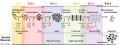
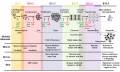
- Mouse Stages: E1 | E2.5 | E3.0 | E3.5 | E4.5 | E5.0 | E5.5 | E6.0 | E7.0 | E7.5 | E8.0 | E8.5 | E9.0 | E9.5 | E10 | E10.5 | E11 | E11.5 | E12 | E12.5 | E13 | E13.5 | E14 | E14.5 | E15 | E15.5 | E16 | E16.5 | E17 | E17.5 | E18 | E18.5 | E19 | E20 | Timeline | About timed pregnancy
| Carnegie | Stage | |||||||||||||||||||||||
| Human | Days | 1 | 2-3 | 4-5 | 5-6 | 7-12 | 13-15 | 15-17 | 17-19 | 20 | 22 | 24 | 28 | 30 | 33 | 36 | 40 | 42 | 44 | 48 | 52 | 54 | 55 | 58 |
| Mouse | Days | 1 | 2 | 3 | E4.5 | E5.0 | E6.0 | E7.0 | E8.0 | E9.0 | E9.5 | E10 | E10.5 | E11 | E11.5 | E12 | E12.5 | E13 | E13.5 | E14 | E14.5 | E15 | E15.5 | E16 |
| Rat | Days | 1 | 3.5 | 4-5 | 5 | 6 | 7.5 | 8.5 | 9 | 10.5 | 11 | 11.5 | 12 | 12.5 | 13 | 13.5 | 14 | 14.5 | 15 | 15.5 | 16 | 16.5 | 17 | 17.5 |
| Note these Carnegie stages are only approximate day timings for average of embryos. Links: Carnegie Stage Comparison | ||||||||||||||||||||||||
| ||||||||||||||||||||||||
| Timeline Links: human timeline | mouse timeline | mouse detailed timeline | chicken timeline | rat timeline | Medaka | Category:Timeline |
Search Pubmed: Mouse E13.5
Events
- Musculoskeletal System - Abdominal wall all five the principal muscles observed (rectus abdominis, external oblique, internal oblique, transversus abdominis, and panniculus carnosus). Rectus abdominis not completely segregated from the transversus abdominis and obliques. The panniculus carnosus was formed in the dorsal half of the abdominal wall adjacent to the skin. The myoblasts have migrated approximately three fourths of the distance to the ventral midline. Connective tissue between the muscle layers and dermis comprised the majority of the thickness of the secondary abdominal wall with the layer between the panniculus carnosus and external oblique being the thickest of these layers (470 μm). Directional organization of the myoblasts in the external oblique, internal oblique, and transverses abdominis.[1]
- taste - Pax9 expression in tongue circumvallate papilla and foliate papillae Image | Pax
- Pancreas - day 13 to 14 (E13, E13.5, E14) stomach rotation brings the smaller ventral pancreatic bud dorsally to fuse with the larger dorsal pancreatic bud.
References
Search Pubmed: Mouse E13.5
Pages in category 'Mouse E13.5'
The following 3 pages are in this category, out of 3 total.
Media in category 'Mouse E13.5'
The following 37 files are in this category, out of 37 total.
 Anderson2016-fig26a.jpg 800 × 800; 126 KB
 Anderson2016-fig26b.jpg 800 × 800; 103 KB
 Embryonic Trachea.png 468 × 326; 205 KB
 Mouse - forebrain Robo3 expression.jpg 675 × 1,280; 289 KB
 Mouse - palate MMP-25 expression.jpg 1,000 × 818; 243 KB
 Mouse - stomach 01.png 599 × 600; 1.45 MB
 Mouse Bmp4 expression face 01.jpg 1,200 × 322; 58 KB
 Mouse Bmp4 expression limb and face 01.jpg 1,200 × 513; 91 KB
 Mouse cochlea development cartoon.jpg 1,000 × 280; 53 KB
 Mouse cochlea gene expression.jpg 1,000 × 346; 75 KB
 Mouse cornea development 01.jpg 1,200 × 880; 325 KB
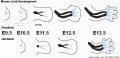
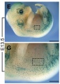
 Mouse cornea E13.5.jpg 700 × 550; 103 KB
 Mouse E13.5 Bmp7 palate 1.jpg 1,200 × 993; 156 KB
 Mouse E13.5 Bmp7 palate 2.jpg 917 × 800; 88 KB
 Mouse E13.5 Bmp7 palate 3.jpg 800 × 488; 40 KB
 Mouse E13.5 gene expression.jpg 1,764 × 1,100; 266 KB
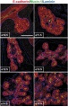
 Mouse eye cell proliferation E13.5.jpg 875 × 1,244; 227 KB
 Mouse eye neural crest.jpg 1,086 × 1,509; 483 KB
 Mouse face Bmp4.mp4 ; 480 KB
 Mouse gonad Gcnf expression 01.jpg 1,947 × 843; 304 KB
 Mouse gonad Gcnf expression E13.5.jpg 332 × 784; 60 KB
 Mouse limb bone development timeline.jpg 1,256 × 469; 107 KB
 Mouse limb skeleton cartoon.jpg 1,000 × 487; 64 KB
 Mouse limb tissue development.jpg 1,280 × 767; 161 KB
 Mouse melanoblast distribution 01.jpg 697 × 1,000; 192 KB
 Mouse melanoblast distribution 03.jpg 751 × 1,051; 150 KB
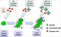 Mouse model of ovarian cord formation 01.jpg 800 × 491; 85 KB
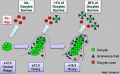 Mouse model of ovarian cord formation.jpg 800 × 491; 85 KB
 Mouse pancreas development.jpg 600 × 939; 261 KB
 Mouse respiratory 36 to 60 somites.jpg 1,200 × 383; 58 KB
 Mouse spleen capsulin expression.jpg 960 × 670; 226 KB
 Mouse thyroid Hes1 model.jpg 600 × 364; 33 KB
 Mouse tongue Pax9 expression 01.jpg 1,200 × 687; 259 KB
 Mouse tongue Pax9 expression 02.jpg 1,200 × 833; 299 KB
 Mouse tongue Pax9 expression 03.jpg 1,091 × 1,000; 145 KB
 Mouse- hindlimb Bmp4 expression.jpg 374 × 241; 19 KB
 Mouse-pancreas duct formation.jpg 1,000 × 709; 154 KB


