Neural - Cerebellum Development: Difference between revisions
| Line 82: | Line 82: | ||
</gallery> | </gallery> | ||
==Abnormalities== | |||
[[File:Mouse cerebellar foliation defects.jpg]] | |||
== References == | == References == | ||
Revision as of 23:57, 22 September 2011
Introduction


Neural development is one of the earliest systems to begin and the last to be completed after birth. This development generates the most complex structure within the embryo and the long time period of development means in utero insult during pregnancy may have consequences to development of the nervous system.
The adult cerebellum anatomy consists of three parts, the vermis (median) and the two hemispheres (lateral), which are continuous with each other.
Within the neural tube stem cells generate the 2 major classes of cells that make the majority of the nervous system : neurons and glia. Both these classes of cells differentiate into many different types generated with highly specialized functions and shapes. This section covers the establishment of neural populations, the inductive influences of surrounding tissues and the sequential generation of neurons establishing the layered structure seen in the brain and spinal cord.
- Neural development beginnings quite early, therefore also look at notes covering Week 3- neural tube and Week 4-early nervous system.
- Development of the neural crest and sensory systems (hearing/vision/smell) are only introduced in these notes and are covered in other notes sections.
- Links: Historic - The Cerebellum
Some Recent Findings
|
Development Overview

Neuralation begins at the trilaminar embryo with formation of the notochord and somites, both of which underly the ectoderm and do not contribute to the nervous system, but are involved with patterning its initial formation. The central portion of the ectoderm then forms the neural plate that folds to form the neural tube, that will eventually form the entire central nervous system.
- Early developmental sequence: Epiblast - Ectoderm - Neural Plate - Neural groove and Neural Crest - Neural Tube and Neural Crest
| Neural Tube | Primary Vesicles | Secondary Vesicles | Adult Structures |
|---|---|---|---|
| week 3 | week 4 | week 5 | adult |
| prosencephalon (forebrain) | telencephalon | Rhinencephalon, Amygdala, hippocampus, cerebrum (cortex), hypothalamus, pituitary | Basal Ganglia, lateral ventricles | |
| diencephalon | epithalamus, thalamus, Subthalamus, pineal, posterior commissure, pretectum, third ventricle | ||
| mesencephalon (midbrain) | mesencephalon | tectum, Cerebral peduncle, cerebral aqueduct, pons | |
| rhombencephalon (hindbrain) | metencephalon | cerebellum | |
| myelencephalon | medulla oblongata, isthmus | ||
| spinal cord, pyramidal decussation, central canal | |||
Early Brain Vesicles
Primary Vesicles

Secondary Vesicles

Fetal Cerebellum (Week 10)

|

|
| Plane A (midline) | Plane B (medial) |

|

|
| Plane C (lateral) | Plane D (most lateral) |
- Links: 10 week Fetal | Fetal Development
Third Trimester

Developing human cerebellum preterm[1]
A greater EGL cell density and reduced EGL thickness were reported in preterms with ex-utero exposure, as compared to their age matched stillborn controls.
Cerebellum Images
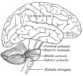
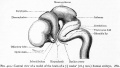
Scheme showing the connections of the several parts of the brain. (After Schwalbe)
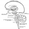
Schematic representation of the chief ganglionic categories (I to V). (Spitzka)
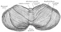
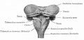
Upper surface of the cerebellum. (Schäfer.)
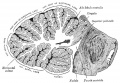
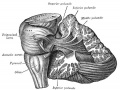
Dissection showing the projection fibers of the cerebellum. (After E. B. Jamieson)
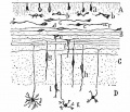
Transverse section of a cerebellar folium. (Diagrammatic, after Cajal and Kölliker)
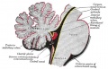
The cerebellum is shown in black.
Lateral view of a model of the brain of a 7.5 weeks (18.5 mm.) human embryo.
Dorsal view of the cerebellum and medulla of a 5 months' human fetus. (Kollmann)
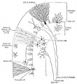
various stages of position and form in the differentiation of granule cells from the outer granular layer. (Cajal)
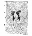
cerebellar cortex of a newborn dog showing dendrites of two Purkinje cells. (Cajal.)


Abnormalities

References
Reviews
<pubmed>19732611</pubmed> <pubmed>17408845</pubmed> <pubmed>16243598</pubmed> <pubmed>15610138</pubmed> <pubmed>12843872</pubmed>
Articles
<pubmed>20460306</pubmed> <pubmed>15496441</pubmed>
Search PubMed
Search Pubmed: Cerebellum Embryology | Cerebellum Development
Glossary Links
- Glossary: A | B | C | D | E | F | G | H | I | J | K | L | M | N | O | P | Q | R | S | T | U | V | W | X | Y | Z | Numbers | Symbols | Term Link
Cite this page: Hill, M.A. (2024, June 18) Embryology Neural - Cerebellum Development. Retrieved from https://embryology.med.unsw.edu.au/embryology/index.php/Neural_-_Cerebellum_Development
- © Dr Mark Hill 2024, UNSW Embryology ISBN: 978 0 7334 2609 4 - UNSW CRICOS Provider Code No. 00098G