Gastrointestinal Tract - Stomach Development: Difference between revisions
mNo edit summary |
|||
| (49 intermediate revisions by 2 users not shown) | |||
| Line 1: | Line 1: | ||
{{Header}} | |||
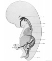
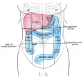
[[File:Gray0982a.jpg|right]] | |||
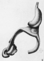
[[File:Stage14 stomach.jpg|thumb|Developing stomach mid embryonic period (Carnegie stage {{CS13}})]] | |||
==Introduction== | |||
This section of notes gives an overview of how the {{stomach}} and duodenum develops. The GIT is best imagined as a simple tube, the upper part being the foregut diverticulum, which is further divided into oesophagus and stomach. | |||
During week 4 at the level where the stomach will form the tube begins to dilate, forming an enlarged lumen. The dorsal border grows more rapidly than ventral, which establishes the '''greater curvature''' of the stomach.{{#pmid:28242610|PMID28242610}} A second rotation (of 90 degrees) occurs on the longitudinal axis establishing the adult orientation of the stomach. | |||
{{Gastrointestinal Tract Links}} | |||
::[[Historic Embryology_Papers|'''Historic Embryology''']]: [[Book_-_Manual_of_Human_Embryology_17-4|1912 Stomach]] | |||
==Some Recent Findings== | ==Some Recent Findings== | ||
{| | {| | ||
|-bgcolor="F5FAFF" | |-bgcolor="F5FAFF" | ||
| | | | ||
* '''FGF10 is required for cell proliferation and gland formation in the stomach epithelium of the chicken embryo.''' | * '''Stomach curvature is generated by left-right asymmetric gut morphogenesis'''{{#pmid:28242610|PMID28242610}} "Left-right (LR) asymmetry is a fundamental feature of internal anatomy, yet the emergence of morphological asymmetry remains one of the least understood phases of organogenesis. Asymmetric rotation of the intestine is directed by forces outside the gut, but the morphogenetic events that generate anatomical asymmetry in other regions of the digestive tract remain unknown. Here, we show in mouse and Xenopus that the mechanisms that drive the curvature of the stomach are intrinsic to the gut tube itself. The left wall of the primitive stomach expands more than the right wall, as the left epithelium becomes more polarized and undergoes radial rearrangement. These asymmetries exist across several species, and are dependent on LR patterning genes, including Foxj1, Nodal and Pitx2 Our findings have implications for how LR patterning manifests distinct types of morphological asymmetries in different contexts." | ||
* '''FGF4 and retinoic acid direct differentiation of hESCs into PDX1-expressing foregut endoderm in a time- and concentration-dependent manner.'''{{#pmid:19277121|PMID19277121}} "Retinoic acid (RA) and fibroblast growth factor 4 (FGF4) signaling control endoderm patterning and pancreas induction/expansion.[[Developmental Signals - Fibroblast Growth Factor|FGF]] | |||
* '''FGF10 is required for cell proliferation and gland formation in the stomach epithelium of the chicken embryo.''' {{#pmid:16616737|PMID16616737}} "The development of digestive organs in vertebrates involves active epithelial-mesenchymal interactions. In the chicken proventriculus (glandular stomach), the morphogenesis and cytodifferentiation of the epithelium are controlled by the inductive signaling factors that are secreted from the underlying mesenchyme. ... FGF10 signaling, mediated by FGFR1b and/or FGFR2b, is required for proliferation and gland formation in the epithelium in the developing chick embryo." [[Developmental Signals - Fibroblast Growth Factor|FGF]] | |||
|} | |||
{| class="wikitable mw-collapsible mw-collapsed" | |||
! More recent papers | |||
|- | |||
| [[File:Mark_Hill.jpg|90px|left]] {{Most_Recent_Refs}} | |||
Search term: [http://www.ncbi.nlm.nih.gov/pubmed/?term=Stomach+Embryology ''Stomach Embryology''] | [http://www.ncbi.nlm.nih.gov/pubmed/?term=Stomach+Development ''Stomach Development''] | |||
|} | |} | ||
== Components of Stomach Formation == | == Components of Stomach Formation == | ||
'''primitive endoderm''' | '''primitive endoderm''' | ||
| Line 42: | Line 55: | ||
Modified from <ref>Kaufman & Bard, '''The Anatomical Basis of Mouse Development''', 1999 Academic Press</ref> | Modified from <ref>Kaufman & Bard, '''The Anatomical Basis of Mouse Development''', 1999 Academic Press</ref> | ||
==Movies== | |||
{| | |||
| valign="bottom"|{{Endoderm movie}} | |||
| valign="bottom"|{{Amnion movie}} | |||
| valign="bottom"|{{GIT growth movie}} | |||
| valign="bottom"|{{Stomach rotation movie}} | |||
| valign="bottom"|{{Lesser sac movie}} | |||
| valign="bottom"|{{Greater omentum movie}} | |||
|} | |||
==Stage 13== | ==Stage 13== | ||
The images below link to larger cross-sections of the mid-embryonic period (end week 4) [[Carnegie stage 13|stage 13]] embryo starting just above the level of the stomach and then in sequence through the stomach to the level of the duodenum. | The images below link to larger cross-sections of the mid-embryonic period (end week 4) [[Carnegie stage 13|stage 13]] embryo starting just above the level of the stomach and then in sequence through the stomach to the level of the duodenum. | ||
{| | {| | ||
|- | |- | ||
| [[File:Stage 13 image 072.jpg|200px]] | |||
| [[File:Stage 13 image 072.jpg| | | [[File:Stage 13 image 073.jpg|200px]] | ||
| [[File:Stage 13 image 073.jpg| | | [[File:Stage 13 image 074.jpg|200px]] | ||
| [[File:Stage 13 image 074.jpg| | |||
|- | |- | ||
| [[:File:Stage 13 image | | [[:File:Stage 13 image 072.jpg|D2 Cardio-oesophageal junction]] | ||
| [[:File:Stage 13 image | | [[:File:Stage 13 image 073.jpg|D3 Stomach body]] | ||
| [[:File:Stage 13 image | | [[:File:Stage 13 image 074.jpg|D4 Stomach body]] | ||
| [[:File:Stage 13 image | |- | ||
| [[:File:Stage 13 image 075.jpg| | | [[File:Stage 13 image 075.jpg|200px]] | ||
| [[:File:Stage 13 image 076.jpg| | | [[File:Stage 13 image 076.jpg|200px]] | ||
| [[:File:Stage 13 image 077.jpg| | | [[File:Stage 13 image 077.jpg|200px]] | ||
|- | |||
| [[:File:Stage 13 image 075.jpg|D5 Stomach body]] | |||
| [[:File:Stage 13 image 076.jpg|D6 Pyloric junction]] | |||
| [[:File:Stage 13 image 077.jpg|D7 Duodenum]] | |||
|} | |} | ||
{| | {| | ||
| | | valign="bottom"|{{Gastrointestinal stage 13 movie}} | ||
| | | This is an animation based on a reconstruction of the above embryo entire [[Carnegie stage 13|stage 13]] gastrointestinal tract. | ||
This is an animation based on a reconstruction of the above embryo entire [[Carnegie stage 13|stage 13]] gastrointestinal tract. | |||
|} | |} | ||
:'''Links:''' [[Carnegie stage 13 - serial sections]] | [[Embryo Serial Sections]] | [[ | :'''Links:''' [[Carnegie stage 13 - serial sections]] | [[Embryo Serial Sections]] | [[Movies]] | ||
==Stage 22== | ==Stage 22== | ||
[[ | The images below link to larger cross-sections of the end of the embryonic period ([[week 8]]) [[Carnegie stage 22|stage 22]] embryo starting just above the level of the stomach and then in sequence through the stomach to the level of the duodenum. Note how the stomach is "embedded" within the large developing liver. | ||
{| | {| | ||
|- | |- | ||
| [[File:Stage_22 image 082.jpg| | | [[File:Stage_22 image 082.jpg|200px]] | ||
| [[File:Stage_22 image 083.jpg| | | [[File:Stage_22 image 083.jpg|200px]] | ||
| [[File:Stage_22 image 084.jpg| | | [[File:Stage_22 image 084.jpg|200px]] | ||
| [[File:Stage_22 image 085.jpg|200px]] | |||
|- | |- | ||
| [[:File:Stage_22 image 082.jpg| | | [[:File:Stage_22 image 082.jpg|E5 Oesophagus]] | ||
| [[:File:Stage_22 image 083.jpg| | | [[:File:Stage_22 image 083.jpg|E6 Cardio-oesophageal junction]] | ||
| [[:File:Stage_22 image 084.jpg| | | [[:File:Stage_22 image 084.jpg|E7 Stomach body Pyloric junction]] | ||
| [[:File:Stage_22 image 085.jpg|F1 Stomach body Pylorus]] | |||
|- | |- | ||
| [[File:Stage_22 image 086.jpg|200px]] | |||
| [[File:Stage_22 image 086.jpg| | | [[File:Stage_22 image 087.jpg|200px]] | ||
| [[File:Stage_22 image 087.jpg| | | [[File:Stage_22 image 088.jpg|200px]] | ||
| [[File:Stage_22 image 088.jpg| | | [[File:Stage_22 image 089.jpg|200px]] | ||
| [[File:Stage_22 image 089.jpg| | |||
|- | |- | ||
| [[:File:Stage_22 image 086.jpg|F2 Stomach body]] | |||
| [[:File:Stage_22 image 086.jpg| | | [[:File:Stage_22 image 087.jpg|F3 Stomach body Duodenum]] | ||
| [[:File:Stage_22 image 087.jpg| | | [[:File:Stage_22 image 088.jpg|F4 Duodenal-Jejunal junction]] | ||
| [[:File:Stage_22 image 088.jpg| | | [[:File:Stage_22 image 089.jpg|F5 Duodenum Jejunum]] | ||
| [[:File:Stage_22 image 089.jpg| | |||
|} | |} | ||
| Line 110: | Line 126: | ||
{| | {| | ||
| | | valign="bottom"|{{Gastrointestinal stage 22 movie}} | ||
| | | This is an animation based on a reconstruction of the above embryo entire [[Carnegie stage 22|stage 22]] gastrointestinal tract. | ||
This is an animation based on a reconstruction of the above embryo entire [[Carnegie stage 22|stage 22]] gastrointestinal tract. | |||
|} | |||
:'''Links:''' [[Carnegie stage 22 - serial sections]] | [[Embryo Serial Sections]] | [[Movies]] | |||
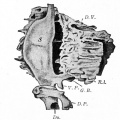
==Stomach Mesentery== | |||
In the second trimester, the ventral and dorsal mesenteries associated with the stomach are still anatomically different from the newborn. The figure shows a lateral view of this process comparing the early second trimester arrangement with the newborn structure. | |||
===Ventral Mesogastrium=== | |||
Attached to the superior end of the stomach will form the [[L#lesser omentum|lesser omentum]]. This structure will connect the lesser curvature of the stomach to the liver as a ligamentous structure. | |||
===Dorsal Mesogastrium=== | |||
Attached to the inferior end of the stomach initially as an extended fold, this later fuses as a single "apron-like" structure, the [[G#greater omentum|greater omentum]]. Fusion will also incorporate the transverse colon part of the large intestine. This will also contribute the gastrosplenic ligament (gastrolienal ligament). | |||
{| | {| | ||
| [[File:Greater-omentum.jpg]] | | [[File:Greater-omentum.jpg]] | ||
| Line 139: | Line 160: | ||
This diagram shows the rotation with spinal cord at the top, vertebral body then dorsal aorta then pertioneal wall and cavity. | This diagram shows the rotation with spinal cord at the top, vertebral body then dorsal aorta then pertioneal wall and cavity. | ||
|} | |} | ||
==Glands== | |||
The data below comes from a historic study by Johnson (1910)<ref name=Johnson1910>{{Ref-Johnson1910}}</ref> | |||
# Early - A few vacuoles, similar to those of the oesophagus, are found in corresponding stages in the stomach. | |||
# '''16 mm''' - the smooth epithelium begins to show a number of pit-like depressions, the first appearances of the gastric pits. These rapidly increase in number and many become elongated to form grooves. At first the basal surface of the epithelium shows no irregularities due to the gastric pits. Then appear slight swellings into the mesenchyma. Still later depressions or furrows, alternating with the gastric pits, extend inward into the epithelium from the mesenchymal surface. This brings about a readjustment of the cells, and causes the heretofore 2 to 3 layered epithelium to become single layered. | |||
# The groove-like pits anastomose with one another to form a network. This network marks out irregular areas of the surface epithelium, which have been described as villi. | |||
# Growth of the mucous membrane is accompanied by an increase in the number of pits and by an increase in their size. The additional pits develop in between those already formed. | |||
# '''120 mm''' - Glands first seen bud out from the bottoms of the pits. These rapidly increase in number and give off branches. Parietal cells are first distinct at 120 mm | |||
# Large longitudinal folds of the mucous membrane occur in the stomach, but are variable in position, number, and size. Their occurrence is probably due to the contraction of the muscularis. | |||
== Stomach Hormonal Development == | == Stomach Hormonal Development == | ||
| Line 152: | Line 184: | ||
'''11 weeks''' - Serotonin containing cells in both the antrum and the fundus. | '''11 weeks''' - Serotonin containing cells in both the antrum and the fundus. | ||
Expression data based upon | Expression data based upon{{#pmid:6136542|PMID6136542}} | ||
===Other Gut Peptides=== | ===Other Gut Peptides=== | ||
* Cholecystokinin (CCK), pancreatic polypeptide, peptide YY, glucagon-like peptide-1 (GLP-1), oxyntomodulin (increase satiety and decrease food intake) and ghrelin | * Cholecystokinin (CCK), pancreatic polypeptide, peptide YY, glucagon-like peptide-1 (GLP-1), oxyntomodulin (increase satiety and decrease food intake) and ghrelin | ||
==Mouse== | |||
The images below show differential gene expression of some selected markers during development (E10.5 and E13.5) of the mouse gastrointestinal tract.{{#pmid:19300477|PMID19300477}} | |||
[[File:Mouse-Gastrointestinal-tract-E10.5-01.jpg|600px]] | |||
[[File:Mouse-Gastrointestinal-tract-E13.5-01.jpg|600px]] | |||
:'''Links:''' [[Mouse Development]] | [[:File:Mouse_-_stomach_01.png|Full original figure]] | [[:File:Mouse-Gastrointestinal-tract-E10.5-01.jpg|E10.5]] | [[:File:Mouse-Gastrointestinal-tract-E13.5-01.jpg|E13.5]] | |||
==References== | ==References== | ||
<references/> | <references/> | ||
===Reviews=== | |||
{{#pmid:28238948}} | |||
{{#pmid:26884394}} | |||
{{#pmid:19575677}} | |||
{{#pmid:16109035}} | |||
{{#pmid:7526882}} | |||
{{#pmid:3922287}} | |||
{{#pmid:4923462}} | |||
===Articles=== | |||
{{#pmid:16616737}} | |||
{{#pmid:11329933}} | |||
| Line 166: | Line 228: | ||
'''Search Pubmed Now:''' [http://www.ncbi.nlm.nih.gov/sites/entrez?db=pubmed&cmd=search&term=Stomach%20Development Stomach Development] | '''Search Pubmed Now:''' [http://www.ncbi.nlm.nih.gov/sites/entrez?db=pubmed&cmd=search&term=Stomach%20Development Stomach Development] | ||
===Historic References=== | |||
{{Ref-GrosserLewisMcmurrich1912}} | |||
{{Ref-Bardeen1914}} | |||
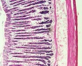
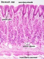
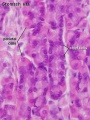
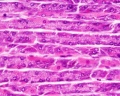
==Images== | ==Images== | ||
[[File: | <gallery caption="[[Gastrointestinal_Tract_-_Histology#Stomach|Stomach Histology]]"> | ||
File:Stomach gastric gland distribution.jpg|adult stomach gastric gland distribution | |||
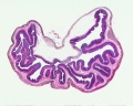
File:Stomach histology 008.jpg|rat stomach overview | |||
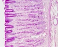
File:Stomach_histology_001.jpg|stomach labeled overview | |||
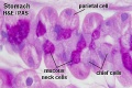
File:Stomach_histology_002.jpg|parietal cells - chief cells | |||
File:Stomach_histology_003.jpg|mucus neck - parietal cells - chief cells | |||
File:Stomach_histology_004.jpg|stomach overview | |||
File:Stomach_histology_005.jpg|stomach mucosa | |||
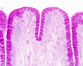
File:Stomach_histology_006.jpg|mucosa - secretory epithelial sheath - goblet cell | |||
File:Stomach_histology_007.jpg|gastric glands - parietal cells - chief cells | |||
</gallery> | |||
===Historic Images=== | |||
{{Historic Disclaimer}} | |||
<gallery> | |||
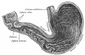
File:Keith1902 fig217.jpg|1902 transverse section mesogastrium | |||
File:Keith1902 fig218.jpg|1902 4th week human embryo | |||
File:Keith1902 fig220.jpg|1902 lesser sac of peritoneum | |||
File:Keibel Mall 2 239.jpg|1912 human embryo 4.9 mm | |||
File:Keibel Mall 2 240.jpg|1912 human embryo 7.5 mm | |||
File:Keibel Mall 2 242.jpg|1912 human embryo 9.4 mm | |||
File:Keibel Mall 2 243.jpg|1912 human embryo 22.8 mm | |||
File:Keibel Mall 2 246.jpg|1912 human embryo 42 mm | |||
File:Bardeen1914-fig02.jpg|1914 lateral view human embryo 27 mm | |||
File:Gray0990.jpg|1918 greater omentum | |||
File:Gray1050.jpg|1918 interior adult stomach | |||
File:Gray1223.png|1918 adult stomach position | |||
File:Bailey275.jpg|1921 human embryo 8 mm | |||
File:Bailey263.jpg|1921 human embryo 6 weeks | |||
File:Bailey265.jpg|1921 human embryo 28 mm | |||
</gallery> | |||
{{Glossary}} | |||
{{ | {{Footer}} | ||
[[Category:Stomach]] | |||
Latest revision as of 13:19, 16 April 2019
| Embryology - 14 Jun 2024 |
|---|
| Google Translate - select your language from the list shown below (this will open a new external page) |
|
العربية | català | 中文 | 中國傳統的 | français | Deutsche | עִברִית | हिंदी | bahasa Indonesia | italiano | 日本語 | 한국어 | မြန်မာ | Pilipino | Polskie | português | ਪੰਜਾਬੀ ਦੇ | Română | русский | Español | Swahili | Svensk | ไทย | Türkçe | اردو | ייִדיש | Tiếng Việt These external translations are automated and may not be accurate. (More? About Translations) |


Introduction
This section of notes gives an overview of how the stomach and duodenum develops. The GIT is best imagined as a simple tube, the upper part being the foregut diverticulum, which is further divided into oesophagus and stomach.
During week 4 at the level where the stomach will form the tube begins to dilate, forming an enlarged lumen. The dorsal border grows more rapidly than ventral, which establishes the greater curvature of the stomach.[1] A second rotation (of 90 degrees) occurs on the longitudinal axis establishing the adult orientation of the stomach.
Some Recent Findings
|
| More recent papers |
|---|
 This table allows an automated computer search of the external PubMed database using the listed "Search term" text link.
More? References | Discussion Page | Journal Searches | 2019 References | 2020 References Search term: Stomach Embryology | Stomach Development |
Components of Stomach Formation
primitive endoderm
- foregut diverticulum (pocket)
- pharyngeal region of foregut
- laryngo-tracheal groove (see respiratory tract)
- oesophageal region of foregut
- oesophagus
- stomach
- glandular/proventricular/pyloric stenosis
- fundus/pyloric antrum
- pyloric sphincter
- fundus/pyloric antrum
- dorsal mesogastrium
- lieno-renal ligament
- splenic primordium
- spleen
- gastro-splenic ligament
- duodenum (rostral half)
- splenic primordium
- lieno-renal ligament
- glandular/proventricular/pyloric stenosis
- stomach
- oesophagus
- pharyngeal region of foregut
- foregut-midgut junction
- midgut region
- hindgut diverticulum (pocket)
Modified from [4]
Movies
|
|
|
|
|
|
Stage 13
The images below link to larger cross-sections of the mid-embryonic period (end week 4) stage 13 embryo starting just above the level of the stomach and then in sequence through the stomach to the level of the duodenum.

|

|

|
| D2 Cardio-oesophageal junction | D3 Stomach body | D4 Stomach body |

|

|

|
| D5 Stomach body | D6 Pyloric junction | D7 Duodenum |
|
This is an animation based on a reconstruction of the above embryo entire stage 13 gastrointestinal tract. |
Stage 22
The images below link to larger cross-sections of the end of the embryonic period (week 8) stage 22 embryo starting just above the level of the stomach and then in sequence through the stomach to the level of the duodenum. Note how the stomach is "embedded" within the large developing liver.

|

|

|

|
| E5 Oesophagus | E6 Cardio-oesophageal junction | E7 Stomach body Pyloric junction | F1 Stomach body Pylorus |

|

|

|

|
| F2 Stomach body | F3 Stomach body Duodenum | F4 Duodenal-Jejunal junction | F5 Duodenum Jejunum |
|
This is an animation based on a reconstruction of the above embryo entire stage 22 gastrointestinal tract. |
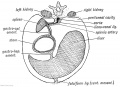
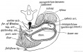
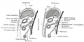
Stomach Mesentery
In the second trimester, the ventral and dorsal mesenteries associated with the stomach are still anatomically different from the newborn. The figure shows a lateral view of this process comparing the early second trimester arrangement with the newborn structure.
Ventral Mesogastrium
Attached to the superior end of the stomach will form the lesser omentum. This structure will connect the lesser curvature of the stomach to the liver as a ligamentous structure.
Dorsal Mesogastrium
Attached to the inferior end of the stomach initially as an extended fold, this later fuses as a single "apron-like" structure, the greater omentum. Fusion will also incorporate the transverse colon part of the large intestine. This will also contribute the gastrosplenic ligament (gastrolienal ligament).

|
The greater omentum hangs like an apron over the small intestine and transverse colon. It begins attacted to the inferior end of the stomach as a fold of the dorsal mesogastrium which later fuses to form the structure we recognise anatomically. The figure below shows a lateral view of this process comparing the early second trimester arrangement with the newborn structure. |
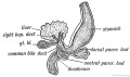
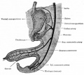
Duodenum/Pancreas Rotation

|
After the stomach the initial portion of the gastrointestinal tract tube is the duodenum which initially lies in the midline within the peritoneal cavity.
This region, along with the attached pancreas, undergoes rotation to become a retroperitoneal structure. This diagram shows the rotation with spinal cord at the top, vertebral body then dorsal aorta then pertioneal wall and cavity. |
Glands
The data below comes from a historic study by Johnson (1910)[5]
- Early - A few vacuoles, similar to those of the oesophagus, are found in corresponding stages in the stomach.
- 16 mm - the smooth epithelium begins to show a number of pit-like depressions, the first appearances of the gastric pits. These rapidly increase in number and many become elongated to form grooves. At first the basal surface of the epithelium shows no irregularities due to the gastric pits. Then appear slight swellings into the mesenchyma. Still later depressions or furrows, alternating with the gastric pits, extend inward into the epithelium from the mesenchymal surface. This brings about a readjustment of the cells, and causes the heretofore 2 to 3 layered epithelium to become single layered.
- The groove-like pits anastomose with one another to form a network. This network marks out irregular areas of the surface epithelium, which have been described as villi.
- Growth of the mucous membrane is accompanied by an increase in the number of pits and by an increase in their size. The additional pits develop in between those already formed.
- 120 mm - Glands first seen bud out from the bottoms of the pits. These rapidly increase in number and give off branches. Parietal cells are first distinct at 120 mm
- Large longitudinal folds of the mucous membrane occur in the stomach, but are variable in position, number, and size. Their occurrence is probably due to the contraction of the muscularis.
Stomach Hormonal Development
The gastrointestinal tract has its own complex entero-endocrine system (enterohormones) that regulates many regional tract functions.
Cells within the stomach express a range of peptide hormones known to regulate a range of gastric functions including secretion of digestive enzymes, mucous and the movement of the luminal contents. The list below shows the earliest detectible presence of specific hormone-containing cells in regions of the developing human stomach.
Hormonal Timecourse
8 weeks - Gastrin containing cells in stomach antrum. Somatostatin cells in both the antrum and the fundus.
10 weeks - Glucagon containing cells in stomach fundus.
11 weeks - Serotonin containing cells in both the antrum and the fundus.
Expression data based upon[6]
Other Gut Peptides
- Cholecystokinin (CCK), pancreatic polypeptide, peptide YY, glucagon-like peptide-1 (GLP-1), oxyntomodulin (increase satiety and decrease food intake) and ghrelin
Mouse
The images below show differential gene expression of some selected markers during development (E10.5 and E13.5) of the mouse gastrointestinal tract.[7]


- Links: Mouse Development | Full original figure | E10.5 | E13.5
References
- ↑ 1.0 1.1 Davis A, Amin NM, Johnson C, Bagley K, Ghashghaei HT & Nascone-Yoder N. (2017). Stomach curvature is generated by left-right asymmetric gut morphogenesis. Development , 144, 1477-1483. PMID: 28242610 DOI.
- ↑ Johannesson M, Ståhlberg A, Ameri J, Sand FW, Norrman K & Semb H. (2009). FGF4 and retinoic acid direct differentiation of hESCs into PDX1-expressing foregut endoderm in a time- and concentration-dependent manner. PLoS ONE , 4, e4794. PMID: 19277121 DOI.
- ↑ Shin M, Noji S, Neubüser A & Yasugi S. (2006). FGF10 is required for cell proliferation and gland formation in the stomach epithelium of the chicken embryo. Dev. Biol. , 294, 11-23. PMID: 16616737 DOI.
- ↑ Kaufman & Bard, The Anatomical Basis of Mouse Development, 1999 Academic Press
- ↑ Johnson FP. The development of the mucous membrane of the oesophagus, stomach and small intestine in the human embryo. (1910) Amer. J Anat., 10: 521-559.
- ↑ Stein BA, Buchan AM, Morris J & Polak JM. (1983). The ontogeny of regulatory peptide-containing cells in the human fetal stomach: an immunocytochemical study. J. Histochem. Cytochem. , 31, 1117-25. PMID: 6136542 DOI.
- ↑ Matsuyama M, Aizawa S & Shimono A. (2009). Sfrp controls apicobasal polarity and oriented cell division in developing gut epithelium. PLoS Genet. , 5, e1000427. PMID: 19300477 DOI.
Reviews
McCracken KW & Wells JM. (2017). Mechanisms of embryonic stomach development. Semin. Cell Dev. Biol. , 66, 36-42. PMID: 28238948 DOI.
Kim TH & Shivdasani RA. (2016). Stomach development, stem cells and disease. Development , 143, 554-65. PMID: 26884394 DOI.
Zorn AM & Wells JM. (2009). Vertebrate endoderm development and organ formation. Annu. Rev. Cell Dev. Biol. , 25, 221-51. PMID: 19575677 DOI.
Fukuda K & Yasugi S. (2005). The molecular mechanisms of stomach development in vertebrates. Dev. Growth Differ. , 47, 375-82. PMID: 16109035 DOI.
Yasugi S. (1994). Regulation of pepsinogen gene expression in epithelial cells of vertebrate stomach during development. Int. J. Dev. Biol. , 38, 273-9. PMID: 7526882
Johnson LR. (1985). Functional development of the stomach. Annu. Rev. Physiol. , 47, 199-215. PMID: 3922287 DOI.
Deren JS. (1971). Development of structure and function in the fetal and newborn stomach. Am. J. Clin. Nutr. , 24, 144-59. PMID: 4923462
Articles
Shin M, Noji S, Neubüser A & Yasugi S. (2006). FGF10 is required for cell proliferation and gland formation in the stomach epithelium of the chicken embryo. Dev. Biol. , 294, 11-23. PMID: 16616737 DOI.
Yasugi S. (2000). Epithelial cell differentiation during stomach development. Hum. Cell , 13, 177-84. PMID: 11329933
Search Pubmed
Search Bookshelf: Stomach Development
Search Pubmed Now: Stomach Development
Historic References
Template:Ref-GrosserLewisMcmurrich1912
Bardeen CR. The critical period in the development of the intestines. (1914) Amer. J Anat. 16: 427 – 445.
Images
- Stomach Histology
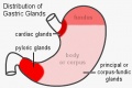
adult stomach gastric gland distribution
rat stomach overview
stomach labeled overview
parietal cells - chief cells
mucus neck - parietal cells - chief cells
stomach overview
stomach mucosa
mucosa - secretory epithelial sheath - goblet cell
gastric glands - parietal cells - chief cells


Historic Images
| Historic Disclaimer - information about historic embryology pages |
|---|
|
1902 transverse section mesogastrium
1902 4th week human embryo
1902 lesser sac of peritoneum
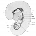
1912 human embryo 4.9 mm
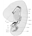
1912 human embryo 7.5 mm
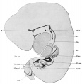
1912 human embryo 9.4 mm
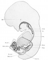
1912 human embryo 22.8 mm
1912 human embryo 42 mm
1914 lateral view human embryo 27 mm
1918 greater omentum
1918 interior adult stomach
1918 adult stomach position
1921 human embryo 8 mm
1921 human embryo 6 weeks
1921 human embryo 28 mm


Glossary Links
- Glossary: A | B | C | D | E | F | G | H | I | J | K | L | M | N | O | P | Q | R | S | T | U | V | W | X | Y | Z | Numbers | Symbols | Term Link
Cite this page: Hill, M.A. (2024, June 14) Embryology Gastrointestinal Tract - Stomach Development. Retrieved from https://embryology.med.unsw.edu.au/embryology/index.php/Gastrointestinal_Tract_-_Stomach_Development
- © Dr Mark Hill 2024, UNSW Embryology ISBN: 978 0 7334 2609 4 - UNSW CRICOS Provider Code No. 00098G