2010 Lab 5: Difference between revisions
No edit summary |
|||
| (39 intermediate revisions by the same user not shown) | |||
| Line 1: | Line 1: | ||
==Introduction== | ==Introduction== | ||
This laboratory will allow time to study both gastrointestinal tract and respiratory development. The class will study features and events of development occurring: early-embryonic, mid-embryonic, late-embryonic and fetal. | This laboratory will allow time to study both gastrointestinal tract and respiratory development. The class will study features and events of development occurring: early-embryonic, mid-embryonic, late-embryonic and fetal. | ||
Both these systems do not carry out their postnatal function before birth. Though during the fetal period both these systems "prepare" for their postnatal functions. Clinically even in normal development the respiratory system only matures towards the end of the third trimester, leading to respiratory problems with premature infants. | |||
In this lab we will use animations to observe some developmental events and then look within the embryo and fetus at different periods of development. | |||
The materials used in this class can also be seen at the following links: [[Gastrointestinal Tract - Carnegie Stage 13]] | [[Gastrointestinal Tract - Carnegie Stage 22|Stage 22]] | [[Respiratory System - Carnegie Stage 13]] | [[Respiratory System - Carnegie Stage 22]] | [[Fetal Development - 10 Weeks]] | [[2010_Lecture_9|Gastrointestinal Tract Lecture]] | [[2010_Lecture_10|Respiratory Lecture]] | [[Flash_Movies#Gastrointestinal_Tract|Flash movies]] | [[Quicktime_Movies#Gastrointestinal_Tract|Quicktime movies]] | |||
Related notes pages | |||
{{Template:Gastrointestinal Tract Links}} | |||
{{Template:Respiratory Links}} | |||
==Gastrointestinal Tract Movies== | ==Gastrointestinal Tract Movies== | ||
| Line 18: | Line 25: | ||
| [[File:Lesser sac_01_icon.jpg|120px|link=Development_Animation_-_Lesser_Sac]] | | [[File:Lesser sac_01_icon.jpg|120px|link=Development_Animation_-_Lesser_Sac]] | ||
| [[File:Greater_omentum_001_icon.jpg|120px|link=Development_Animation_-_Greater_Omentum]] | | [[File:Greater_omentum_001_icon.jpg|120px|link=Development_Animation_-_Greater_Omentum]] | ||
| [[File:Urogenital_septum_001 icon.jpg|90px|link=Development_Animation_-_Urogenital_Septum]] | |||
|-bgcolor="a3bfb1" | |-bgcolor="a3bfb1" | ||
| [[Development_Animation_-_Endoderm|Endoderm]] | | [[Development_Animation_-_Endoderm|Endoderm]] | ||
| Line 25: | Line 33: | ||
| [[Development_Animation_-_Lesser_Sac|Lesser Sac]] | | [[Development_Animation_-_Lesser_Sac|Lesser Sac]] | ||
| [[Development_Animation_-_Greater_Omentum|Greater Omentum]] | | [[Development_Animation_-_Greater_Omentum|Greater Omentum]] | ||
| [[Development_Animation_-_Urogenital_Septum|Urogenital Septum]] | |||
|-bgcolor="F5FFFA" | |-bgcolor="F5FFFA" | ||
| [[Quicktime Development_Animation_-_Endoderm|Quicktime version]] | | [[Quicktime Development_Animation_-_Endoderm|Quicktime version]] | ||
| Line 32: | Line 41: | ||
| [[Quicktime Development_Animation_-_Lesser_Sac|Quicktime version]] | | [[Quicktime Development_Animation_-_Lesser_Sac|Quicktime version]] | ||
| [[Quicktime Development_Animation_-_Greater_Omentum|Quicktime version]] | | [[Quicktime Development_Animation_-_Greater_Omentum|Quicktime version]] | ||
| [[Quicktime Development_Animation_-_Urogenital_Septum|Quicktime version]] | |||
|- | |- | ||
|} | |} | ||
== Gastrointestinal Tract - Mid-Embryonic (Stage 13)== | == Gastrointestinal Tract and Respiratory - Mid-Embryonic (Stage 13)== | ||
'''Begin''' by studying the 3d model and getting an idea of the position within the embryo of the gastrointestinal tract, its associated organs and the early lung buds. | |||
'''Then''' look through the selected sections and identify these structures in relation to the surrounding organs and tissues. | |||
'''Finally''' compare this mid-embryonic organisation and structure with that seen at the end of the embryonic period (Stage 22). What has changed? | |||
(MH - Note it is best to open each image in a new browser Tab) | |||
{| | |||
| [[File:Stage14 respiratory tract.jpg]] | |||
| [[File:Stage13-GIT-icon.jpg|250px|link=Movie_-_Gastrointestinal_Tract_3D_stage_13]] | |||
[[Movie_-_Gastrointestinal_Tract_3D_stage_13|Gastrointestinal and Respiratory]] | |||
The individual serial slices below have been incorporated into a 3D model of this embryo. | |||
|} | |||
:{{Template:Respiratory Links}} | |||
{| class="wikitable" border="1" | {| class="wikitable" border="1" | ||
|+ Stage 13 | |+ [[Carnegie stage 13 - serial sections|Stage 13]] '''- Gastrointestinal Tract''' | ||
! Section | ! Section | ||
! Name | ! Name | ||
| Line 56: | Line 78: | ||
[[:File:Stage 13 image 058.jpg|B2L]] | [[:File:Stage 13 image 058.jpg|B2L]] | ||
| Pharynx | | '''[[P#pharynx|Pharynx]]''' | ||
[[File:Head arches cartoon.jpg|200px]] Note how in this region it is arched over and cut twice in this section. | |||
The righthand side towards the buccopharyngeal membrane, the lefthand side descending into the embryo body. [[File:Stage13 B2 excerpt.gif]] | |||
Central region is the floor of [[P#pharynx|pharynx]] formed by fusion of 3rd pharyngeal arches = [[H#hypopharyngeal eminence|hypopharyngeal eminence]] (precursor of root of tongue). | |||
Rathke's pouch forming the rudimentary [[A#adenohypophysis|adenohypophysis]] (anterior pituitary). | Rathke's pouch forming the rudimentary [[A#adenohypophysis|adenohypophysis]] (anterior pituitary). | ||
| Line 64: | Line 90: | ||
| [[File:Stage 13 image 059.jpg|200px]] | | [[File:Stage 13 image 059.jpg|200px]] | ||
| [[:File:Stage 13 image 059.jpg|B3L]] | | [[:File:Stage 13 image 059.jpg|B3L]] | ||
| Rudimentary thyroid ventral to aortic sac (also seen in [[:File:Stage 13 image 058.jpg|B2]], ventral to the hypopharyngeal eminence). | | '''Laryngeal tracheal groove''' - beginning of ventral compression, at 90 degrees to the lateral plane of the pharynx above this point. | ||
Rudimentary thyroid ventral to aortic sac (also seen in [[:File:Stage 13 image 058.jpg|B2]], ventral to the hypopharyngeal eminence). | |||
|- | |- | ||
| [[File:Stage 13 image 060.jpg|200px]] | | [[File:Stage 13 image 060.jpg|200px]] | ||
| [[:File:Stage 13 image 060.jpg|B4L]] | | [[:File:Stage 13 image 060.jpg|B4L]] | ||
| Caudal pharynx compressed dorsoventrally. | | '''Laryngeal tracheal groove''' - Caudal pharynx compressed dorsoventrally. | ||
Note that it lies between the aortic sac (ventrally) directly above the heart and the paired vessels of arch artery 6 and the dorsal aortas. The pale staining region behind these blood vessels is where the vertebral column will form. | |||
|- | |||
| [[File:Stage 13 image 061.jpg|200px]] | |||
| [[:File:Stage 13 image 061.jpg|B5L]] | |||
| '''Laryngopharynx''' - now compressed dorsoventrally between the paired arch artery 6 vessels. | |||
|- | |- | ||
| [[File:Stage 13 image 063.jpg|200px]] | | [[File:Stage 13 image 063.jpg|200px]] | ||
| [[:File:Stage 13 image 063.jpg|B7L]] | | [[:File:Stage 13 image 063.jpg|B7L]] | ||
| Glottis | | '''Glottis''' - initial separation of the oesophagus (dorsal) from the trachea (ventral). | ||
Note that this is occurring at the level of the heart atria. | |||
Nasal placodes. Pulmonary arteries. | Nasal placodes. Pulmonary arteries. | ||
|- | |- | ||
| [[File:Stage 13 image 064.jpg|200px]] | | [[File:Stage 13 image 064.jpg|200px]] | ||
| [[:File:Stage 13 image 064.jpg|C1L]] | | [[:File:Stage 13 image 064.jpg|C1L]] | ||
| Gastrointestinal tract oesophagus (dorsal) is now separate from the respiratory trachea (ventral). | |||
|- | |||
[[:File:Stage 13 image 065.jpg|C2L]] | | [[File:Stage 13 image 065.jpg|200px]] | ||
| | | [[:File:Stage 13 image 065.jpg|C2L]] | ||
| Oesophagus and trachea both surrounded by dense mesenchyme. | |||
Right nasal pit. | Right nasal pit. | ||
| Line 91: | Line 128: | ||
| [[:File:Stage 13 image 066.jpg|C3L]] | | [[:File:Stage 13 image 066.jpg|C3L]] | ||
[[:File:Stage 13 image 067.jpg|C4L]] | |||
| Oesophagus and trachea both surrounded by dense mesenchyme. | |||
Common cardinal vein in the posterior wall of the [[I#intraembryonic coelom|intraembryonic coelom]]. | |||
The pleuropericardial folds which contribute later to the formation of the pleura and pericardium. | The pleuropericardial folds which contribute later to the formation of the pleura and pericardium. | ||
| Line 103: | Line 141: | ||
| [[File:Stage 13 image 068.jpg|200px]] | | [[File:Stage 13 image 068.jpg|200px]] | ||
| [[:File:Stage 13 image 068.jpg|C5L]] | | [[:File:Stage 13 image 068.jpg|C5L]] | ||
| Smaller oesophagus | | Smaller oesophagus and expanding trachea, this is also the upper region of the lung buds. | ||
The ventral anchoring of attachment site is at the most cranial extension of the septum transversum. This attachment now divides the intraembryonic coelom around the trachea into two canals, the left and right pleuro (pericardio-peritoneal) canals. | |||
Canals are lined by coelomic mesothelium and are continuous with whole intraembryonic coelom (they will be referred to hereafter simply as coelomic canals). | |||
The pleuroperitoneal fold on the medial side of the right common cardinal vein will form part of the diaphragm. | |||
|- | |- | ||
| [[File:Stage 13 image | | [[File:Stage 13 image 069.jpg|200px]] | ||
[[File:Stage 13 image 069.jpg| | | [[:File:Stage 13 image 069.jpg|C6L]] | ||
| | | Trachea expanded and beginning to bifurcate to the major bronchial branches for each lung. | ||
Lateral extension of pulmonary mesenchyme is moulded to shape of coelomic canals. Oesophagus lumen obliterated (common site of oesophageal atresia and/or tracheo-oesophageal fistula). Prominent R pleuroperitoneal fold. | |||
|- | |- | ||
| [[File:Stage 13 image 070.jpg|200px]] | | [[File:Stage 13 image 070.jpg|200px]] | ||
| [[:File:Stage 13 image 070.jpg|C7L]] | | [[:File:Stage 13 image 070.jpg|C7L]] | ||
| Note dorsal extent of coelomic canals. | | Trachea bifurcated to the major bronchial branches for each lung. | ||
Note dorsal extent of coelomic canals. | |||
Oesophagus lumen reappears caudal to bifurcation. | Oesophagus lumen reappears caudal to bifurcation. | ||
| Line 128: | Line 169: | ||
| [[File:Stage 13 image 071.jpg|200px]] | | [[File:Stage 13 image 071.jpg|200px]] | ||
| [[:File:Stage 13 image 071.jpg|D1L]] | | [[:File:Stage 13 image 071.jpg|D1L]] | ||
| Oesophagus/stomach junction. Coelomic canals. | | Oesophagus/stomach junction. | ||
Right lung bud bronchus still visible, left bronchus ends above this section. Note the oesophagus now lies in the midline between the 2 bronchi. | |||
Coelomic canals. | |||
|- | |- | ||
| [[File:Stage 13 image 072.jpg|200px]] | | [[File:Stage 13 image 072.jpg|200px]] | ||
| Line 146: | Line 191: | ||
Liver embedded in septum transversum (ventral border of septum transversum contributes to diaphragm). | Liver embedded in septum transversum (ventral border of septum transversum contributes to diaphragm). | ||
|- | |- | ||
| [[File:Stage 13 image | | [[File:Stage 13 image 074.jpg|200px]] | ||
| [[:File:Stage 13 image | | [[:File:Stage 13 image 074.jpg|D4L]] | ||
| | | Rotation of stomach (seen from above) to right side. | ||
Ventral mesogastrium - Stomach is attached ventrally to the liver. (note the position of the ductus venosus) | |||
Dorsal mesogastrium - within this structure the spleen will begin to form and later the greater omentum. | |||
Peritoneal spaces - identify greater and lesser sac. | |||
|- | |- | ||
| [[File:Stage 13 image 076.jpg|200px]] | |||
| [[File:Stage 13 image 076.jpg| | |||
| [[:File:Stage 13 image 076.jpg|D6L]] | | [[:File:Stage 13 image 076.jpg|D6L]] | ||
| | | Pyloric region of stomach. | ||
Ventral mesogastrium - Stomach is closely attached ventrally to the liver. | |||
Dorsal mesogastrium - within this structure the spleen will begin to form and later the greater omentum. | |||
Peritoneal spaces - identify greater and lesser sac. | |||
|- | |- | ||
| [[ | | [[File:Stage 13 image 081.jpg|200px]] | ||
| [[:File:Stage 13 image 081.jpg|E4L]] | | [[:File:Stage 13 image 081.jpg|E4L]] | ||
| | | Midgut. | ||
Region close to the umbilicus. Note the close associated portal vein and the paired placental (umbilical) veins. | |||
|- | |- | ||
| [[File:Stage 13 image 085.jpg|200px]] | |||
| [[:File:Stage 13 image 085.jpg|F1L]] | | [[:File:Stage 13 image 085.jpg|F1L]] | ||
| | | Midgut. | ||
Looping out of body wall ventrally (cut tangentially). | |||
Also note the righthand side hindgut region. | |||
|- | |- | ||
| [[File:Stage 13 image | | [[File:Stage 13 image 098.jpg|200px]] | ||
| [[ | | [[:File:Stage 13 image 098.jpg|G7L]] | ||
| Caudal pharynx (extending laterally, ventral to dorsal aorta - cf B4). Stomach, mesentery | |||
| | |||
|- | |- | ||
| [[ | | [[File:Stage 13 image 097.jpg|200px]] | ||
| [[:File:Stage 13 image 097.jpg|G6L]] | | [[:File:Stage 13 image 097.jpg|G6L]] | ||
| | | Narrow oesophagus. Tracheal bifurcation dorsal to sinus venosus. | ||
|} | |} | ||
== Gastrointestinal Tract - Late-Embryonic (Stage 22)== | == Gastrointestinal Tract - Late-Embryonic (Stage 22)== | ||
[[File:Stage22-GIT-icon.jpg| | {| | ||
| [[File:Stage22-GIT-icon.jpg|200px|link=Movie_-_Gastrointestinal_Tract_3D_stage_22]] | |||
[[Movie_-_Gastrointestinal_Tract_3D_stage_22|Gastrointestinal]] | [[Movie_-_Gastrointestinal_Tract_3D_stage_22|Gastrointestinal]] | ||
| The individual serial slices have also been incorporated into a 3D model of this embryo. | |||
|} | |||
{| class="wikitable" border="1" | {| class="wikitable" border="1" | ||
|+ Stage 22 | |+ [[Carnegie stage 22 - serial sections|Stage 22]] '''- Gastrointestinal Tract''' | ||
! Section | ! Section | ||
! Name | ! Name | ||
| Line 401: | Line 364: | ||
[[:File:Stage_22 image 098.jpg|G7L]] | [[:File:Stage_22 image 098.jpg|G7L]] | ||
| Anal canal with triangular lumen. | | Anal canal with triangular lumen. | ||
|} | |} | ||
| Line 759: | Line 607: | ||
|} | |} | ||
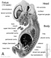
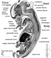
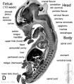
==Early Fetal (Week 10)== | |||
The fetal period is a time of extensive growth in size and mass as well as differentiation of organ systems established in the embryonic period. In particular, the brain continues to grow and develop, the respiratory system differentiates, the urogenital system further differentiates between male/female, endocrine and gastrointestinal tract begins to function. | |||
These 4 images are from a 10 week female fetus approximately 40 mm in size. This stage of development is after the embryonic period (up to week 8), but only 2 weeks into early fetal development. | |||
Compare this 10 week fetus with the earlier Carnegie stage embryos in relation to gastrointestinal tract and respiratory development. | |||
'''Note:''' | |||
# The structure, size and position of the early fetal lungs. | |||
# The relatively underdeveloped diaphragm. | |||
# The position of the stomach and liver. | |||
# The midgut herniated at the umbilicus and will only be taken into the peritoneal cavity on further body wall growth. | |||
# The herniated midgut remains attached to the posterior body wall by its mesentery. | |||
# The hindgut rectum is now separated from the ventral urogenital region. | |||
:{{Template:Fetal Links}} | |||
There are 4 sections taken in the sagittal plane (moving from the right at Plane A towards the midline at Plane D). Click on the small images (or the text below) to open the linked large image pages. | |||
<gallery> | |||
File:Human- fetal week 10 sagittal plane A.jpg|Plane A | |||
File:Human- fetal week 10 sagittal plane B.jpg|Plane B | |||
File:Human- fetal week 10 sagittal plane C.jpg|Plane C | |||
File:Human- fetal week 10 sagittal plane D.jpg|Plane D | |||
</gallery> | |||
{{Template:10wkFetus}} | |||
===Fetal Tract Development=== | |||
'''Week 11''' - villi begin to appear in small intestine, goblet cells present | |||
'''Week 16''' - villi apparent in entire intestine | |||
Adult small intestine will be divided into 3 regions with the same basic histological organization: duodenum (25-30 cm), jejunum (about first two-fifths of the rest), ileum | |||
'''Week 20''' - Peyer's patches appear in small intestine | |||
The adult intestinal immune system includes: Peyer's patches, isolated lymphoid follicles, cryptopatches, mesenteric lymph nodes | |||
[[Category:Human Fetus]] [[Category:Week 10]] | |||
[[Category:Carnegie Stage 22]] | [[Category:Carnegie Stage 22]] | ||
==Group Project== | |||
* There should now be major sub-headings on the project page and commencing to populate each section. | |||
* The discussion page should have images and resources either uploaded or linked for your project page. | |||
* What is the student drawn figure(s) and who is doing it? | |||
* Do you have your references collected and have you identified the PMID for each reference so that it will format correctly on the project page. | |||
Latest revision as of 08:04, 26 August 2010
Introduction
This laboratory will allow time to study both gastrointestinal tract and respiratory development. The class will study features and events of development occurring: early-embryonic, mid-embryonic, late-embryonic and fetal. Both these systems do not carry out their postnatal function before birth. Though during the fetal period both these systems "prepare" for their postnatal functions. Clinically even in normal development the respiratory system only matures towards the end of the third trimester, leading to respiratory problems with premature infants.
In this lab we will use animations to observe some developmental events and then look within the embryo and fetus at different periods of development.
The materials used in this class can also be seen at the following links: Gastrointestinal Tract - Carnegie Stage 13 | Stage 22 | Respiratory System - Carnegie Stage 13 | Respiratory System - Carnegie Stage 22 | Fetal Development - 10 Weeks | Gastrointestinal Tract Lecture | Respiratory Lecture | Flash movies | Quicktime movies
Related notes pages
| Respiratory Links: respiratory | Science Lecture | Lecture Movie | Med Lecture | Stage 13 | Stage 22 | upper respiratory tract | diaphragm | Histology | Postnatal | respiratory abnormalities | Respiratory Quiz | Respiratory terms | Category:Respiratory | ||
|
Gastrointestinal Tract Movies
| Endoderm | Yolk Sac | Tract Growth | Stomach Rotation | Lesser Sac | Greater Omentum | Urogenital Septum |
| Quicktime version | Quicktime version | Quicktime version | Quicktime version | Quicktime version | Quicktime version | Quicktime version |
Gastrointestinal Tract and Respiratory - Mid-Embryonic (Stage 13)
Begin by studying the 3d model and getting an idea of the position within the embryo of the gastrointestinal tract, its associated organs and the early lung buds.
Then look through the selected sections and identify these structures in relation to the surrounding organs and tissues.
Finally compare this mid-embryonic organisation and structure with that seen at the end of the embryonic period (Stage 22). What has changed?
(MH - Note it is best to open each image in a new browser Tab)

|
Gastrointestinal and Respiratory The individual serial slices below have been incorporated into a 3D model of this embryo. |
| Respiratory Links: respiratory | Science Lecture | Lecture Movie | Med Lecture | Stage 13 | Stage 22 | upper respiratory tract | diaphragm | Histology | Postnatal | respiratory abnormalities | Respiratory Quiz | Respiratory terms | Category:Respiratory | ||
|
| Section | Name | Description |
|---|---|---|

|
B1L | Pharynx
The righthand side towards the buccopharyngeal membrane, the lefthand side descending into the embryo body. Central region is the floor of pharynx formed by fusion of 3rd pharyngeal arches = hypopharyngeal eminence (precursor of root of tongue). Rathke's pouch forming the rudimentary adenohypophysis (anterior pituitary). |

|
B3L | Laryngeal tracheal groove - beginning of ventral compression, at 90 degrees to the lateral plane of the pharynx above this point.
Rudimentary thyroid ventral to aortic sac (also seen in B2, ventral to the hypopharyngeal eminence). |

|
B4L | Laryngeal tracheal groove - Caudal pharynx compressed dorsoventrally.
Note that it lies between the aortic sac (ventrally) directly above the heart and the paired vessels of arch artery 6 and the dorsal aortas. The pale staining region behind these blood vessels is where the vertebral column will form. |

|
B5L | Laryngopharynx - now compressed dorsoventrally between the paired arch artery 6 vessels. |

|
B7L | Glottis - initial separation of the oesophagus (dorsal) from the trachea (ventral).
Note that this is occurring at the level of the heart atria. Nasal placodes. Pulmonary arteries. |

|
C1L | Gastrointestinal tract oesophagus (dorsal) is now separate from the respiratory trachea (ventral). |

|
C2L | Oesophagus and trachea both surrounded by dense mesenchyme.
Right nasal pit. |

|
C3L | Oesophagus and trachea both surrounded by dense mesenchyme.
Common cardinal vein in the posterior wall of the intraembryonic coelom. The pleuropericardial folds which contribute later to the formation of the pleura and pericardium. In C4, junction of right common cardinal vein with dorsal wall of sinus venosus. Left nasal pit. |

|
C5L | Smaller oesophagus and expanding trachea, this is also the upper region of the lung buds.
The ventral anchoring of attachment site is at the most cranial extension of the septum transversum. This attachment now divides the intraembryonic coelom around the trachea into two canals, the left and right pleuro (pericardio-peritoneal) canals. Canals are lined by coelomic mesothelium and are continuous with whole intraembryonic coelom (they will be referred to hereafter simply as coelomic canals). The pleuroperitoneal fold on the medial side of the right common cardinal vein will form part of the diaphragm. |

|
C6L | Trachea expanded and beginning to bifurcate to the major bronchial branches for each lung.
Lateral extension of pulmonary mesenchyme is moulded to shape of coelomic canals. Oesophagus lumen obliterated (common site of oesophageal atresia and/or tracheo-oesophageal fistula). Prominent R pleuroperitoneal fold. |

|
C7L | Trachea bifurcated to the major bronchial branches for each lung.
Note dorsal extent of coelomic canals. Oesophagus lumen reappears caudal to bifurcation. Distinct R (smaller on L) pleuroperitoneal fold below the common cardinal vein. |

|
D1L | Oesophagus/stomach junction.
Right lung bud bronchus still visible, left bronchus ends above this section. Note the oesophagus now lies in the midline between the 2 bronchi. Coelomic canals. |

|
D2L | Ovoid stomach with developing space of the lesser sac on R.
Dorsal and ventral attachments of the mesenchyme are now known as dorsal and ventral mesogastria. Coelomic canals. |

|
D3L | Rotation of stomach (seen from above) to right side.
Note change in outline of coelomic canals due to presence of liver. Lesser sac. Note thick mesothelium lining the coelom along left edge of stomach, the primordium of the spleen and greater omentum along greater curvature. Liver embedded in septum transversum (ventral border of septum transversum contributes to diaphragm). |

|
D4L | Rotation of stomach (seen from above) to right side.
Ventral mesogastrium - Stomach is attached ventrally to the liver. (note the position of the ductus venosus) Dorsal mesogastrium - within this structure the spleen will begin to form and later the greater omentum. Peritoneal spaces - identify greater and lesser sac. |

|
D6L | Pyloric region of stomach.
Ventral mesogastrium - Stomach is closely attached ventrally to the liver. Dorsal mesogastrium - within this structure the spleen will begin to form and later the greater omentum. Peritoneal spaces - identify greater and lesser sac. |

|
E4L | Midgut.
Region close to the umbilicus. Note the close associated portal vein and the paired placental (umbilical) veins. |

|
F1L | Midgut.
Looping out of body wall ventrally (cut tangentially). Also note the righthand side hindgut region. |

|
G7L | Caudal pharynx (extending laterally, ventral to dorsal aorta - cf B4). Stomach, mesentery |

|
G6L | Narrow oesophagus. Tracheal bifurcation dorsal to sinus venosus. |


Gastrointestinal Tract - Late-Embryonic (Stage 22)
| The individual serial slices have also been incorporated into a 3D model of this embryo. |
| Section | Name | Description |
|---|---|---|

|
E6L | Liver. Ductus venosus.
Cardio-oesophageal junction (cf. E5). Inferior vena cava. |

|
E7L | Stomach body, with mucosa, submucosa and muscularis externa.
Lesser sac. Lesser omentum. Pyloroduodenal junction. Folded duodenal mucosa. Inferior vena cava. Portal vein. Hepatic ducts. Gallbladder. |

|
F1L | Stomach body. Spleen. Pyloric canal. Duodenum.
Pancreas. Small intestine loop (jejunum) cut tangentially, ventral to liver. Portal vein. |

|
F2L | Stomach, spleen. Superior mesenteric artery.
Superior mesenteric vein crossing cranial to body of pancreas. Tail of pancreas. Duodenum. Small intestinal loop herniating from abdominal cavity into the coelom of the umbilical cord (remnant of extra-embryonic coelom). |

|
F4L | Greater curvature of stomach (tangential section). Lesser sac. Greater omentum. Duodenal/jejunal junction.
Note colon (small lumen, darkly-staining wall) and its mesocolon. Note the sections of small and large intestine within the umbilical cord coelom and their mesenteries. Note the thickened jelly to one side of the umbilical cord, containing umbilical vein and R umbilical artery. |

|
F5L | Lesser sac. Greater omentum. Duodenum. Jejunum (cut twice with mesentery in between). Colon and mesocolon. |

|
F6L | Greater omentum and lesser sac.
Jejunum with mesentery. Colon with mesocolon. Three layers of abdominal muscles. Both umbilical arteries now inside abdominal cavity with urachus between them. |

|
F7L | In abdominal cavity - colon with mesocolon, jejunum. Greater omentum and lesser sac.
Umbilical cord - containing umbilical arteries and small dark allantois. Umbilical cord coelom containing mainly, small intestinal loops with their mesentery. |

|
G1L
|
Umbilical cord and coelom containing small intestine loops.
Colon and mesocolon. Jejunum (G1 only). Bladder with umbilical arteries either side. Knees. |

|
G3L | Rectum.
Bladder. Umbilical arteries arising from common iliac arteries. |

|
G4L | Rectum. |

|
G5L | Recto-anal junction with rectovesical pouch of peritoneal cavity. |

|
G6L | Anal canal with triangular lumen. |


Respiratory - Late-Embryonic (Stage 22)
| The individual serial slices have also been incorporated into a 3D model of this embryo.
The respiratory system is endodermal in origin, initially "budding off" the foregut during week 3. This bud forms the respiratory diverticulum, at the level of the glottis between the adult oesophagus and trachea. It continues to bud in week 4, forming a pair of lung buds. |
| Respiratory Links: respiratory | Science Lecture | Lecture Movie | Med Lecture | Stage 13 | Stage 22 | upper respiratory tract | diaphragm | Histology | Postnatal | respiratory abnormalities | Respiratory Quiz | Respiratory terms | Category:Respiratory | ||
|
| Section | Name | Description |
|---|---|---|

|
A5L | Bridge of nose.
R and L olfactory bulbs from forebrain. |

|
A6L | Nose. Nasal septum. Nasal capsule.
Olfactory epithelium lining roof of nasal cavity. Orbital part of the developing sphenoid bone (intramembranous ossification). |

|
A7L | Conchae. Nasal capsule and septum. |

|
B1L | Conchae. Optic nerve. |

|
B2L | Description |

|
B3L | Perpendicular plate of ethmoid cartilage. Adenohypophysis. Neurohypophysis. Ant. and post. walls of hypopophysial fossa. Lesser wings of sphenoid cartilage. Internal carotid arteries. |

|
B4L | Dorsum of tongue. Oropharynx communicating with naso-pharynx (cf. B3L - palatal processes not fused). |

|
B5L | Tongue with palatal processes at either side. Transverse (intrinsic) muscle of tongue.
Pharyngotympanic tubes. |

|
B6L | Tongue with transverse muscle, genioglossus muscle (medial) and hyoglossus muscle (lateral).
Palatal processes. Meckel's cartilage. Note teeth enamel organs (dark masses at sides of tongue attachment). |

|
B7L | Transverse caudal pharynx. epiglottis. Hyoid musculature. Pharyngeal constrictor muscle. Submandibular gland. |

|
C1L | Pharynx. Pharyngeal constrictor muscle. laryngeal caecum (ventral). Arytenoid swellings in contact.
Thyroid cartilage laminae (anterolateral), with superior horns (posterolateral). Hyoid cartilage. Internal jugular veins. "Muz's cheshire cat" |

|
C2L | Pharynx. Thyroid cartilage. Smaller laryngeal caecum (cf.C1). Carotid neurovascular bundle. |

|
C3L | Pharynx with its inferior constrictor muscle. Glottis region. |

|
C4L | Oesophagus with muscle layer and trachea with thyroid gland laterally.
Common carotid arteries. Vagus nerve. Internal jugular veins. (Section damaged) |

|
C5L | Oesophagus, smaller than in C4. Trachea.
Thyroid gland (isthmus). Clavicle. Small dark masses near posterolateral borders of thyroid gland are the parathyroid glands from the caudal part of 3rd pharyngeal pouch. |

|
C6L | Trachea.
Clavicles. Dark connecting stalk between parathyroid gland and thymus (rostral end of 3rd pharyngeal pouch). Common carotid artery. |

|
C7L | Trachea.
Oesphagus. Apex of R lung in pleural cavity Sternum. Thymus gland. L brachiocephalic vein. Brachiocephalic trunk. |

|
D1L | Lungs. Visceral and parietal pleurae. Pleural cavities.
Sternum. Thymus. Other contents of superior mediastinum. |

|
D2L | Lungs. |

|
D3L | Tracheal bifurcation. |

|
D4L | Right primary bronchus (torn) and right superior lobe bronchus.
Left primary bronchus. Left and right pulmonary arteries. Ribs joining to sternum. |

|
D5L | R, L primary bronchi. R anterior and posterior segmental bronchi coming off R superior lobe bronchus. L, R pulmonary arteries. Hilar attachments of lungs to mediastinal tissues - note extent of R, L pleural cavities. |

|
D6L | R, L primary bronchi (note left still has not branched). R pulmonary artery. |

|
D7L | R, L primary bronchi: note distinct horizontal course of L, vertical course of R, L pulmonary veins (L empty). R pulmonary artery. |

|
E1L | Pulmonary veins. Azygos, hemiazygos veins. Ribs. Intercostal muscles. |

|
E2L | Pulmonary veins. Azygos, hemiazygos veins. Ribs. Intercostal muscles. |

|
E3L | R dome of diaphragm. R long middle and inferior lobes. L long superior and inferior lobes. Xiphoid process.
Liver. |

|
E4L | Diaphragm (note costal attachment). R lung inferior lobe.
Inferior vena cava, dorsal to diaphragm. |

|
E5L | Inferior lobes of lungs.
Diaphragm with sternal attachments. Inferior vena cava, now ventral to diaphragm (vena caval foramen). Liver. |
|
|
E6L | Liver. Thoracic aorta. Large adrenal glands. |
|
|
E7L | Lumbar diaphragm. Thoracic aorta. Note ribs 11 and 12 on L and three layers of abdominal muscles extending ventrally. |
|
|
F1L | Lumbar diaphragm. Thoracic aorta. |
|
|
F2L | Attachment of lumbar diaphragm near L 1 on R with psoas muscle dorsal to it. Note abdominal aorta giving rise to superior mesenteric artery. |
Early Fetal (Week 10)
The fetal period is a time of extensive growth in size and mass as well as differentiation of organ systems established in the embryonic period. In particular, the brain continues to grow and develop, the respiratory system differentiates, the urogenital system further differentiates between male/female, endocrine and gastrointestinal tract begins to function.
These 4 images are from a 10 week female fetus approximately 40 mm in size. This stage of development is after the embryonic period (up to week 8), but only 2 weeks into early fetal development.
Compare this 10 week fetus with the earlier Carnegie stage embryos in relation to gastrointestinal tract and respiratory development.
Note:
- The structure, size and position of the early fetal lungs.
- The relatively underdeveloped diaphragm.
- The position of the stomach and liver.
- The midgut herniated at the umbilicus and will only be taken into the peritoneal cavity on further body wall growth.
- The herniated midgut remains attached to the posterior body wall by its mesentery.
- The hindgut rectum is now separated from the ventral urogenital region.
| Fetal Links: fetal | Week 10 | Week 12 | second trimester | third trimester | fetal neural | Fetal Blood Sampling | fetal growth restriction | birth | birth weight | preterm birth | Developmental Origins of Health and Disease | macrosomia | BGD Practical | Medicine Lecture | Science Lecture | Lecture Movie | Category:Human Fetus | Category:Fetal | |||
|
There are 4 sections taken in the sagittal plane (moving from the right at Plane A towards the midline at Plane D). Click on the small images (or the text below) to open the linked large image pages.
Plane A

Plane B
Plane C
Plane D


Related Images
Fetus (week 10) Planes A (most lateral), B (lateral), C (medial) and D (midline) from lateral towards the midline.
- Human Fetus - most lateral | lateral | medial | midline
- Head - most lateral | lateral | medial | midline
- Cerebellum - most lateral | lateral | medial | midline
- Urogenital Unlabelled - most lateral | lateral | medial | midline
- Urogenital Labelled - most lateral | lateral | medial | midline
- Large Images - midline
- Image Source: UNSW Embryology, no reproduction without permission.
Fetal Tract Development
Week 11 - villi begin to appear in small intestine, goblet cells present
Week 16 - villi apparent in entire intestine
Adult small intestine will be divided into 3 regions with the same basic histological organization: duodenum (25-30 cm), jejunum (about first two-fifths of the rest), ileum
Week 20 - Peyer's patches appear in small intestine
The adult intestinal immune system includes: Peyer's patches, isolated lymphoid follicles, cryptopatches, mesenteric lymph nodes
Group Project
- There should now be major sub-headings on the project page and commencing to populate each section.
- The discussion page should have images and resources either uploaded or linked for your project page.
- What is the student drawn figure(s) and who is doing it?
- Do you have your references collected and have you identified the PMID for each reference so that it will format correctly on the project page.