|
|
| Line 14: |
Line 14: |
|
| |
|
| == Development of the Rat == | | == Development of the Rat == |
|
| |
| {| class="prettytable"
| |
| | <center>'''Standard Stages'''
| |
|
| |
| (Witschi)</center>
| |
| | <center>'''Age''' (days)</center>
| |
| | <center>'''Size''' (mm)</center>
| |
| | <center>'''Identification of Stages'''</center>
| |
|
| |
| |-
| |
| | colspan="4" | <center>'''Cleavage and Blastula'''</center>
| |
|
| |
| |-
| |
| | 1
| |
| | 1
| |
| | 0.07
| |
| | 1 cell (in oviduct)
| |
|
| |
| |-
| |
| | 2
| |
| | 2
| |
| | 0.08 x 0.06
| |
| | 2 cells (in oviduct)
| |
|
| |
| |-
| |
| | 3
| |
| | 3
| |
| | 0.08 x 0.05
| |
| | 4 cells (in oviduct)
| |
|
| |
| |-
| |
| | 4
| |
| | 3.5
| |
| |
| |
| | 8-12 cells (in oviduct)
| |
|
| |
| |-
| |
| | 5
| |
| | 3.25
| |
| | 0.08 x 0.04
| |
| | Morula (in uterus)
| |
|
| |
| |-
| |
| | 6
| |
| | 4
| |
| | (0.08 x 0.03)
| |
| | Early blastocyst (in uterus)
| |
|
| |
| |-
| |
| | 7
| |
| | 5
| |
| | (0.12 x 0.05)
| |
| | Free blastocyst (in uterus)
| |
|
| |
| |-
| |
| | colspan="4" | <center>'''Gastrula'''</center>
| |
|
| |
| |-
| |
| | 8
| |
| | 6
| |
| | (0.28 x 0.07)
| |
| | Implanting blastocyst, with trophoblastic cone and inner cell mass; outgrowth of endoderm (hypoblast)
| |
|
| |
| |-
| |
| | 9
| |
| | 6.75
| |
| |
| |
| | Diplotrophoblast; inner cell mass (pendant), covered with endoderm
| |
|
| |
| |-
| |
| | 10
| |
| | 7.25
| |
| | (0.3 x 0.1)
| |
| | Near complete implantation; pendant begins differentiation into embryonic and extra-embryonic parts
| |
|
| |
| |-
| |
| | 11
| |
| | 7.75
| |
| | (0.5 x 0.1)
| |
| | Completion of implantation; primary amniotic cyst; ectoplacental cone
| |
|
| |
| |-
| |
| | colspan="4" | <center>'''Primitive Streak'''</center>
| |
|
| |
| |-
| |
| | 12
| |
| | 8.5
| |
| | (1.04 x 0.26)
| |
| | Connecting ectochorionic and amniotic cavities; rudiments of amniotic folds; primitive streak; start of 3rd layer formation; blastemas of heart and pericardium
| |
|
| |
| |-
| |
| | colspan="4" | <center>'''Neurula'''</center>
| |
|
| |
| |-
| |
| | 13
| |
| | 9
| |
| | 1.0
| |
| | Presomite neurula; fusion of chorio-amniotic folds, chorio-amniotic stalk; neural plate; embryo bent dorsally; bud of allantoic stalk
| |
|
| |
| |-
| |
| | 14
| |
| | 9.5
| |
| | 1.5
| |
| | Somites 1-4 (occipital); pendant with 3 cavities: ectochorionic cyst, exocoelom, and amniotic cavity; ectochorionic cyst collapsing; allantoic stalk projects into exocoelom; embryo bent dorsally
| |
|
| |
| |-
| |
| | 15
| |
| | 10
| |
| | 2
| |
| | Somites 5-12 (cervical); 1st visceral arch; ectochorionic cyst fused with ectoplacenta and with allantoic stalk; regression of peripheral (distal) yolk sac and trophectoderm (diplotrophoblast); Reichert's membrane; gonia in endoderm; embryo bent dorsally
| |
|
| |
| |-
| |
| | 16
| |
| | 10.5
| |
| | 2.4
| |
| | Somites 13-20 (upper thoracic); 2 visceral arches; disc and yolk sac placentas; appendicular folds; embryo reverses, curves ventrally
| |
|
| |
| |-
| |
| | 17
| |
| | 11
| |
| | 3.3
| |
| | Somites 21-25 (lower thoracic); yolk stalk closes at level of 15th somite; primary gonia in mesentery; primitive streak disappears; tail bud becomes organized; arm and leg buds recognizable
| |
|
| |
| |-
| |
| | colspan="4" | <center>'''Tail Bud Embryo'''</center>
| |
|
| |
| |-
| |
| | 18
| |
| | 11.5
| |
| | 3.8
| |
| | Somites 26-28 (upper lumbar); 3 visceral arches; arm buds recognizable
| |
|
| |
| |-
| |
| | 19
| |
| | 11.75
| |
| | 4.2
| |
| | Somites 29-31 (lower lumbar); visceral arches I-IV; cervical folds; appendicular folds and buds
| |
|
| |
| |-
| |
| | 20
| |
| | 11.875
| |
| | 5
| |
| | Somites 32-33 (upper sacral)
| |
|
| |
| |-
| |
| | 21
| |
| | 12
| |
| | 5.1
| |
| | Somites 34-35 (lower sacral); deep cervical sinuses
| |
|
| |
| |-
| |
| | 22
| |
| | 12.125
| |
| | 5.2
| |
| | Somite 36 (1st caudal); olfactory pits
| |
|
| |
| |-
| |
| | 23
| |
| | 12.25
| |
| | 5.6
| |
| | Somites 37-38 (caudal); start of umbilical herniation
| |
|
| |
| |-
| |
| | 24
| |
| | 12.375
| |
| | 6
| |
| | Somites 39-40 (caudal)
| |
|
| |
| |-
| |
| | colspan="4" | <center>'''Complete Embryo'''</center>
| |
|
| |
| |-
| |
| | 25
| |
| | 12.5
| |
| | 6.2
| |
| | Somites 41-42 (caudal); occipital somites dispersing; 4 visceral arches; deep cervical sinuses; arm buds at somite levels 8-14, about as high as long; leg buds at somite levels 28-31, smaller; body forms a spiral of about 11/2 turns, the left face and trunk applied to yolk sac, the right side turned toward placenta; tail and allantoic stalk rise to the placenta
| |
|
| |
| |-
| |
| | colspan="4" | <center>'''Metamorphosing Embryo'''</center>
| |
|
| |
| |-
| |
| | 26
| |
| | 12.75
| |
| | 7
| |
| | Somites 43-45 (caudal); mandibular, maxillary, and frontonasal processes; cervical sinuses closing; mammary welts; differentiaion of handplates; arm buds vascularized, brachial nerves entering; beginning of umbilical hernia
| |
|
| |
| |-
| |
| | 27
| |
| | 13
| |
| | 8
| |
| | Somites 46-48 (caudal); prominent facial processes and clefts; nose-snout projecting; cervical sinuses closed; primordia of mammary glands; round handplates and footplates; larger umbilical hernia
| |
|
| |
| |-
| |
| | 28
| |
| | 13.5
| |
| | 8.5
| |
| | Somites 49-51 (caudal); 1st visceral cleft transforms into external ear duct; precartilaginous condensations in handplates
| |
|
| |
| |-
| |
| | 29
| |
| | 14
| |
| | 9.5
| |
| | Somites 52-55 (caudal); auricular hillocks on visceral arches I and II
| |
|
| |
| |-
| |
| | 30
| |
| | 14.5
| |
| | 10.5
| |
| | Somites 56-60 (caudal); body uncoils; mandibular precartilage; nearly round opening of external ear duct; pleuroperitoneal canal has become very narrow
| |
|
| |
| |-
| |
| | 31
| |
| | 15
| |
| | 12
| |
| | Somites 61-63 (caudal); facial clefts closed; pleuroperitoneal canal closed; complete diaphragm
| |
|
| |
| |-
| |
| | 32
| |
| | 15.5
| |
| | 14.2
| |
| | Somite 64 (caudal); pinna turns forward; maximal size of umbilical hernia
| |
|
| |
| |-
| |
| | 33
| |
| | 16
| |
| | 15.5
| |
| | Somite 65 (usually this is last caudal); snout lifts off chest; last stage of metamorphosis
| |
|
| |
| |-
| |
| | colspan="4" | <center>'''Fetus'''</center>
| |
|
| |
| |-
| |
| | 34
| |
| | 17-18
| |
| | 16-20
| |
|
| |
|
| |
| | 1st fetal stage: rapid growth of eyelids (eyes entirely covered at end of 18th day); palate complete; pinna covers ear duct; umbilical hernia withdraws
| |
|
| |
| |-
| |
| | 35
| |
|
| |
| ante-natal
| |
| | 19-22
| |
| | 20-40
| |
|
| |
|
| |
| | 2nd fetal stage: sealed eyelids; fetal membranes and placentas reach peak of development; tail grows to 10mm;
| |
|
| |
| '''Birth''' occurs (22nd day in rat, 19th day in mouse)
| |
|
| |
| |-
| |
| | 35
| |
|
| |
| post-natal
| |
| | 1-16 post-partum
| |
| | 40-1001
| |
| | After birth, fetus becomes a breathing and suckling nestling2<nowiki>; during 1st 16 days (22 to 38 days total age), eyelids remain sealed and external ear ducts plugged with periderm</nowiki>
| |
|
| |
| |-
| |
| | 36
| |
|
| |
| post-natal
| |
| | 17+ post-partum
| |
| | 100+1
| |
| | Periderm seals of ears and eyelids vanish; active feeding begins within next 3 days and weaning after 1 week (total weaning age, 45-48 days for rats and mice)
| |
|
| |
| |}
| |
| ''' '''
| |
|
| |
| '''<nowiki>* Age (days)-</nowiki>''' days after fertilization
| |
|
| |
| '''<nowiki>* Size (mm)-</nowiki>''' largest and smallest dimensions
| |
|
| |
| === Table Data ===
| |
| The main table data is modified from <ref name="Witschi 1962">Witschi, E. (1962) Development: Rat. In: '''Growth Including Reproduction and Morphological Development.''' Altman, P. L. , and D. S. Dittmer, ed. Fed. Am. Soc. Exp. Biol., Washington DC, pp. 304-314.</ref> and other sources as listed within the table.
| |
|
| |
|
| ==Species Stages Comparison == | | ==Species Stages Comparison == |
Introduction
Rats (taxon- rattus) are readily available as inbred, outbred and mutant strains. They have been generally beaten as a model by their mice brethren, as the molecular tools that became available (stem cells, knockout genes, etc).
Rat embryos do have the advantage of being much larger than mouse embryos and easy to breed. Another advantage of rats is in vision development studies, as that retinal development continues postnatally (most vertebrate neurological systems are difficult to access during periods of development). Rat development is also generally 1 day behind that of mouse. (The table below gives details relating to the staging of rat development). Below this are a list of internet resources relating to the rat.
- rat | original Rat page
Some Recent Findings
- Comparison of staging systems for the gastrulation and early neurulation period in rodents[1]"Because there is no standard developmental staging system for the early postimplantation period of rodent embryos, investigators must now choose between a variety of systems that differ significantly. We have reviewed many of these staging systems and have summarized the ambiguities within them and the inconsistencies among them. In order to compare systems, we first obtained a consensus of the order of developmental events from the literature, and then attempted to fit existing systems into this order taking into account inconsistencies in terminology and blurred borderlines between stages."
Development of the Rat
Species Stages Comparison
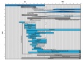
The table below gives an approximate comparison of human, mouse and rat embryos based upon Carnegie staging.
| Species
|
Stage
|
9
|
10
|
11
|
12
|
13
|
14
|
15
|
16
|
17
|
18
|
19
|
20
|
21
|
22
|
23
|
| Human [2]
|
Days
|
20
|
22
|
24
|
28
|
30
|
33
|
36
|
40
|
42
|
44
|
48
|
52
|
54
|
55
|
58
|
| Mouse [3]
|
Days
|
9
|
9.5
|
10
|
10.5
|
11
|
11.5
|
12
|
12.5
|
13
|
13.5
|
14
|
14.5
|
15
|
15.5
|
16
|
| Rat [4]
|
Days
|
10.5
|
11
|
11.5
|
12
|
12.5
|
13
|
13.5
|
14
|
14.5
|
15
|
15.5
|
16
|
16.5
|
17
|
17.5
|
References
- ↑ <pubmed>1440421</pubmed>
- ↑ <pubmed>400868</pubmed>
- ↑ The House Mouse: Atlas of Mouse Development by Theiler Springer-Verlag, NY (1972, 1989). | online book
- ↑ Cite error: Invalid
<ref> tag; no text was provided for refs named Witschi 1962
Search Pubmed: Rat Development
Additional Images
Reconstructed early rat embryos
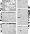
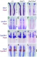
Rat hindbrain E11.5, down-regulated genes and Pax6
Rat thyroid system and neural development
External Links
- Rat Genome Database RGD <pubmed>17151068</pubmed>
Glossary Links
- Glossary: A | B | C | D | E | F | G | H | I | J | K | L | M | N | O | P | Q | R | S | T | U | V | W | X | Y | Z | Numbers | Symbols | Term Link
Cite this page: Hill, M.A. (2024, June 26) Embryology Rat Development. Retrieved from https://embryology.med.unsw.edu.au/embryology/index.php/Rat_Development
- What Links Here?
- © Dr Mark Hill 2024, UNSW Embryology ISBN: 978 0 7334 2609 4 - UNSW CRICOS Provider Code No. 00098G