Category:Human: Difference between revisions
From Embryology
mNo edit summary |
mNo edit summary |
||
| Line 1: | Line 1: | ||
This {{Embryology}} category shows pages and media related | This {{Embryology}} category shows pages and media related to human development. | ||
[[Category:Human]] | [[Category:Human]] | ||
Latest revision as of 09:58, 30 October 2015
This Embryology category shows pages and media related to human development.
Subcategories
This category has the following 72 subcategories, out of 72 total.
C
- Carnegie Embryo
- Carnegie Embryo 1
- Carnegie Embryo 112
- Carnegie Embryo 1134B
- Carnegie Embryo 116
- Carnegie Embryo 1266
- Carnegie Embryo 1455
- Carnegie Embryo 148
- Carnegie Embryo 172
- Carnegie Embryo 19
- Carnegie Embryo 239
- Carnegie Embryo 2393
- Carnegie Embryo 240
- Carnegie Embryo 248
- Carnegie Embryo 256
- Carnegie Embryo 296
- Carnegie Embryo 3527
- Carnegie Embryo 3956
- Carnegie Embryo 4046
- Carnegie Embryo 4059
- Carnegie Embryo 407
- Carnegie Embryo 4148
- Carnegie Embryo 43
- Carnegie Embryo 4405
- Carnegie Embryo 460
- Carnegie Embryo 463
- Carnegie Embryo 523
- Carnegie Embryo 5541
- Carnegie Embryo 5609
- Carnegie Embryo 5652
- Carnegie Embryo 5682
- Carnegie Embryo 5874
- Carnegie Embryo 6032
- Carnegie Embryo 625
- Carnegie Embryo 6426
- Carnegie Embryo 6581
- Carnegie Embryo 7618
- Carnegie Embryo 7669
- Carnegie Embryo 786
- Carnegie Embryo 808
- Carnegie Embryo 8147
- Carnegie Embryo 8239
- Carnegie Embryo 8370
- Carnegie Embryo 858
- Carnegie Embryo 8630
- Carnegie Embryo 8967
- Carnegie Embryo 9296
- Carnegie Embryo 96
- Carnegie Embryo 963
- Carnegie Embryo 966
- Carnegie Embryo 9697
D
F
H
Pages in category 'Human'
The following 200 pages are in this category, out of 332 total.
(previous page) (next page)A
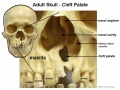
- Abnormal Development - Cleft Lip and Palate
- Abnormal Development - Cleft Palate
- Abnormal Development - Toxoplasmosis
- Template:Abnormal Newborn Neural Exam Table
- Template:Adipose Timeline table
- Template:Adrenal GA32 Links
- Template:Anderson2016 collapsetable1
- Template:Anderson2016 table1
- Template:AnsonBlack1934 figures
- Aschheim-Zondek Test 1928 Movie
- Template:Australian GIT abnormalities 2002-2003
- Template:Australian Palate abnormalities 2002-2003
B
- Template:B050966
- Template:B100658
- Template:B220849
- Template:Bardeen1906 figures
- Template:Barniville1914 figures
- Template:Bartelmez1922 figures
- Template:BaxterBoyd1939 figures
- BGDA Practical 7 - Week 6
- Birth MRI Movie
- Book - Contributions to Embryology Carnegie Institution No.10
- Book - Contributions to Embryology Carnegie Institution No.112
- Book - Contributions to Embryology Carnegie Institution No.131
- Book - Contributions to Embryology Carnegie Institution No.159
- Book - Contributions to Embryology Carnegie Institution No.21
- Book - Contributions to Embryology Carnegie Institution No.22
- Book - Contributions to Embryology Carnegie Institution No.27
- Book - Contributions to Embryology Carnegie Institution No.29
- Book - Contributions to Embryology Carnegie Institution No.30
- Book - Contributions to Embryology Carnegie Institution No.32
- Book - Contributions to Embryology Carnegie Institution No.33
- Book - Contributions to Embryology Carnegie Institution No.34
- Book - Contributions to Embryology Carnegie Institution No.35
- Book - Contributions to Embryology Carnegie Institution No.38
- Book - Contributions to Embryology Carnegie Institution No.40
- Book - Contributions to Embryology Carnegie Institution No.42
- Book - Contributions to Embryology Carnegie Institution No.43
- Book - Contributions to Embryology Carnegie Institution No.44
- Book - Contributions to Embryology Carnegie Institution No.46
- Book - Contributions to Embryology Carnegie Institution No.47
- Book - Contributions to Embryology Carnegie Institution No.48
- Book - Contributions to Embryology Carnegie Institution No.52
- Book - Contributions to Embryology Carnegie Institution No.55
- Book - Contributions to Embryology Carnegie Institution No.59
- Book - Contributions to Embryology Carnegie Institution No.61
- Book - Contributions to Embryology Carnegie Institution No.65
- Book - Contributions to Embryology Carnegie Institution No.69
- Book - Contributions to Embryology Carnegie Institution No.72
- Book - Contributions to the Development of the Human Brain (1919)
- History:Book - Contributions to the Development of the Human Brain (1919)
- Book - Human embryos of different ages examined in median sections - a contribution to the mechanics of development
- Template:Braune 1877 Plate 2
C
E
F
H
- Template:Hamilton1944 figures
- Harvard Collection
- Template:Hearing EAM timeline
- Hela Apoptosis Movie
- Template:HillH12inks
- Template:HillH13 links
- Template:HillH145 links
- Template:HillH159 links
- Template:HillH202 links
- Template:HillH257 links
- Template:HillH4 links
- Template:HillH5 links
- Template:HillH52
- Template:HillH58 links
- Template:HillH6 links
- Template:HillH8 links
- Template:Hochstadter plates
- Template:Huber1905 table1
- Template:Human 7.5mm Embryo links
- Human Adult Brain Movie
- Human Development Timeline Movie
- Human Embryo - Scanning electron microscopy
- Template:Human embryo neck links
- Human Embryo SEM
- Human Fertilization Detail Movie
- Human Fertilization Movie
- Template:Human Fertilization Movie 1 frame table
- Template:Human Fertilization Movie 1 frames
- Template:Human Fertilization Movie 2 frame table
- Template:Human follicles lm and em links
- Template:Human ovary - corpus luteum links
- Template:Human Spermatozoa Statistics collapse table
- Template:Human Spermatozoa Statistics table
- Human Sylvian Fissure Movie
- Template:Human timeline
- Hutchinson-Gilford Progeria Syndrome
J
K
L
M
- Template:Macklin1921 figures
- Template:Mall1912 figures
- Template:Mall1916 figures
- Template:Mall1917 figures
- Menstrual Cycle - Histology
- Model Embryo 1.6mm Movie 1
- Model Embryo 10mm Movie 1
- Model Embryo 3.1mm Movie 1
- Model Embryo 7.5mm Movie 1
- Monosomic Embryo Movie 1
- Template:Morton1949 figures
- Template:Mouse Human lung table
- Movie - Neural Sylvian Fissure
N
P
- Palate Development
- Paper - A human embryo before the appearance of the myotomes (1918)
- Paper - A Human Embryo of Twenty-five Somites
- Paper - A Human Embryo of Twenty-seven Pairs of Somites, Embedded in Decidua
- Paper - A human embryo with head-process and commencing arch enteric canal
- Paper - A Human Embryo with Seven Pairs of Somites Measuring about 2 mm in Length
- Paper - A human embryo with seventeen pairs of somites (1930)
- Paper - A morphological study of testicular descent
- Paper - A Note on the Development of the Septum Transversum and the Liver
- Paper - A presomite human embryo (Shaw) - the implantation
- Paper - A presomite human embryo (Shaw) - the implantation (1942)
- Paper - A presomite human embryo showing a yolk-sac duct
- Paper - A presomite human embryo showing an early stage of the primitive streak
- Paper - A presomite human embryo with a neurenteric canal (embryo R.S.)
- Paper - A study of a 7 mm human embryo with special reference to its peculiar spirally twisted form, and its large aortic cell-clusters
- Paper - A study of the development of certain features of the cerebellum (1920)
- Paper - A very Young Human Embryo found embedded in a "Decidual Cast" of the Uterus
- Paper - A well-preserved human embryo of 10 somites (1929)
- Paper - A Young Human Embryo (Embryo Dobbin) with Head-Process and Prochordal Plate
- Paper - An early human embryo (no. 1285, Manchester Collection) with capsular attachment of the connecting stalk (1935)
- Paper - An Early Human Embryo (No. 1285, Manchester Collection), with Capsular Attachment of the Connecting Stalk
- Paper - An Early Human Embryo, with 0.55 mm long Embryonic Shield
- Paper - An Early Human Ovum (Thomson) in situ
- Paper - An iconometrographic representation of the growth of the central nervous system in man
- Paper - Breech fused twin monster (1934)
- Paper - Changes in fetuses due to formalin preservation
- Special:Badtitle/NS501:Paper - Description of a Human Embryo of 13-14 Mesodermic Somites
- Paper - Description of a Human Embryo of Twenty-three Paired Somites
- History:Paper - Description of a Human Embryo of Twenty-two paired Somites
- Paper - Description of a reconstruction of the head of a thirty-millimetre embryo (1910)
- Paper - Development and variation of the nerves and the musculature of the inferior extremity and of the neighboring regions of the trunk in man
- Paper - Development of the human heart from its earliest appearance to the stage found in embryos of twenty paired somites (1927)
- Paper - Developmental Changes in the Pericardium, the Mesocardia, and the Pleural Sacs in the Human Embryo
Media in category 'Human'
The following 200 files are in this category, out of 2,421 total.
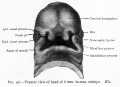
(previous page) (next page) 220px-Patauhand.PNG 220 × 200; 69 KB
 226.jpg 565 × 705; 71 KB
 231.jpg 747 × 493; 69 KB
 3D Human pancreatic islet.jpg 1,088 × 1,280; 295 KB
 7.5mm Embryo movie 1 icon.jpg 299 × 400; 38 KB
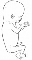
 9 Week Human Embryo.jpg 400 × 600; 41 KB
 Abbott 191.jpg 1,034 × 1,000; 234 KB
 Abbott 201.jpg 1,150 × 1,000; 203 KB
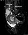 Abdominal ectopic pregnancy MRI.jpg 553 × 700; 45 KB
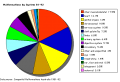 Abnormal AusData81-92.png 523 × 358; 10 KB
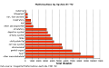 Abnormal AusData81-92Graph.png 509 × 320; 7 KB
 Abnormal81-92-heart.png 481 × 344; 6 KB
 Abnormal81-92-neuron.png 481 × 344; 9 KB
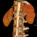 Accessory renal artery.jpg 800 × 798; 103 KB
 Adrenal and gonad steroidogenic factor 1 expression.jpg 1,000 × 636; 88 KB
 Adult brain 01.mov ; 1.59 MB
- Adult brain 02.mov ; 434 KB
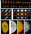 Adult female fibroblast and lymphocyte nuclear DNA 01.jpg 1,095 × 1,200; 197 KB
 Adult female fibroblast nuclear DNA 01.jpg 2,186 × 1,000; 219 KB
 Adult gastrointestinal tract cartoon01.jpg 745 × 698; 55 KB
 Adult gastrointestinal tract cartoon02.jpg 541 × 738; 42 KB
 Adult human brain MRI01.jpg 700 × 607; 81 KB
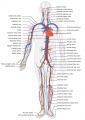 Adult human cardiovascular system.jpg 707 × 1,000; 151 KB
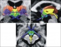 Adult human hypothalamus 01.jpg 917 × 700; 124 KB
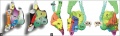 Adult human hypothalamus 02.jpg 1,151 × 343; 100 KB
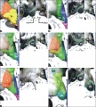 Adult human hypothalamus 03.jpg 767 × 854; 176 KB
 Adult human hypothalamus 04.jpg 660 × 500; 69 KB
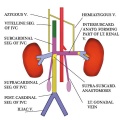 Adult renal venous cartoon.jpg 600 × 600; 62 KB
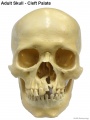 Adult skull cleft palate 01.jpg 750 × 1,000; 89 KB
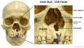 Adult skull cleft palate 02.jpg 1,280 × 720; 139 KB
 Adult skull cleft palate 03.jpg 904 × 678; 104 KB
 Advanced Heart Development Timeline GA.jpg 1,000 × 434; 97 KB
 Advanced Heart Development Timeline.jpg 1,772 × 769; 158 KB
 Agenesis of left lung.jpg 600 × 424; 28 KB
 Anderson2016-fig07a.jpg 800 × 800; 193 KB
 Anderson2016-fig07b.jpg 800 × 800; 304 KB
 Anderson2016-fig09a.jpg 800 × 800; 106 KB
 Anderson2016-fig09b.jpg 800 × 800; 90 KB
 Anderson2016-fig10.jpg 800 × 800; 109 KB
 Anderson2016-fig11a.jpg 800 × 800; 108 KB
 Anderson2016-fig11b.jpg 800 × 800; 98 KB
 Anderson2016-fig12b.jpg 800 × 800; 138 KB
 Anderson2016-fig13b.jpg 800 × 800; 191 KB
 Anderson2016-fig14.jpg 800 × 800; 168 KB
 Anderson2016-fig15a.jpg 800 × 800; 142 KB
 Anderson2016-fig15b.jpg 800 × 800; 148 KB
 Anderson2016-fig16a.jpg 800 × 800; 193 KB
 Anderson2016-fig16b.jpg 800 × 800; 167 KB
 Anderson2016-fig18.jpg 800 × 800; 105 KB
 Anderson2016-fig19.jpg 800 × 800; 99 KB
 Anderson2016-fig20.jpg 800 × 800; 101 KB
 Anderson2016-fig21.jpg 762 × 800; 74 KB
 Anderson2016-fig22.jpg 800 × 779; 90 KB
 Anderson2016-fig23.jpg 783 × 800; 97 KB
 Anderson2016-fig34b.jpg 800 × 800; 92 KB
 Anderson2016-fig35b.jpg 800 × 800; 84 KB
 Anderson2016-fig36.jpg 800 × 800; 112 KB
 Anderson2016-fig37.jpg 800 × 800; 69 KB
 Anderson2016-fig41a.jpg 800 × 755; 92 KB
 Anderson2016-fig43a.jpg 800 × 800; 60 KB
 Anderson2016-fig43b.jpg 800 × 800; 48 KB
 Anderson2016-fig44a.jpg 800 × 800; 83 KB
 Anderson2016-fig44b.jpg 800 × 800; 76 KB
 Anderson2016-fig45a.jpg 800 × 800; 62 KB
 Anderson2016-fig45b.jpg 800 × 800; 57 KB
 Anderson2016-fig47.jpg 800 × 800; 83 KB
 Anderson2016-fig48.jpg 800 × 800; 203 KB
 Anderson2016-fig49.jpg 800 × 800; 90 KB
 Anencephaly ultrasound.jpg 900 × 658; 108 KB
 Aneuploidy model based on fragmentation 1.jpg 946 × 907; 154 KB
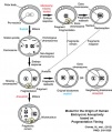 Aneuploidy model based on fragmentation.jpg 668 × 790; 105 KB
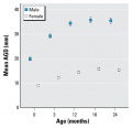 Anogenital distance from birth to 2 years.jpg 565 × 545; 34 KB
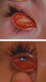 Anophthalmia and microphthalmia.jpg 273 × 477; 30 KB
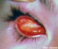 Anophthalmia.jpg 600 × 504; 46 KB
 Anotia 01.jpg 796 × 660; 61 KB
 Anson1934 fig01-8.jpg 1,337 × 888; 126 KB
 Anson1934 fig09.jpg 546 × 272; 16 KB
 Anson1934 fig10.jpg 520 × 612; 31 KB
 Anson1934 fig11.jpg 758 × 870; 52 KB
 Anson1934 fig12.jpg 545 × 968; 44 KB
 Anson1934 fig13.jpg 761 × 1,323; 74 KB
 Anson1934 fig14.jpg 761 × 1,323; 81 KB
 Anson1934 fig15.jpg 693 × 1,003; 62 KB
 Anson1934 fig16.jpg 691 × 985; 47 KB
 Anson1934 fig17.jpg 605 × 1,143; 57 KB
 Anson1934 fig18.jpg 418 × 1,161; 34 KB
 Anson1934 fig19.jpg 524 × 1,218; 48 KB
 Anson1934 plate01.jpg 1,557 × 2,279; 288 KB
 Anson1934 plate02.jpg 1,464 × 2,311; 259 KB
 Arthrogryposis.jpg 800 × 503; 39 KB
 Australian abnormalities 81-92 git.jpg 481 × 344; 43 KB
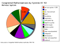
 Australian abnormalities pie skmus.png 481 × 344; 9 KB
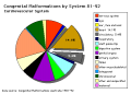
 Australian abnormalities pie urogen.png 481 × 344; 6 KB
 B050966-01.jpg 2,000 × 992; 681 KB
 B100658-01.jpg 1,388 × 1,040; 677 KB
 B100658-02.jpg 1,068 × 800; 458 KB
 B220849-01.jpg 1,311 × 849; 325 KB
 B220849-02.jpg 1,311 × 849; 358 KB
 B220849-03.jpg 1,388 × 1,040; 346 KB
 Bailey001.jpg 850 × 794; 155 KB
 Bailey004.jpg 364 × 1,013; 40 KB
 Bailey008.jpg 838 × 815; 89 KB
 Bailey009.jpg 774 × 766; 68 KB
 Bailey014.jpg 704 × 587; 116 KB
 Bailey073.jpg 705 × 501; 122 KB
 Bailey074.jpg 885 × 700; 176 KB
 Bailey075.jpg 920 × 590; 110 KB
 Bailey076.jpg 603 × 644; 63 KB
 Bailey077.jpg 775 × 686; 86 KB
 Bailey081.jpg 782 × 755; 118 KB
 Bailey082.jpg 879 × 756; 137 KB
 Bailey083.jpg 924 × 774; 129 KB
 Bailey084.jpg 569 × 590; 56 KB
 Bailey085.jpg 505 × 425; 59 KB
 Bailey086.jpg 720 × 662; 85 KB
 Bailey087.jpg 928 × 803; 132 KB
 Bailey088.jpg 888 × 701; 102 KB
 Bailey089.jpg 482 × 690; 55 KB
 Bailey090.jpg 716 × 478; 55 KB
 Bailey091.jpg 884 × 549; 81 KB
 Bailey094.jpg 376 × 689; 27 KB
 Bailey095.jpg 409 × 635; 52 KB
 Bailey096.jpg 693 × 501; 59 KB
 Bailey097.jpg 776 × 674; 71 KB
 Bailey098.jpg 680 × 432; 47 KB
 Bailey099.jpg 704 × 464; 52 KB
 Bailey103.jpg 739 × 738; 89 KB
 Bailey104.jpg 878 × 638; 107 KB
 Bailey105.jpg 545 × 567; 61 KB
 Bailey106.jpg 601 × 610; 86 KB
 Bailey109.jpg 710 × 669; 124 KB
 Bailey113.jpg 813 × 553; 128 KB
 Bailey114.jpg 690 × 433; 63 KB
 Bailey121.jpg 800 × 279; 51 KB
 Bailey122.jpg 514 × 440; 48 KB
 Bailey123.jpg 885 × 618; 78 KB
 Bailey125.jpg 496 × 429; 40 KB
 Bailey127.jpg 597 × 270; 43 KB
 Bailey128.jpg 805 × 356; 46 KB
 Bailey129.jpg 560 × 464; 44 KB
 Bailey130.jpg 714 × 439; 59 KB
 Bailey131.jpg 235 × 560; 39 KB
 Bailey132+133.jpg 940 × 570; 101 KB
 Bailey132.jpg 466 × 413; 43 KB
 Bailey133.jpg 806 × 655; 85 KB
 Bailey135.jpg 940 × 965; 216 KB
 Bailey137.jpg 672 × 539; 73 KB
 Bailey138.jpg 831 × 400; 62 KB
 Bailey139.jpg 961 × 671; 96 KB
 Bailey140.jpg 793 × 505; 58 KB
 Bailey141.jpg 761 × 323; 66 KB
 Bailey142.jpg 778 × 479; 72 KB
 Bailey143.jpg 911 × 673; 114 KB
 Bailey144.jpg 491 × 398; 39 KB
 Bailey145.jpg 777 × 654; 80 KB
 Bailey146.jpg 609 × 476; 42 KB
 Bailey147.jpg 660 × 632; 54 KB
 Bailey148.jpg 574 × 459; 38 KB
 Bailey149.jpg 576 × 520; 38 KB
 Bailey150.jpg 406 × 596; 42 KB
 Bailey151.jpg 585 × 631; 73 KB
 Bailey152.jpg 900 × 803; 300 KB
 Bailey153.jpg 799 × 585; 175 KB
 Bailey154.jpg 898 × 563; 191 KB
 Bailey155.jpg 894 × 833; 209 KB
 Bailey162.jpg 799 × 642; 102 KB
 Bailey163.jpg 757 × 691; 107 KB
 Bailey164.jpg 928 × 862; 149 KB
 Bailey166.jpg 787 × 656; 90 KB
 Bailey167.jpg 610 × 458; 58 KB
 Bailey169.jpg 504 × 264; 27 KB
 Bailey170.jpg 709 × 457; 67 KB
 Bailey171.jpg 954 × 507; 81 KB
 Bailey174.jpg 955 × 542; 88 KB
 Bailey175.jpg 885 × 306; 51 KB
 Bailey176.jpg 918 × 352; 62 KB
 Bailey178.jpg 913 × 653; 125 KB
 Bailey179.jpg 892 × 794; 114 KB
 Bailey180.jpg 938 × 431; 70 KB
 Bailey182.jpg 913 × 718; 103 KB
 Bailey183.jpg 847 × 448; 57 KB
 Bailey184.jpg 625 × 462; 48 KB
 Bailey185.jpg 944 × 499; 98 KB
 Bailey187.jpg 817 × 732; 70 KB
 Bailey188.jpg 906 × 538; 64 KB
 Bailey189.jpg 810 × 632; 63 KB
 Bailey190.jpg 801 × 584; 69 KB
 Bailey191.jpg 863 × 509; 109 KB
 Bailey192.jpg 960 × 806; 133 KB
 Bailey193.jpg 747 × 848; 94 KB
 Bailey194.jpg 841 × 638; 80 KB
 Bailey195.jpg 924 × 781; 79 KB
 Bailey196.jpg 760 × 510; 47 KB
 Bailey197.jpg 878 × 705; 122 KB
 Bailey198.jpg 931 × 623; 83 KB
 Bailey199.jpg 924 × 451; 62 KB
 Bailey200.jpg 898 × 671; 149 KB
 Bailey201.jpg 1,059 × 1,033; 259 KB
 Bailey202.jpg 906 × 848; 138 KB
 Bailey206.jpg 973 × 854; 162 KB


