Carnegie stage 18: Difference between revisions
mNo edit summary |
|||
| Line 61: | Line 61: | ||
Image source: [http://embryology.med.unsw.edu.au/wwwhuman/Stages/stage18.htm Embryology page Created: 19.03.1999] | Image source: [http://embryology.med.unsw.edu.au/wwwhuman/Stages/stage18.htm Embryology page Created: 19.03.1999] | ||
[[File:Human stage18 face 01.jpg]] | |||
Ventral view of head region (1 mm scale). | |||
{{Kyoto collection}} | {{Kyoto collection}} | ||
Revision as of 18:29, 15 May 2013
Introduction

Facts
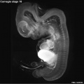
Week 7, 44 - 48 days, 13 - 17 mm
Gestational Age - week 9
Events
Ectoderm: sensory placodes, lens pit, otocyst,nasal pits moved ventrally, fourth ventricle of brain
Mesoderm: heart prominence
Head: 1st, 2nd and 3rd pharyngeal arch, forebrain, eye, auricular hillocks
Body: heart, liver, umbilical cord
Limb: upper and lower limb buds, foot plate, wrist, hand plate with digital rays
Features
Development indices: number of semicircular ducts (1-3) and length of the paramesonephric duct.
Identify: pigmented eye, eyelid, nasolacrimal groove, external acoustic meatus, heart, digital rays, liver prominance, thigh, ankle, foot plate, umbilical cord
- Links: Week 7 | System Development | Lecture - Limb | Lecture - Head Development | Lecture - Sensory | Science Practical - Head | Science Practical - Sensory | Science Practical - Urogenital | Category:Carnegie Stage 18 | Stage 19
- Carnegie Stages: 1 | 2 | 3 | 4 | 5 | 6 | 7 | 8 | 9 | 10 | 11 | 12 | 13 | 14 | 15 | 16 | 17 | 18 | 19 | 20 | 21 | 22 | 23 | About Stages | Timeline
Bright Field

|

|
| Embryo in gestational sac | Embryo open sac |

|

|
| Embryo with placentation (ectopic) | Embryo in amniotic sac |
- Stage 18 Links: Embryo in gestational sac | Embryo open sac | Embryo 2 and gestational sac | Embryo 2 | Carnegie stage 18
Scanning EM

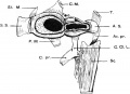
Ventral view of head showing upper lip, maxilla and nasal region.
Image Source: Prof Virginia Diewert
Kyoto Collection

View: This is a dorsolateral view of embryo. Amniotic membrane removed.
Image source: Embryology page Created: 19.03.1999

Ventral view of head region (1 mm scale).
Image source: The Kyoto Collection images are reproduced with the permission of Prof. Kohei Shiota and Prof. Shigehito Yamada, Anatomy and Developmental Biology, Kyoto University Graduate School of Medicine, Kyoto, Japan for educational purposes only and cannot be reproduced electronically or in writing without permission.
Carnegie Collection


| iBook - Carnegie Embryos | |
|---|---|

|
|
Additional Images
Stage 18 Optical Projection Tomography

External ear Stages 14-23 and adult
Human embryonic shoulder girdle

17 mm Embryo


- Carnegie Stages: 1 | 2 | 3 | 4 | 5 | 6 | 7 | 8 | 9 | 10 | 11 | 12 | 13 | 14 | 15 | 16 | 17 | 18 | 19 | 20 | 21 | 22 | 23 | About Stages | Timeline
Cite this page: Hill, M.A. (2024, June 14) Embryology Carnegie stage 18. Retrieved from https://embryology.med.unsw.edu.au/embryology/index.php/Carnegie_stage_18
- © Dr Mark Hill 2024, UNSW Embryology ISBN: 978 0 7334 2609 4 - UNSW CRICOS Provider Code No. 00098G