User:Z5117343
| Embryology - 26 Apr 2024 |
|---|
| Google Translate - select your language from the list shown below (this will open a new external page) |
|
العربية | català | 中文 | 中國傳統的 | français | Deutsche | עִברִית | हिंदी | bahasa Indonesia | italiano | 日本語 | 한국어 | မြန်မာ | Pilipino | Polskie | português | ਪੰਜਾਬੀ ਦੇ | Română | русский | Español | Swahili | Svensk | ไทย | Türkçe | اردو | ייִדיש | Tiếng Việt These external translations are automated and may not be accurate. (More? About Translations) |
Z5117343 16:47, 10 August 2017 (AEST)
Peer Reviews
Group 1: CEREBRAL CORTEX
The chosen headings for the development of the cerebral cortex were very suitable to highlight the key topics in providing a page of summarised information. It was then easy to navigate through the page using the shortcuts and finding information. Although, there was one sub sub heading “Timeline of Corticogenesis” that was formatted to be in bold while the rest were not.
The disorders listed seems to be really interesting and it covers the whole spectrum of the case abnormalities. But I suggest to get rid of the letter bullets (e.g. A), B), C) ) for the breakdown of the abnormalities.
The introduction had a quick and concise text, however, an image of the cerebellum would be suitable in this section on the side. While the sub sub heading stated that the introduction section will talk about the features of a cerebellum, a paragraph about the development and its stages were written down in this section as well. This could be moved into the ‘Early Development of the Brain’ subheading underneath. Bullet points of the brain layers as well as a diagram would be helpful for the visualisation of the brain.
For the sections that explain the development in specific weeks, a table would be advisable to make it neater and easier to look at. Also, an image was left inside the table grids and it was confusing whether it was meant to be there or not. Perhaps adding a photo gallery showing the stages at the bottom of the table would be better.
Hand drawn diagrams were really precise, neat and was very visually appealing. It was taking up all the space and unless it is intentional, I suggest to resize the drawing into a smaller one that fits the page as well as the accompanying text and content of the drawing.
The variety of visual aids were really entertaining and were referenced properly.
Finally, the reference list at the bottom of the page did not have a consistent format. It was mostly APA format however the others looked like a different format.
Group 2: KIDNEY
This page is really impressive for its organisation and balanced ratio of texts to images. There is a nice structure and flow in each different sections, this caught my attention and I read through most of the sections without any problems. All of the images were also labelled appropriately, the key words were formatted in bold and certain definitions were stated. These all helped in keeping the page really interesting and organised. A list of abnormalities and its causes were also stated in a very neat and informative matter with bullet points and images. It was nice to see that the research question was relevant and thought provoking.
Some paragraphs were not referenced especially the first paragraphs in each section. In-text citations should be changed into superscripts in some sections. This page contained really visually appealing images however, some images were not referenced and/or it didn't state the copyright message that states it can be reused with no issues. Some of the headings (e.g. 'Stages in nephron formation' and 'Common congenital kidney defects') were in an italics format, this could be changed into another sub-sub heading or maybe increase its font size. Blood supply section should be reviewed, summarised and referenced appropriately.
Information about the kidney development were mostly sourced from reputable journals articles that was published quite recently. However, the reference list section should be reviewed to keep the referencing format consistent. At the moment, it has APA format and some have different format I am not familiar with.
Group 3: HEART
The headings were all neat, concise and impressive. It successfully highlighted and sectioned the key topics in the development of the heart. The addition of the technical signalling pathways and the details of the development were well summarised with appropriate references in superscript format. There was a nice variety of visual resources, both hand drawn and externally sourced. Most images have their copyright approval and reference included perfectly, except "Figure 1 Morphological defects in CTCF mutant embryonic hearts" and "Figure 2 - defects of mitochondria in CTCF mutant hearts". There was a nice flow throughout the page through the use of effective paragraph sectioning. The table for the glossary of terms was really useful and neat.
Some of the images didn't have a box around it and these figures were not labelled, this should be easily changed in the edit mode. Some of the hand drawn images were somewhat unclear, due to the writing as well as the rough outline of the heart. Signatures should also be removed. The references were also retained in the bottom of the sections. It was a confusing because it wasn't next to any paragraphs that needed to be referenced. A reference was also repeated in this section.
For example: "This image is based upon Robert H Anderson, Sandra Webb, Nigel A Brown, Wouter Lamers, Antoon Moorman Development of the heart: (3) formation of the ventricular outflow tracts, arterial valves, and intrapericardial arterial trunks. Heart: 2003, 89(9);1110-8 PubMed 12923046
Robert H Anderson, Sandra Webb, Nigel A Brown, Wouter Lamers, Antoon Moorman Development of the heart: (3) formation of the ventricular outflow tracts, arterial valves, and intrapericardial arterial trunks. Heart: 2003, 89(9);1110-8 PubMed 12923046
Marc Sylva, Maurice J B van den Hoff, Antoon F M Moorman Development of the human heart. Am. J. Med. Genet. A: 2014, 164A(6);1347-71 PubMed 23633400"
Finally, there is a great variety of reputable sources of information. The only thing that needs changing is that the reference list should be revised. Some were left as a link and the list were inconsistent with its reference format.
Group 5: LUNG
This page is really impressive when the hand drawn images caught my eye as well as the balanced text-to-images ratio. It is well organised and there was a decent flow throughout the page. It is useful that keywords were formatted to be in bold formatting to draw the attention of the readers to the main terms. The development timeline is very fascinating, it had a description as well as images. Summaries are well-informative as well as brief in some sections. Some images were reference properly and copyright approval was provided. Abnormal development was neatly organised into sections and appropriate journal articles for evidence. However, there are a few abnormalities that did not feature an image to provide more visual aid to the readers.
The 'Alveolus' was left in bold format while the rest were in normal format, this could be easily changed in the edit page. The hand drawn images did not provide a reference where it was based off. Also, one of the images has a very low resolution ("This image is a stylised typical developmental branching pattern over time in a lung bud."). The images should be encased in boxes and a label underneath would be neater. Laboratory results from the animal models would be useful to see. The lung histology section didn't provide any references. The movies section disrupts the flow of the sections, it might be best to place them at the bottom of the page.
This page seems like it is almost complete.
Group 6: CEREBELLUM
It was really good that the structure and function of the cerebellum was explained in a succinct way in the beginning. The introduction repeated the word 'hence' a few times, maybe it's better to modify it into bullet points, in a similar way when lecturers provide a slide on the lecture overview. Appropriate images were added as well as figure labeling. Copyright approval was also provided for the images and were referenced appropriately. The use of tables was also appropriate in some of the topic sections. Images were also in appropriate sizes that avoided covering the while page. The page was very detailed as well. Some sections like "Cell Signaling" was a bit lengthy, images would be nice. It was good that reputable journal articles were used for the project, proper in text citations superscripts were also done properly. However, revise the reference list because some were left as links and the list did not have a consistent reference format. But overall, the page looks almost complete.
Search Databases
Embryo Search
Embryo
Notochord Search
Notochord
Salmon Reference
<pubmed>28786202</pubmed>
Referenced Images
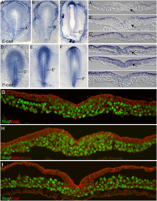
 Chicken embryo E-cadherin and P-cadherin in gastrulation[1]
Links
Student Page
2017 Group Project 4
References
| 2017 Project Groups | |||||
|---|---|---|---|---|---|
| Group 1 | Group 2 | Group 3 | Group 4 | Group 5 | Group 6 |
| Mark Hill - Lab 1 page | |||||