Sheep Development
| Embryology - 27 Apr 2024 |
|---|
| Google Translate - select your language from the list shown below (this will open a new external page) |
|
العربية | català | 中文 | 中國傳統的 | français | Deutsche | עִברִית | हिंदी | bahasa Indonesia | italiano | 日本語 | 한국어 | မြန်မာ | Pilipino | Polskie | português | ਪੰਜਾਬੀ ਦੇ | Română | русский | Español | Swahili | Svensk | ไทย | Türkçe | اردو | ייִדיש | Tiếng Việt These external translations are automated and may not be accurate. (More? About Translations) |
Introduction
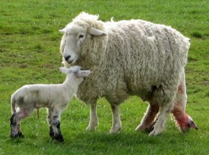
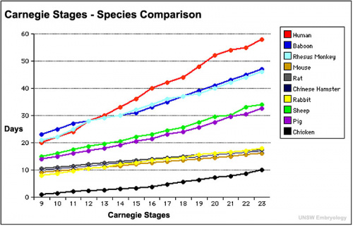
The domestic sheep (Ovis aries) has been used as a mammalian model of development with a term gestational period of 145 - 150 days.
Category:Sheep
Historic Embryology: 1940 respiratory muscles nerve-endings
| Animal Development Time | ||||||||||||||||||||||||||||||||||||||||||||||||||||||||||||||||||||||||||||||||||||||||||||||||||||||||||||||||||||||||||||||||||||||||||||||
|---|---|---|---|---|---|---|---|---|---|---|---|---|---|---|---|---|---|---|---|---|---|---|---|---|---|---|---|---|---|---|---|---|---|---|---|---|---|---|---|---|---|---|---|---|---|---|---|---|---|---|---|---|---|---|---|---|---|---|---|---|---|---|---|---|---|---|---|---|---|---|---|---|---|---|---|---|---|---|---|---|---|---|---|---|---|---|---|---|---|---|---|---|---|---|---|---|---|---|---|---|---|---|---|---|---|---|---|---|---|---|---|---|---|---|---|---|---|---|---|---|---|---|---|---|---|---|---|---|---|---|---|---|---|---|---|---|---|---|---|---|---|---|
| ||||||||||||||||||||||||||||||||||||||||||||||||||||||||||||||||||||||||||||||||||||||||||||||||||||||||||||||||||||||||||||||||||||||||||||||
Animal Notes and Table Data Sources
|
Some Recent Findings
|
| More recent papers |
|---|
|
This table allows an automated computer search of the external PubMed database using the listed "Search term" text link.
More? References | Discussion Page | Journal Searches | 2019 References | 2020 References Search term: Sheep Development | Sheep Embryology | Ovine Development |
| Older papers |
|---|
| These papers originally appeared in the Some Recent Findings table, but as that list grew in length have now been shuffled down to this collapsible table.
See also the Discussion Page for other references listed by year and References on this current page.
|
Development Overview
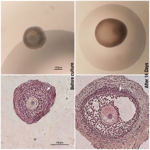
| Day | Event |
|---|---|
| 4 | embryo enters the uterus at the morula stage |
| 6 | blastocyst stage |
| 8 | blastocyst hatches from zona pellucida |
| 11-16 | elongates to a filamentous form |
| 14 - 16 | binucleate cells begin to differentiate in the trophoblast |
| 16 | adplantation |
See also Implantation mechanisms: insights from the sheep[5]
Dolly
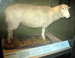
A female domestic sheep remarkable in being the first mammal to be cloned from an adult somatic cell, using the process of nuclear transfer.[6]
Cloned by Ian Wilmut, Keith Campbell and colleagues at the Roslin Institute near Edinburgh in Scotland, born on 5 July 1996 and she lived until the age of six (5 July 1996 – 14 February 2003). The cell used as the donor for the cloning of Dolly was taken from a mammary gland, and the production of a healthy clone therefore proved that a cell taken from a specific part of the body could recreate a whole individual. As Dolly was cloned from part of a mammary gland, she was named after the famously curvaceous country western singer Dolly Parton.
Oocyte Development
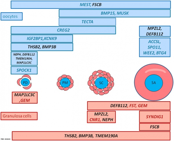
| Sheep follicle gene expression[7] |
|
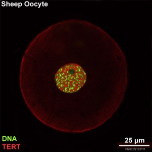
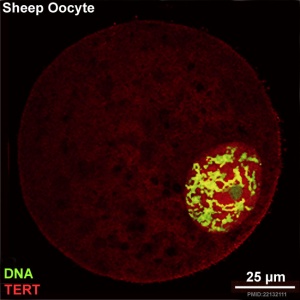
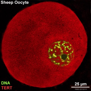
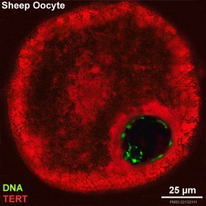
Sheep Oocyte Distribution of Telomerase reverse transcriptase
The following oocyte images are from a recent study of sheep in vitro follicle development.[4]
|
|
| preantral | early antral |
|
|
| early antral | preovulatory follicle |
- TERT - Red (Cy3-conjugated secondary antibody) (telomerase reverse transcriptase, TERT)
- DNA - Green (SYBR Green 14/I)
- Sheep Oocyte TERT: preantral | early antral | early antral | preovulatory follicle | Oocyte Development | Sheep Development
Respiratory
Phases of fetal lung development:[8]
- embryonic - 0 to 40 days.
- pseudoglandular - 40 to 80 days.
- canalicular - 80 to 120 days.
- saccular - 120 to term 148 days gestation.
- Links: Respiratory Development
Immune
Lymphocytes development has been characterised by an immunohistology study of T lymphocytes in the sheep fetal spleen.[9]
| Day (days of gestation) | Event |
|---|---|
| 43 - 44 | SBU-T1- and SBU-T8-positive lymphocytes were present in low numbers. |
| 45 - 50 | Surface immunoglobulin (sIg) was first detected on fetal spleen cells |
| 50 - 55 | SBU-T4, 20.96-, 25.69-, 38.38-, or 46.66-positive lymphocytes present. |
| 57 | SBU-T19 lymphocytes appeared. |
- Links: Immune Development
References
- ↑ Sinclair KD, Corr SA, Gutierrez CG, Fisher PA, Lee JH, Rathbone AJ, Choi I, Campbell KH & Gardner DS. (2016). Healthy ageing of cloned sheep. Nat Commun , 7, 12359. PMID: 27459299 DOI.
- ↑ Arunakumari G, Shanmugasundaram N & Rao VH. (2010). Development of morulae from the oocytes of cultured sheep preantral follicles. Theriogenology , 74, 884-94. PMID: 20615540 DOI.
- ↑ Black SG, Arnaud F, Burghardt RC, Satterfield MC, Fleming JA, Long CR, Hanna C, Murphy L, Biek R, Palmarini M & Spencer TE. (2010). Viral particles of endogenous betaretroviruses are released in the sheep uterus and infect the conceptus trophectoderm in a transspecies embryo transfer model. J. Virol. , 84, 9078-85. PMID: 20610723 DOI.
- ↑ 4.0 4.1 Barboni B, Russo V, Cecconi S, Curini V, Colosimo A, Garofalo ML, Capacchietti G, Di Giacinto O & Mattioli M. (2011). In vitro grown sheep preantral follicles yield oocytes with normal nuclear-epigenetic maturation. PLoS ONE , 6, e27550. PMID: 22132111 DOI.
- ↑ Spencer TE, Johnson GA, Bazer FW & Burghardt RC. (2004). Implantation mechanisms: insights from the sheep. Reproduction , 128, 657-68. PMID: 15579583 DOI.
- ↑ Wilmut I, Schnieke AE, McWhir J, Kind AJ & Campbell KH. (1997). Viable offspring derived from fetal and adult mammalian cells. Nature , 385, 810-3. PMID: 9039911 DOI.
- ↑ Bonnet A, Servin B, Mulsant P & Mandon-Pepin B. (2015). Spatio-Temporal Gene Expression Profiling during In Vivo Early Ovarian Folliculogenesis: Integrated Transcriptomic Study and Molecular Signature of Early Follicular Growth. PLoS ONE , 10, e0141482. PMID: 26540452 DOI.
- ↑ Gnanalingham MG, Mostyn A, Dandrea J, Yakubu DP, Symonds ME & Stephenson T. (2005). Ontogeny and nutritional programming of uncoupling protein-2 and glucocorticoid receptor mRNA in the ovine lung. J. Physiol. (Lond.) , 565, 159-69. PMID: 15774522 DOI.
- ↑ Maddox JF, Mackay CR & Brandon MR. (1987). Ontogeny of ovine lymphocytes. II. An immunohistological study on the development of T lymphocytes in the sheep fetal spleen. Immunology , 62, 107-12. PMID: 3308689
Reviews
Thompson RP, Nilsson E & Skinner MK. (2020). Environmental epigenetics and epigenetic inheritance in domestic farm animals. Anim. Reprod. Sci. , , 106316. PMID: 32094003 DOI.
Gebreselassie G, Berihulay H, Jiang L & Ma Y. (2019). Review on Genomic Regions and Candidate Genes Associated with Economically Important Production and Reproduction Traits in Sheep (Ovies aries). Animals (Basel) , 10, . PMID: 31877963 DOI.
Lazzari G, Colleoni S, Lagutina I, Crotti G, Turini P, Tessaro I, Brunetti D, Duchi R & Galli C. (2010). Short-term and long-term effects of embryo culture in the surrogate sheep oviduct versus in vitro culture for different domestic species. Theriogenology , 73, 748-57. PMID: 19726075 DOI.
Campbell KH, Alberio R, Choi I, Fisher P, Kelly RD, Lee JH & Maalouf W. (2005). Cloning: eight years after Dolly. Reprod. Domest. Anim. , 40, 256-68. PMID: 16008756 DOI.
Articles
Wang J, Guillomot M & Hue I. (2009). Cellular organization of the trophoblastic epithelium in elongating conceptuses of ruminants. C. R. Biol. , 332, 986-97. PMID: 19909921 DOI.
Guillomot M, Turbe A, Hue I & Renard JP. (2004). Staging of ovine embryos and expression of the T-box genes Brachyury and Eomesodermin around gastrulation. Reproduction , 127, 491-501. PMID: 15047940 DOI.
Search PubMed
Search Pubmed: sheep development | ovine development | ovine embryo development
External Links
External Links Notice - The dynamic nature of the internet may mean that some of these listed links may no longer function. If the link no longer works search the web with the link text or name. Links to any external commercial sites are provided for information purposes only and should never be considered an endorsement. UNSW Embryology is provided as an educational resource with no clinical information or commercial affiliation.
| Animal Development: axolotl | bat | cat | chicken | cow | dog | dolphin | echidna | fly | frog | goat | grasshopper | guinea pig | hamster | horse | kangaroo | koala | lizard | medaka | mouse | opossum | pig | platypus | rabbit | rat | salamander | sea squirt | sea urchin | sheep | worm | zebrafish | life cycles | development timetable | development models | K12 |
Glossary Links
- Glossary: A | B | C | D | E | F | G | H | I | J | K | L | M | N | O | P | Q | R | S | T | U | V | W | X | Y | Z | Numbers | Symbols | Term Link
Cite this page: Hill, M.A. (2024, April 27) Embryology Sheep Development. Retrieved from https://embryology.med.unsw.edu.au/embryology/index.php/Sheep_Development
- © Dr Mark Hill 2024, UNSW Embryology ISBN: 978 0 7334 2609 4 - UNSW CRICOS Provider Code No. 00098G
